 10.1007/s40204-019-00121-3
A novel polycaprolactone/carbon nanofiber composite as a conductive neural guidance channel: an in vitro and in vivo study
- Saeed Farzamfar
 1
1 - Majid Salehi2
- Seyed Mohammad Tavangar3
- Javad Verdi1
- Korosh Mansouri4
- Arman Ai5
- Ziba Veisi Malekshahi1
- Jafar Ai*1
- Department of Tissue Engineering and Applied Cell Sciences, School of Advanced Technologies in Medicine, Tehran University of Medical Sciences, Tehran, IR
- Department of Tissue Engineering, School of Medicine, Shahroud University of Medical Sciences, Shahroud, IR Tissue Engineering and Stem Cells Research Center, Shahroud University of Medical Sciences, Shahroud, IR
- Department of Pathology, Shariati Hospital, Tehran University of Medical Sciences, Tehran, IR
- Neuromusculoskletal Research Centre Firozgar Hospital, Iran University of Medical Sciences, Tehran, IR
- School of Medicine, Tehran University of Medical Sciences, Tehran, 141556447, IR

Published in Issue 2019-12-12
How to Cite
Farzamfar, S., Salehi, M., Tavangar, S. M., Verdi, J., Mansouri, K., Ai, A., Malekshahi, Z. V., & Ai, J. (2019). A novel polycaprolactone/carbon nanofiber composite as a conductive neural guidance channel: an in vitro and in vivo study. Progress in Biomaterials, 8(4 (December 2019). https://doi.org/10.1007/s40204-019-00121-3


Share:


Abstract
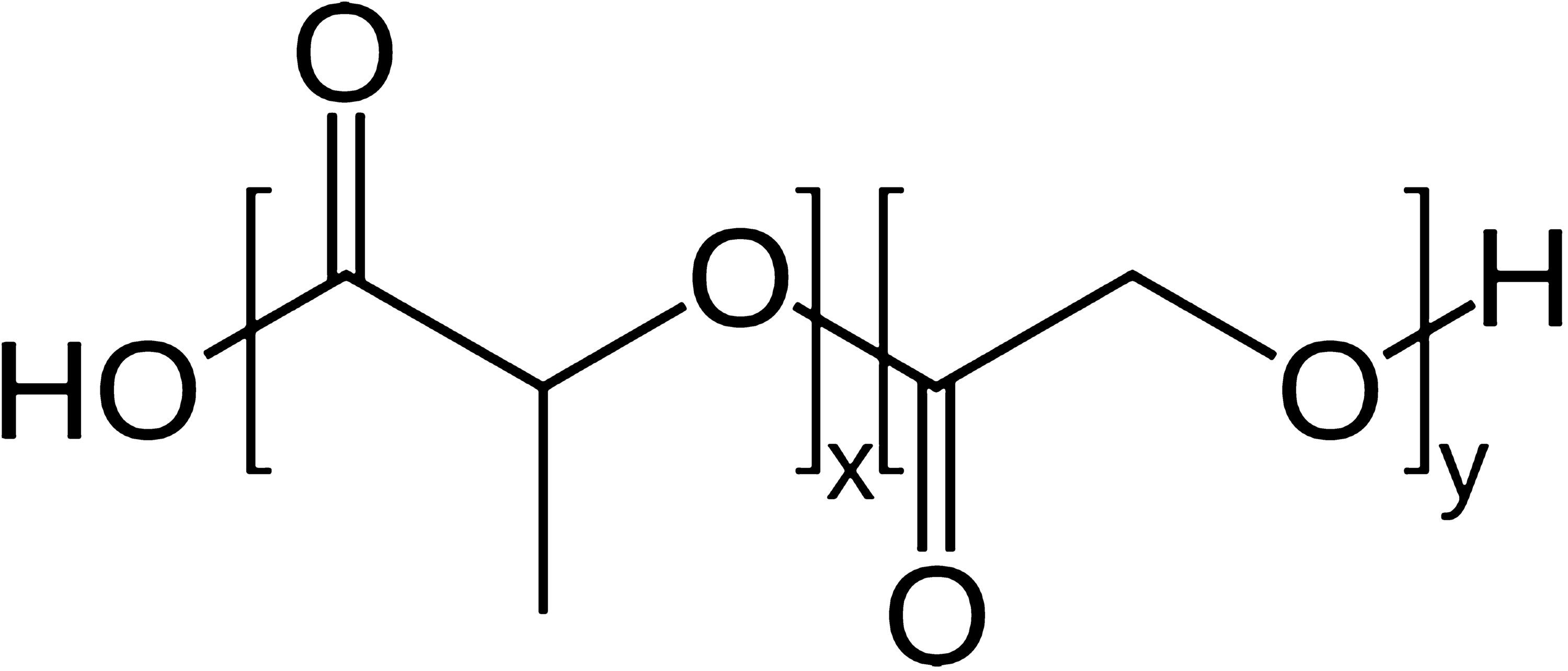
Abstract The current study aimed to investigate the potential of carbon nanofibers to promote peripheral nerve regeneration. The carbon nanofiber-imbedded scaffolds were produced from polycaprolactone and carbon nanofibers using thermally induced phase separation method. Electrospinning technique was utilized to fabricate polycaprolactone/collagen nanofibrous sheets. The incorporation of carbon nanofibers into polycaprolactone’s matrix significantly reduced its electrical resistance from 4.3 × 10 9 ± 0.34 × 10 9 Ω to 8.7 × 10 4 ± 1.2 × 10 4 Ω. Further in vitro studies showed that polycaprolactone/carbon nanofiber scaffolds had the porosity of 82.9 ± 3.7% and degradation rate of 1.84 ± 0.37% after 30 days and 3.58 ± 0.39% after 60 days. The fabricated scaffolds were favorable for PC-12 cells attachment and proliferation. Neural guidance channels were produced from the polycaprolactone/carbon nanofiber composites using water jet cutter machine then incorporated with PCL/collagen nanofibrous sheets. The composites were implanted into severed rat sciatic nerve. After 12 weeks, the results of histopathological examinations and functional analysis proved that conductive conduit out-performed the non-conductive type and induced no toxicity or immunogenic reactions, suggesting its potential applicability to treat peripheral nerve damage in the clinic.Keywords
- Carbon nanofiber,
- Polycaprolactone,
- Electrical conductivity,
- Sciatic nerve regeneration
References
- Anderson et al. (2015) Peripheral nerve regeneration strategies: electrically stimulating polymer based nerve growth conduits https://doi.org/10.1615/CritRevBiomedEng.2015014015
- Cao et al. (2009) The application of nanofibrous scaffolds in neural tissue engineering (pp. 1055-1064) https://doi.org/10.1016/j.addr.2009.07.009
- Daly et al. (2011) A biomaterials approach to peripheral nerve regeneration: bridging the peripheral nerve gap and enhancing functional recovery (pp. 202-221) https://doi.org/10.1098/rsif.2011.0438
- Das et al. (2015) In vivo studies of silk based gold nano-composite conduits for functional peripheral nerve regeneration (pp. 66-75) https://doi.org/10.1016/j.biomaterials.2015.04.047
- de Ruiter et al. (2009) Designing ideal conduits for peripheral nerve repair https://doi.org/10.3171/FOC.2009.26.2.E5
- Dong Z (2015) Electrospinning and characterization of composite membranes for biomedical applications. Ph.D. Thesis, University of Rochester
- Dong (2015) A dual role of graphene oxide sheet deposition on titanate nanowire scaffolds for osteo-implantation: mechanical hardener and surface activity regulator https://doi.org/10.1038/srep18266
- Evans (2001) Peripheral nerve injury: a review and approach to tissue engineered constructs (pp. 396-404) https://doi.org/10.1002/ar.1120
- Faroni et al. (2015) Peripheral nerve regeneration: experimental strategies and future perspectives (pp. 160-167) https://doi.org/10.1016/j.addr.2014.11.010
- Farzamfar (2018) Neural tissue regeneration by a gabapentin-loaded cellulose acetate/gelatin wet-electrospun scaffold (pp. 1229-1238) https://doi.org/10.1007/s10570-017-1632-z
- Ghasemi-Mobarakeh (2011) Application of conductive polymers, scaffolds and electrical stimulation for nerve tissue engineering (pp. e17-e35) https://doi.org/10.1002/term.383
- Glowacki and Mizuno (2008) Collagen scaffolds for tissue engineering (pp. 338-344)
- Gu et al. (2014) Neural tissue engineering options for peripheral nerve regeneration (pp. 6143-6156) https://doi.org/10.1016/j.biomaterials.2014.04.064
- Habre et al. (2018) The surgical management of nerve gaps: present and future (pp. 252-261) https://doi.org/10.1097/SAP.0000000000001252
- Hu et al. (1997) Neurologic evaluation of infant and adult rats before and after sciatic nerve blockade (pp. 957-965)
- Keane and Badylak (2014) Biomaterials for tissue engineering applications (pp. 112-118) https://doi.org/10.1053/j.sempedsurg.2014.06.010
- Ku et al. (2013) Carbon-based nanomaterials for tissue engineering (pp. 244-260) https://doi.org/10.1002/adhm.201200307
- Lannutti et al. (2007) Electrospinning for tissue engineering scaffolds (pp. 504-509) https://doi.org/10.1016/j.msec.2006.05.019
- Lanza et al. (2011) Academic Press
- Lee et al. (2012) End-to-side neurorrhaphy using an electrospun PCL/collagen nerve conduit for complex peripheral motor nerve regeneration (pp. 9027-9036) https://doi.org/10.1016/j.biomaterials.2012.09.008
- Lee et al. (2013) Functional evaluation in the rat sciatic nerve defect model: a comparison of the sciatic functional index, ankle angles, and isometric tetanic force (pp. 1173-1180) https://doi.org/10.1097/PRS.0b013e3182a3bfeb
- Mirzaei et al. (2016) The differentiation of human endometrial stem cells into neuron-like cells on electrospun PAN-derived carbon nanofibers with random and aligned topographies (pp. 4798-4808) https://doi.org/10.1007/s12035-015-9410-0
- Muheremu and Ao (2015) Past, present, and future of nerve conduits in the treatment of peripheral nerve injury https://doi.org/10.1155/2015/237507
- Naseri-Nosar et al. (2017) Cerium oxide nanoparticle-containing poly (ε-caprolactone)/gelatin electrospun film as a potential wound dressing material: in vitro and in vivo evaluation (pp. 366-372) https://doi.org/10.1016/j.msec.2017.08.013
- Nectow et al. (2011) Biomaterials for the development of peripheral nerve guidance conduits (pp. 40-50) https://doi.org/10.1089/ten.teb.2011.0240
- Nisbet et al. (2009) A review of the cellular response on electrospun nanofibers for tissue engineering (pp. 7-29) https://doi.org/10.1177/0885328208099086
- Prabhakaran et al. (2008) Surface modified electrospun nanofibrous scaffolds for nerve tissue engineering https://doi.org/10.1088/0957-4484/19/45/455102
- Salehi et al. (2015) Preparation of pure PLLA, pure chitosan, and PLLA/chitosan blend porous tissue engineering scaffolds by thermally induced phase separation method and evaluation of the corresponding mechanical and biological properties (pp. 675-682) https://doi.org/10.1080/00914037.2014.1002093
- Salehi et al. (2018) Sciatic nerve regeneration by transplantation of Schwann cells via erythropoietin controlled-releasing polylactic acid/multiwalled carbon nanotubes/gelatin nanofibrils neural guidance conduit (pp. 1463-1476) https://doi.org/10.1002/jbm.b.33952
- Sarker et al. (2018) Strategic design and fabrication of nerve guidance conduits for peripheral nerve regeneration 13(7) https://doi.org/10.1002/biot.201700635
- Schnell et al. (2007) Guidance of glial cell migration and axonal growth on electrospun nanofibers of poly-ε-caprolactone and a collagen/poly-ε-caprolactone blend (pp. 3012-3025) https://doi.org/10.1016/j.biomaterials.2007.03.009
- Siemionow and Brzezicki (2009) Current techniques and concepts in peripheral nerve repair (pp. 141-172) https://doi.org/10.1016/S0074-7742(09)87008-6
- Smith et al. (2015) Bone tissue engineering challenges in oral & maxillofacial surgery (pp. 57-78) Springer https://doi.org/10.1007/978-3-319-22345-2_4
- Stout et al. (2011) Poly (lactic-co-glycolic acid): carbon nanofiber composites for myocardial tissue engineering applications (pp. 3101-3112) https://doi.org/10.1016/j.actbio.2011.04.028
- Wang et al. (2008) Creation of highly aligned electrospun poly-l-lactic acid fibers for nerve regeneration applications https://doi.org/10.1088/1741-2560/6/1/016001
- Yang et al. (2005) Electrospinning of nano/micro scale poly (L-lactic acid) aligned fibers and their potential in neural tissue engineering (pp. 2603-2610) https://doi.org/10.1016/j.biomaterials.2004.06.051
- Yu (2011) Sciatic nerve regeneration in rats by a promising electrospun collagen/poly (ε-caprolactone) nerve conduit with tailored degradation rate https://doi.org/10.1186/1471-2202-12-68
- Zheng (2012) Endothelialization and patency of RGD-functionalized vascular grafts in a rabbit carotid artery model (pp. 2880-2891) https://doi.org/10.1016/j.biomaterials.2011.12.047