 10.1007/s40204-019-0111-z
Cold atmospheric plasma as a promising approach for gelatin immobilization on poly(ε-caprolactone) electrospun scaffolds
- Marziyeh Meghdadi
 1
1 - Seyed-Mohammad Atyabi2
- Mohamad Pezeshki-Modaress*3
- Shiva Irani1
- Zahra Noormohammadi1
- Mojgan Zandi4
- Department of Biology, School of Basic Sciences, Sciences and Research Branch, Islamic Azad University, Tehran, IR
- Department of Pilot Nanobiotechnology, Pasteur Institute of Iran, Tehran, IR
- Burn Research Center, Iran University of Medical Sciences, Tehran, IR
- Department of Biomaterial, Iran Polymer and Petrochemical Institute, Tehran, IR

Published in Issue 2019-03-27
How to Cite
Meghdadi, M., Atyabi, S.-M., Pezeshki-Modaress, M., Irani, S., Noormohammadi, Z., & Zandi, M. (2019). Cold atmospheric plasma as a promising approach for gelatin immobilization on poly(ε-caprolactone) electrospun scaffolds. Progress in Biomaterials, 8(2 (June 2019). https://doi.org/10.1007/s40204-019-0111-z


Share:


Abstract
Abstract
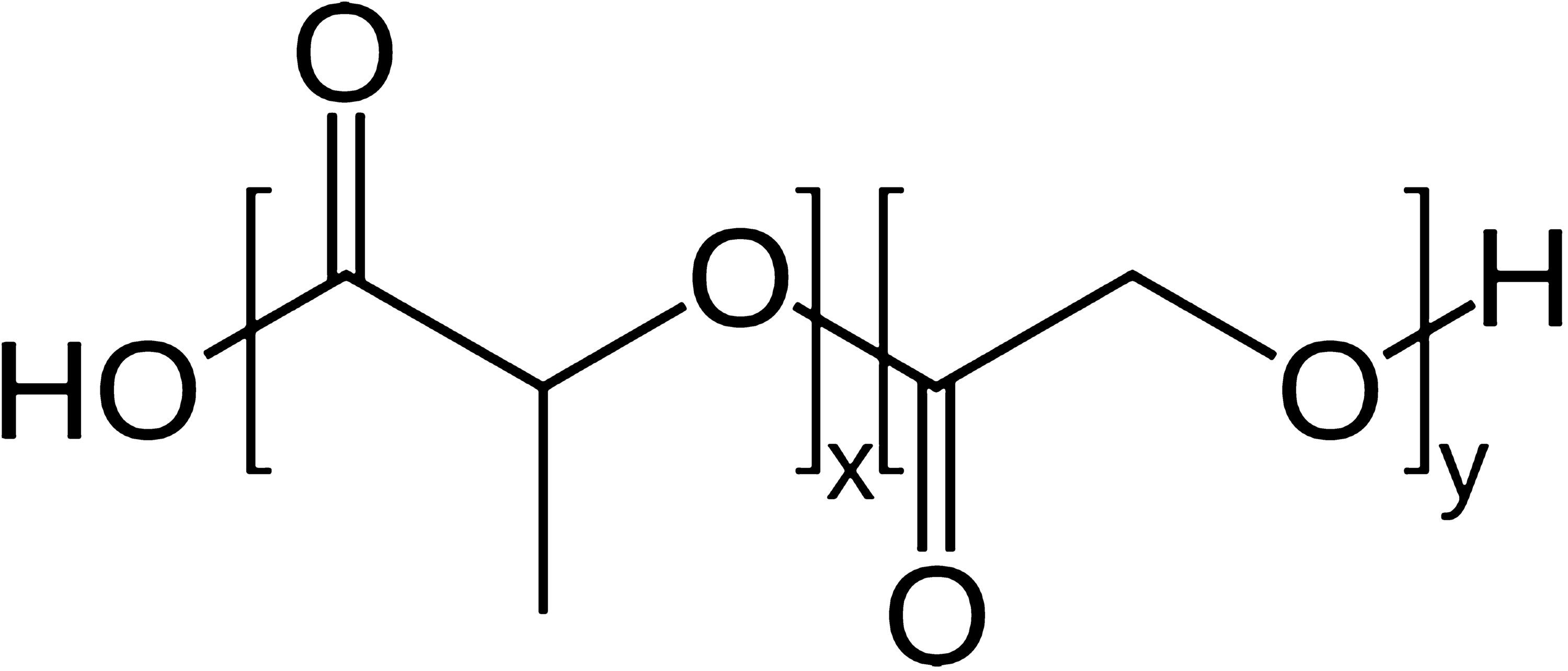
Poly(Ɛ-caprolactone) (PCL) is a biocompatible polymer with a high potential to be used in tissue engineering especially in tight tissues. In the current study, cold atmospheric plasma (CAP) is used as a promising method for immobilization of gelatin as a functional biomacromolecule on PCL nanofibrous substrates. The CAP surface modification leads to oxidation of chemical groups existing on the PCL surface without doing any damage to the bulk properties of biomaterials for gelatin biomacromolecule grafting. The water contact angle (WCA) of the CAP-treated surface and gelatin-grafted PCL using CAP indicates an effective increment in the hydrophilicity of the PCL surface. Also to achieve the highest levels of gelatin grafting on the PCL surface, two different grafting methods and gelatin concentration diversity are utilized in the grafting process. The immobilization of gelatin biomacromolecules onto the CAP surface-modified PCL nanofibers is investigated using scanning electron microscope (SEM) and Fourier transform infrared spectroscopy (FTIR). The gelatin-modified PCL substrates revealed uniform nanofibrous morphology with increased average fiber diameter. The results of FTIR spectra, including hydroxyl groups, NH groups, and amide II of gelatin-grafting peaks, confirm the gelatin immobilization on the surface of nanofibers. The metabolic activity of cultured mesenchymal stem cells (MSCs) on the surface-modified scaffolds is evaluated using MTT analysis (
P ≤
0.05). The results of metabolic activity and also SEM and DAPI staining observations indicate proper attachment on the surface and viability for MSCs on the surface-immobilized nanofibrous scaffolds. Therefore, CAP treatment would be an effective method for biomacromolecule immobilization on nanofibers towards the enhancement of cell behavior.
References
- Atyabi et al. (2016) Cell attachment and viability study of PCL nano-fiber modified by cold atmospheric plasma (pp. 181-190) https://doi.org/10.1007/s12013-015-0718-1
- Bárdos and Baránková (2010) Cold atmospheric plasma: sources, processes, and applications (pp. 6705-6713) https://doi.org/10.1016/j.tsf.2010.07.044
- Beachley and Wen (2010) Polymer nanofibrous structures: fabrication, biofunctionalization, and cell interactions (pp. 868-892) https://doi.org/10.1016/j.progpolymsci.2010.03.003
- Chong et al. (2007) Evaluation of electrospun PCL/gelatin nanofibrous scaffold for wound healing and layered dermal reconstitution (pp. 321-330) https://doi.org/10.1016/j.actbio.2007.01.002
- Cipitria et al. (2011) Design, fabrication and characterization of PCL electrospun scaffolds—a review (pp. 9419-9453) https://doi.org/10.1039/c0jm04502k
- Cools P, Vrekhem Sv, Ghobeira R, Geyterand Nv, Morent R (2016) Non-thermal plasma technology for the improvement of scaffolds for tissue engineering and regenerative medicine: a review. In: Mieno T (ed) Plasma science and technology: progress in physical states and chemical reactions. IntechOPen, pp 173–212
- Courtney et al. (2006) Design and analysis of tissue engineering scaffolds that mimic soft tissue mechanical anisotropy (pp. 3631-3638)
- Dabouian et al. (2018) β-Carotene: a natural osteogen to fabricate osteoinductive electrospun scaffolds (pp. 9941-9945) https://doi.org/10.1039/C7RA13237A
- De Geyter et al. (2008) Influence of ambient conditions on the ageing behaviour of plasma-treated PET surfaces (pp. 3086-3090) https://doi.org/10.1016/j.nimb.2008.03.167
- Dolci (2014) Carboxyl surface functionalization of poly (L-lactic acid) electrospun nanofibers through atmospheric non-thermal plasma affects fibroblast (pp. 203-213) https://doi.org/10.1002/ppap.201300104
- Gümüşderelioğlu and Türkoğlu (2002) Biomodification of non-woven polyester fabrics by insulin and RGD for use in serum-free cultivation of tissue cells (pp. 3927-3935) https://doi.org/10.1016/S0142-9612(02)00128-X
- Koo and Jang (2008) Surface modification of poly (lactic acid) by UV/Ozone irradiation (pp. 674-678) https://doi.org/10.1007/s12221-008-0106-1
- Krok-Borkowicz (2015) Biofunctionalization of poly (l-lactide-co-glycolide) by post-plasma grafting of 2-aminoethyl methacrylate and gelatin immobilization (pp. 344-347) https://doi.org/10.1016/j.matlet.2014.10.100
- Lannutti et al. (2007) Electrospinning for tissue engineering scaffolds (pp. 504-509) https://doi.org/10.1016/j.msec.2006.05.019
- Laroussi (2015) Low-temperature plasma jet for biomedical applications: a review (pp. 703-712) https://doi.org/10.1109/TPS.2015.2403307
- Li (2012) Surface modification of smooth poly (l-lactic acid) films for gelatin immobilization (pp. 687-693) https://doi.org/10.1021/am201795g
- Lu et al. (2014) Effect of non-thermal plasma modified NiAl layered double hydroxides on the removal of fluoride from aqueous solution https://doi.org/10.4236/ajac.2014.59062
- Ma et al. (2005) Cartilage tissue engineering PLLA scaffold with surface immobilized collagen and basic fibroblast growth factor (pp. 1253-1259) https://doi.org/10.1016/j.biomaterials.2004.04.031
- Ma et al. (2005) Grafting of gelatin on electrospun poly (caprolactone) nanofibers to improve endothelial cell spreading and proliferation and to control cell orientation (pp. 1149-1158) https://doi.org/10.1089/ten.2005.11.1149
- Ma et al. (2007) Surface modification and property analysis of biomedical polymers used for tissue engineering (pp. 137-157) https://doi.org/10.1016/j.colsurfb.2007.06.019
- Martins et al. (2009) Surface modification of electrospun polycaprolactone nanofiber meshes by plasma treatment to enhance biological performance (pp. 1195-1206)
- Morent et al. (2008) Comparison between XPS-and FTIR-analysis of plasma-treated polypropylene film surfaces (pp. 597-600) https://doi.org/10.1002/sia.2619
- Oyane et al. (2005) Simple surface modification of poly (ε-caprolactone) for apatite deposition from simulated body fluid (pp. 2407-2413) https://doi.org/10.1016/j.biomaterials.2004.07.048
- Pezeshki-Modaress et al. (2015) Gelatin–GAG electrospun nanofibrous scaffold for skin tissue engineering: fabrication and modeling of process parameters (pp. 704-712) https://doi.org/10.1016/j.msec.2014.12.023
- Pezeshki-Modaress et al. (2015) Fabrication of gelatin/chitosan nanofibrous scaffold: process optimization and empirical modeling (pp. 571-580) https://doi.org/10.1002/pi.4843
- Pezeshki-Modaress et al. (2017) Gelatin/chondroitin sulfate nanofibrous scaffolds for stimulation of wound healing: In-vitro and in-vivo study (pp. 2020-2034) https://doi.org/10.1002/jbm.a.35890
- Pham et al. (2006) Electrospinning of polymeric nanofibers for tissue engineering applications: a review (pp. 1197-1211) https://doi.org/10.1089/ten.2006.12.1197
- Qi et al. (2016) Bioactivity assessment of PLLA/PCL/HAP electrospun nanofibrous scaffolds for bone tissue engineering (pp. 139-144) https://doi.org/10.1016/j.lfs.2016.02.040
- Sadeghi et al. (2018) Electrospun polyvinyl alcohol/gelatin/chondroitin sulfate nanofibrous scaffold: Fabrication and in vitro evaluation (pp. 1248-1256) https://doi.org/10.1016/j.ijbiomac.2018.04.002
- Safaeijavan et al. (2014) Biological behavior study of gelatin coated PCL nanofiberous electrospun scaffolds using fibroblasts (pp. 2008-4987)
- Şaşmazel et al. (2009) Water/O2-plasma-assisted treatment of PCL membranes for biosignal immobilization (pp. 1137-1162) https://doi.org/10.1163/156856209X444475
- Schnell et al. (2007) Guidance of glial cell migration and axonal growth on electrospun nanofibers of poly-ε-caprolactone and a collagen/poly-ε-caprolactone blend (pp. 3012-3025) https://doi.org/10.1016/j.biomaterials.2007.03.009
- Shahmirani et al. (2014) Effect of cold atmospheric pressure plasma and gold nanoparticles on cell viability https://doi.org/10.9734/ARRB/2014/7419
- Shen et al. (2013) Skeletal muscle regeneration on protein-grafted and microchannel-patterned scaffold for hypopharyngeal tissue engineering (pp. 1-8) https://doi.org/10.1155/2013/146953
- Surucu et al. (2016) Atmospheric plasma surface modifications of electrospun PCL/chitosan/PCL hybrid scaffolds by nozzle type plasma jets for usage of cell cultivation (pp. 400-409) https://doi.org/10.1016/j.apsusc.2016.05.123
- Yu et al. (2009) Apatite-mineralized polycaprolactone nanofibrous web as a bone tissue regeneration substrate (pp. 747-754) https://doi.org/10.1002/jbm.a.31709
- Yuan et al. (2012) Immobilization of gelatin onto poly (glycidyl methacrylate)-grafted polycaprolactone substrates for improved cell–material interactions https://doi.org/10.1007/s13758-012-0030-1
- Zelzer et al. (2009) Influence of the plasma sheath on plasma polymer deposition in advance of a mask and down pores (pp. 8487-8494) https://doi.org/10.1021/jp902137y