
 10.1007/s40097-018-0275-4
Preventing the collapse of 3D bacterial cellulose network via citric acid
- A. Meftahi
 1
1 - R. Khajavi*2
- A. Rashidi1
- M. K. Rahimi3
- A. Bahador4

Published in Issue 17-08-2018




HTML views: 83
PDF views: 186





Abstract
Abstract
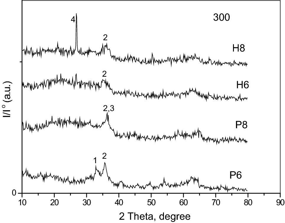
Bacterial cellulose (BC) is a three-dimensional interconnected network of biosynthesized nanofibers. Its rehydration potential would be reduced significantly after its first drying, as a result of entanglement and jamming of cellulose polymer chains. Consequently, its versatility would be also reduced to some limited applications in which repeated water absorbance potential is not of great importance. This study aims to prevent the drawback of carboxylic bridging/cross-linking between cellulose polymer chains. Ten-day-cultured BC pellicles were immersed in various citric acid solutions (as bridging agent) and cured at 160 °C for 5 min. The formation of bridges was confirmed using attenuated total reflection–fourier transform infrared spectroscopy. Scanning electron microscope images showed that there is a different porosity bridged/cross-linked BC specimens (XBC). According to Brunauer–Emmett–Teller analysis, the surface area of XBC (20 w/v % with catalyst) got 87.5 times larger than that of the unbridged/pristine BC (PBC). X-ray diffraction patterns showed no change of crystallinity of XBC in comparison with PBS. The thickness and wettability of XBC samples were 137 and 3.27 times more than PBC samples orderly. Furthermore, the water swelling rate increased significantly for XBC in comparison with PBC. Meanwhile, treated samples had lower elongation and strength than normal BC. The conclusion is that XBC could conserve its repeated absorbency potential after the presented process.
Graphical abstract
Keywords
- Bacterial cellulose (BC),
- Rehydration,
- Cross-linking/bridging agent,
- Citric acid,
- 3D nanostructure of BC
References
- Fu et al. (2013) Present status and applications of bacterial cellulose-based materials for skin tissue repair (pp. 1432-1442) https://doi.org/10.1016/j.carbpol.2012.10.071
- Hu et al. (2014) Functionalized bacterial cellulose derivatives and nanocomposites (pp. 1043-1060) https://doi.org/10.1016/j.carbpol.2013.09.102
- Huang et al. (2014) Recent advances in bacterial cellulose 21(1) (pp. 1-30) https://doi.org/10.1007/s10570-013-0088-z
- Qiu et al. (2016) Bacterial cellulose and bacterial cellulose-vaccarin membranes for wound healing (pp. 303-309) https://doi.org/10.1016/j.msec.2015.10.016
- Rajwade et al. (2015) Applications of bacterial cellulose and its composites in biomedicine (pp. 2491-2511) https://doi.org/10.1007/s00253-015-6426-3
- Stumpf et al. (2013) Enriched glucose and dextrin mannitol-based media modulates fibroblast behavior on bacterial cellulose membranes (pp. 4739-4745) https://doi.org/10.1016/j.msec.2013.07.035
- Sulaeva et al. (2015) Bacterial cellulose as a material for wound treatment: properties and modifications. A review (pp. 1547-1571) https://doi.org/10.1016/j.biotechadv.2015.07.009
- Picheth et al. (2017) Bacterial cellulose in biomedical applications: a review (pp. 97-106) https://doi.org/10.1016/j.ijbiomac.2017.05.171
- Gao et al. (2016) Time-dependent rheological behaviour of bacterial cellulose hydrogel (pp. 153-159) https://doi.org/10.1016/j.msec.2015.08.019
- Gao et al. (2016) Through-thickness stress relaxation in bacterial cellulose hydrogel (pp. 90-98) https://doi.org/10.1016/j.jmbbm.2015.12.021
- Pandey et al. (2017) Microwaved bacterial cellulose-based hydrogel microparticles for the healing of partial thickness burn wounds (pp. 89-99) https://doi.org/10.1007/s13346-016-0341-8
- Yuan et al. (2016) Superior hybrid hydrogels of polyacrylamide enhanced by bacterial cellulose nanofiber clusters (pp. 221-230) https://doi.org/10.1016/j.msec.2016.04.074
- Römling and Galperin (2015) Bacterial cellulose biosynthesis: diversity of operons, subunits, products, and functions 23(9) (pp. 545-557) https://doi.org/10.1016/j.tim.2015.05.005
- Lin et al. (2013) Biosynthesis, production and applications of bacterial cellulose 20(5) (pp. 2191-2219) https://doi.org/10.1007/s10570-013-9994-3
- Ashjaran et al. (2013) Overview of bio nanofabric from bacterial cellulose 104(2) (pp. 121-131) https://doi.org/10.1080/00405000.2012.703796
- Abeer et al. (2014) A review of bacterial cellulose-based drug delivery systems: their biochemistry, current approaches and future prospects 66(8) (pp. 1047-1061)
- Reiniati et al. (2017) Recent developments in the production and applications of bacterial cellulose fibers and nanocrystals 37(4) (pp. 510-524) https://doi.org/10.1080/07388551.2016.1189871
- Ul-Islam et al. (2012) Water holding and release properties of bacterial cellulose obtained by in situ and ex situ modification (pp. 596-603) https://doi.org/10.1016/j.carbpol.2012.01.006
- Ul-Islam et al. (2013) Effect of post-synthetic processing conditions on structural variations and applications of bacterial cellulose (pp. 253-263) https://doi.org/10.1007/s10570-012-9799-9
- Meftahi et al. (2010) The effects of cotton gauze coating with microbial cellulose (pp. 199-204) https://doi.org/10.1007/s10570-009-9377-y
- Brown et al. (2012) Glutaraldehyde treatment of bacterial cellulose/fibrin composites impact on morphology, tensile and viscoelastic properties (pp. 127-137) https://doi.org/10.1007/s10570-011-9617-9
- Brown et al. (2011) Never-dried bacterial cellulose/fibrin composites: preparation, morphology and mechanical properties (pp. 631-641) https://doi.org/10.1007/s10570-011-9500-8
- Cacicedo et al. (2016) Progress in bacterial cellulose matrices for biotechnological applications (pp. 172-180) https://doi.org/10.1016/j.biortech.2016.02.071
- Tang et al. (2010) The influence of fermentation conditions and post-treatment methods on porosity of bacterial cellulose membrane https://doi.org/10.1007/s11274-009-0151-y
- Gao et al. (2011) Preparation and characterization of bacterial cellulose sponge with hierarchical pore structure as tissue engineering scaffold (pp. 139-145) https://doi.org/10.1007/s10934-010-9364-6
- Wang et al. (2012) Immobilization of gelatin on bacterial cellulose nanofibers surface via crosslinking technique (pp. 536-541) https://doi.org/10.1016/j.msec.2011.12.006
- Castro et al. (2015) In-situ glyoxalization during biosynthesis of bacterial cellulose (pp. 32-39) https://doi.org/10.1016/j.carbpol.2015.03.014
- Reddy and Yang (2010) Citric acid cross-linking of starch films (pp. 702-711) https://doi.org/10.1016/j.foodchem.2009.05.050
- Chang et al. (2012) Nano-biomaterials application: morphology and physical properties of bacterial cellulose/gelatin composites via crosslinking (pp. 137-144) https://doi.org/10.1016/j.foodhyd.2011.08.004
- Dahman (2009) Nanostructured biomaterials and biocomposites from bacterial cellulose nanofibers (pp. 5105-5122) https://doi.org/10.1166/jnn.2009.1466
- Fijałkowski et al. (2015) Modification of bacterial cellulose through exposure to the rotating magnetic field (pp. 52-60) https://doi.org/10.1016/j.carbpol.2015.07.011
- Shah et al. (2013) Overview of bacterial cellulose composites: a multipurpose advanced material (pp. 1585-1598) https://doi.org/10.1016/j.carbpol.2013.08.018
- Seifert et al. (2004) Controlling the water content of never dried and reswollen bacterial cellulose by the addition of water-soluble polymers to the culture medium (pp. 463-470) https://doi.org/10.1002/pola.10862
- Xu et al. (2015) Development of tannic acid/chitosan/pullulan composite nanofibers from aqueous solution for potential applications as wound dressing (pp. 16-24) https://doi.org/10.1016/j.carbpol.2014.08.081
- Pinho and Soares (2018) Functionalization of cotton cellulose for improved wound healing 6(13) (pp. 1887-1898) https://doi.org/10.1039/C8TB00052B
- Gyawali et al. (2010) Citric-acid-derived photo-cross-linked biodegradable elastomers 21(13) (pp. 1761-1782) https://doi.org/10.1163/092050609X12567178204169
- Capanema et al. (2018) Superabsorbent crosslinked carboxymethyl cellulose-PEG hydrogels for potential wound dressing applications (pp. 1218-1234) https://doi.org/10.1016/j.ijbiomac.2017.08.124
- El Fawal et al. (2018) Hydroxyethyl cellulose hydrogel for wound dressing: fabrication, characterization and in vitro evaluation (pp. 649-659) https://doi.org/10.1016/j.ijbiomac.2018.01.040
- Ebrahimi et al. (2017) Effects of bio-production process parameters on bacterial cellulose mechanical properties 50(11) (pp. 857-861) https://doi.org/10.1252/jcej.15we301
- Khajavi et al. (2011) Crystalline structure of microbial cellulose compared with native and regenerated cellulose 60(14) (pp. 1178-1192) https://doi.org/10.1080/00914037.2010.551372
- Prabhu et al. (2017) Effect of microwave argon plasma on the glycosidic and hydrogen bonding system of cotton cellulose (pp. 34-44) https://doi.org/10.1016/j.carbpol.2016.08.057
- Awada et al. (2014) Cross-linking of papers based on thermomechanical pulp fibers by polycarboxylic acids: influence on the wet breaking length 53(11) (pp. 4312-4317) https://doi.org/10.1021/ie500101n
- Widsten et al. (2014) Citric acid crosslinking of paper products for improved high-humidity performance (pp. 998-1004) https://doi.org/10.1016/j.carbpol.2013.10.002
- Grande et al. (2009) Development of self-assembled bacterial cellulose–starch nanocomposites (pp. 1098-1104) https://doi.org/10.1016/j.msec.2008.09.024
- Poletto et al. (2014) Native cellulose: structure, characterization and thermal properties 7(9) (pp. 6105-6119) https://doi.org/10.3390/ma7096105
- Ye et al. (2015) Quantitative analysis of citric acid/sodium hypophosphite modified cotton by HPLC and conductometric titration (pp. 92-98) https://doi.org/10.1016/j.carbpol.2014.12.028