 10.1007/s40097-022-00487-0
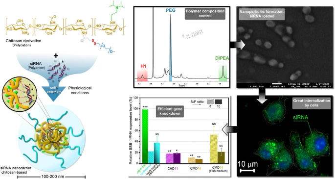
Double-grafted chitosans as siRNA nanocarriers: effects of diisopropylethylamine substitution and labile-PEG coating
- André Miguel Martinez Junior
 1
1 - Ricchard Hallan Felix Viegas de Souza2
- Maicon Segalla Petrônio1
- Grazieli Olinda Martins1
- Júlio Cesar Fernandes3
- Mohamed Benderdour3
- Vera Aparecida Oliveira de Tiera1
- Marcio José Tiera*1
- Department of Chemistry and Environmental Sciences, IBILCE, São Paulo State University-UNESP, R. Cristóvão Colombo, São José do Rio Preto, SP, 2265, 15054-000, BR
- Department of Physics, IBILCE, São Paulo State University-UNESP, São José do Rio Preto, BR
- Orthopedic Research Laboratory, Hôpital du Sacré-Coeur de Montréal, Université de Montréal, Montréal, CA

Published in Issue 31-03-2022
Copyright (c) 2025 André Miguel Martinez Junior, Ricchard Hallan Felix Viegas de Souza, Maicon Segalla Petrônio, Grazieli Olinda Martins, Júlio Cesar Fernandes, Mohamed Benderdour, Vera Aparecida Oliveira de Tiera, Marcio José Tiera (Author)

This work is licensed under a Creative Commons Attribution 4.0 International License.
How to Cite
Martinez Junior, A. M., de Souza, R. H. F. V., Petrônio, M. S., Martins, G. O., Fernandes, J. C., Benderdour, M., Tiera, V. A. O. de, & Tiera, M. J. (2022). Double-grafted chitosans as siRNA nanocarriers: effects of diisopropylethylamine substitution and labile-PEG coating. Journal of Nanostructure in Chemistry, 13(6 (December 2023). https://doi.org/10.1007/s40097-022-00487-0


Share:


Abstract
Abstract The preparation of safe and efficient siRNA carriers remains a challenge that has limited the therapeutic applications of siRNA. In this study, the design of a new small interfering RNA (siRNA) carrier based on diisopropylaminoethyl-chitosan was devised for application in non-viral gene therapy. Polycations having varied proportions (11–32%) of diisopropylethylamine groups (DIPEA) and grafted with polyethylene glycol (1–3%) were synthesized and characterized. The physicochemical and biological properties of the polymers and their nanoparticles were evaluated at pH 6.3 and pH 7.4. The degrees of ionization at pH 7.4 were precisely controlled by the composition and increased from 13% for chitosan to 47% for the more substituted derivative. Nanoparticles with very low toxicities and sizes in the range of 100–200 nm, remained stable up to 24 h after their preparation in both the evaluated pHs under plasma osmolality. As probed by scanning electron and confocal microscopies, an efficient cell uptake of spherical nanoparticles mediated a TNFα knockdown of almost 60% in RAW 264.7 macrophages, and mRNA silence levels higher than the Lipofectamine (up to 90%) in HeLa cells. Overall, the results showed that these derivatives are promising vectors for in vivo studies under physiological conditions. Graphical abstractKeywords
- siRNA,
- DIPEA,
- Nanoparticles,
- Knockdown,
- Non-viral vector,
- Gene therapy,
- TNFα
References
- Mirzaei et al. (2021) Small interfering RNA (siRNA) to target genes and molecular pathways in glioblastoma therapy: current status with an emphasis on delivery systems https://doi.org/10.1016/j.lfs.2021.119368
- Mirzaei et al. (2021) Employing siRNA tool and its delivery platforms in suppressing cisplatin resistance: approaching to a new era of cancer chemotherapy https://doi.org/10.1016/j.lfs.2021.119430
- Rossi and Rossi (2021) siRNA drugs: here to stay https://doi.org/10.1016/j.ymthe.2021.01.015
- Tavakoli et al. (2021) Milk protein-based nanodelivery systems for the cancer treatment (pp. 483-500) https://doi.org/10.1007/s40097-021-00399-5
- Ashrafizadeh et al. (2021) Biomedical application of chitosan-based nanoscale delivery systems: potential usefulness in siRNA delivery for cancer therapy https://doi.org/10.1016/j.carbpol.2021.117809
- Lavertu et al. (2006) High efficiency gene transfer using chitosan/DNA nanoparticles with specific combinations of molecular weight and degree of deacetylation https://doi.org/10.1016/j.biomaterials.2006.04.029
- Jennings and Bumgarden (2017) Woodhead Publishing
- Alameh et al. (2018) siRNA delivery with chitosan: influence of chitosan molecular weight, degree of deacetylation, and amine to phosphate ratio on in vitro silencing efficiency, hemocompatibility, biodistribution, and in vivo efficacy (pp. 112-131) https://doi.org/10.1021/acs.biomac.7b01297
- Dehousse et al. (2010) Comparison of chitosan/siRNA and trimethylchitosan/siRNA complexes behaviour in vitro (pp. 342-349) https://doi.org/10.1016/j.ijbiomac.2010.01.010
- Soliman et al. (2020) Efficiency of chitosan/hyaluronan-based mRNA delivery systems in vitro: influence of composition and structure (pp. 1581-1593) https://doi.org/10.1016/j.xphs.2019.12.020
- Oliveira et al. (2013) Synthesis and evaluation of diethylethylamine-chitosan for gene delivery: composition effects on the in vitro transfection efficiency https://doi.org/10.1088/0957-4484/24/5/055101
- Picola et al. (2016) Chitosan derivatives for gene transfer: effect of phosphorylcholine and diethylaminoethyl grafts on the in vitro transfection efficiency (pp. 1611-1630) https://doi.org/10.1080/09205063.2016.1225333
- De Souza et al. (2018) Diethylaminoethyl-chitosan as an efficient carrier for siRNA delivery: improving the condensation process and the nanoparticles properties (pp. 186-197) https://doi.org/10.1016/j.ijbiomac.2018.07.072
- Jones et al. (2013) Overcoming nonviral gene delivery barriers: perspective and future (pp. 4082-4098) https://doi.org/10.1021/mp400467x
- Giacomelli et al. (2011) pH-triggered block copolymer micelles based on a pH-responsive PDPA (poly[2-(diisopropylamino)ethyl methacrylate]) inner core and a PEO (poly(ethylene oxide)) outer shell as a potential tool for the cancer therapy (pp. 9316-9325) https://doi.org/10.1039/c1sm05992k
- Zhou et al. (2012) Multicolored pH-tunable and activatable fluorescence nanoplatform responsive to physiologic pH stimuli (pp. 7803-7811) https://doi.org/10.1021/ja300176w
- Zhou et al. (2016) Tumor-penetrating peptide modified and pH-sensitive polyplexes for tumor targeted siRNA delivery (pp. 3857-3863) https://doi.org/10.1039/C6PY00427J
- Cao et al. (2019) Recent advances in chitosan-based carriers for gene delivery https://doi.org/10.3390/md17060381
- Tiera et al. (2011) Polycation-based gene therapy: current knowledge and new perspectives (pp. 288-306) https://doi.org/10.2174/156652311796150408
- Blanco et al. (2015) Principles of nanoparticle design for overcoming biological barriers to drug delivery (pp. 941-951) https://doi.org/10.1038/nbt.3330
- Abbaszadeh et al. (2014) Improvement single-wall carbon nanotubes (SWCNTs) based on functionalizing with monomers 2-hydroxyethylmethacryate (HEMA) and N-vinylpyrrolidone (NVP) for pharmaceutical applications as cancer therapy (pp. 2895-2900) https://doi.org/10.1016/j.jiec.2013.11.025
- Partikel et al. (2019) Effect of nanoparticle size and PEGylation on the protein corona of PLGA nanoparticles (pp. 70-80) https://doi.org/10.1016/j.ejpb.2019.05.006
- Tang et al. (2003) Polyethylene glycol modified polyethylenimine for improved CNS gene transfer: effects of PEGylation extent (pp. 2351-2362) https://doi.org/10.1016/S0142-9612(03)00029-2
- Rheiner and Bae (2016) Increased poly(ethylene glycol) density decreases transfection efficacy of siRNA/poly(ethylene imine) complexes (pp. 454-467) https://doi.org/10.3934/bioeng.2016.4.454
- Yang et al. (2017) Impact of PEG chain length on the physical properties and bioactivity of PEGylated chitosan/siRNA nanoparticles in vitro and in vivo (pp. 12203-12216) https://doi.org/10.1021/acsami.6b16556
- Cabral et al. (2018) Block copolymer micelles in nanomedicine applications (pp. 6844-6892) https://doi.org/10.1021/acs.chemrev.8b00199
- Agirre et al. (2014) Low molecular weight chitosan (LMWC)-based polyplexes for pDNA delivery: from bench to bedside (pp. 1727-1755) https://doi.org/10.3390/polym6061727
- Du et al. (2019) Tailoring kidney transport of organic dyes with low-molecular-weight PEGylation (pp. 241-247) https://doi.org/10.1021/acs.bioconjchem.9b00707
- Zhang et al. (2012) In vivo gene delivery by nonviral vectors: overcoming hurdles? (pp. 1298-1304) https://doi.org/10.1038/mt.2012.79
- Tiera et al. (2013) Polymeric systems as nanodevices for siRNA delivery (pp. 358-369) https://doi.org/10.2174/156652321305131212125042
- Tamura and Yui (2014) Lysosomal-specific cholesterol reduction by biocleavable polyrotaxanes for ameliorating niemann-pick type C disease (pp. 1-8) https://doi.org/10.1038/srep04356
- Wang et al. (2013) Glutathione-triggered “off-on” release of anticancer drugs from dendrimer-encapsulated gold nanoparticles (pp. 9805-9810) https://doi.org/10.1021/ja402903h
- Adriaansen et al. (2006) Gene therapy as a therapeutic approach for the treatment of rheumatoid arthritis: innovative vectors and therapeutic genes (pp. 656-668) https://doi.org/10.1093/rheumatology/kel047
- Ta et al. (2013) Origins and hallmarks of macrophages: development, homeostasis, and disease (pp. 445-455) https://doi.org/10.1038/nature12034
- Pandi et al. (2017) Therapeutic approaches for the delivery of TNF-α siRNA (pp. 343-355) https://doi.org/10.4155/tde-2017-0011
- Staudacher et al. (2014) The La antigen is over-expressed in lung cancer and is a selective dead cancer cell target for radioimmunotherapy using the La-specific antibody APOMAB® (pp. 1-13) https://doi.org/10.1186/2191-219X-4-2
- Tiera et al. (2006) Synthesis and characterization of phosphorylcholine-substituted chitosans soluble in physiological pH conditions (pp. 3151-3156) https://doi.org/10.1021/bm060381u
- Martins et al. (2019) Amphipathic chitosans improve the physicochemical properties of siRNA-chitosan nanoparticles at physiological conditions (pp. 332-342) https://doi.org/10.1016/j.carbpol.2019.03.098
- Gunn et al. (2009) A simple and highly sensitive method for magnetic nanoparticle quantitation using 1H-NMR spectroscopy (pp. 2640-2647) https://doi.org/10.1016/j.bpj.2009.08.013
- Gabriel et al. (2015) Synthesis, characterization, and antifungal activities of amphiphilic derivatives of diethylaminoethyl chitosan against Aspergillus flavus (pp. 5725-5731) https://doi.org/10.1021/acs.jafc.5b00278
- Richard et al. (2013) Ionization behavior of chitosan and chitosan-DNA polyplexes indicate that chitosan has a similar capability to induce a proton-sponge effect as PEI (pp. 1732-1740) https://doi.org/10.1021/bm4000713
- Rai et al. (2019) Polymeric nanoparticles in gene therapy: new avenues of design and optimization for delivery applications https://doi.org/10.3390/polym11040745
- Barreto et al. (2019) Development of an automated method to perform a quantitative study of particle size distribution and the effect of a conductive layer in scanning electron microscopy (pp. 447-452)
- Fernandes et al. (2012) Low molecular weight chitosan conjugated with folate for siRNA delivery in vitro: optimization studies
- Fujita and Sakairi (2016) Water soluble EDTA-linked Chitosan as a zwitterionic flocculant for pH sensitive removal of Cu(II) ion (pp. 10385-10392) https://doi.org/10.1039/C5RA24175H
- Fiamingo and Campana-Filho (2016) Structure, morphology and properties of genipin-crosslinked carboxymethylchitosan porous membranes (pp. 155-163) https://doi.org/10.1016/j.carbpol.2016.02.016
- Zhang et al. (2010) NMR and FT Raman characterisation of regioselectively sulfated chitosan regarding the distribution of sulfate groups and the degree of substitution (pp. 4698-4705) https://doi.org/10.1016/j.polymer.2010.08.034
- Zhang et al. (2010) Chitosan modification and pharmaceutical/biomedical applications (pp. 1962-1987) https://doi.org/10.3390/md8071962
- Jain et al. (2013) A new horizon in modifications of chitosan: syntheses and applications https://doi.org/10.1615/CritRevTherDrugCarrierSyst.2013005678
- Thomas et al. (2016) Elsevier
- Vermeulen et al. (2018) The proton sponge hypothesis: fable or fact? (pp. 184-190) https://doi.org/10.1016/j.ejpb.2018.05.034
- Casey et al. (2010) Sensors and regulators of intracellular pH (pp. 50-61) https://doi.org/10.1038/nrm2820
- Doriti et al. (2016) Synthesis of polysarcosine from air and moisture stable N-phenoxycarbonyl-N-methylglycine assisted by tertiary amine base (pp. 3067-3070) https://doi.org/10.1039/C6PY00221H
- Serrano-Sevilla et al. (2019) Natural polysaccharides for siRNA delivery: nanocarriers based on chitosan, hyaluronic acid, and their deerivatives https://doi.org/10.3390/molecules24142570
- Layek et al. (2017) Chitosan for DNA and gene therapy (pp. 209-244) Woodhead Publishing https://doi.org/10.1016/B978-0-08-100228-5.00008-0
- Chen et al. (2015) Grafting chitosan with polyethylenimine in an ionic liquid for efficient gene delivery https://doi.org/10.1371/journal.pone.0121817
- Kean et al. (2005) Trimethylated chitosans as non-viral gene delivery vectors: cytotoxicity and transfection efficiency (pp. 643-653) https://doi.org/10.1016/j.jconrel.2005.01.001
- Layek and Singh (2013) Caproic acid grafted chitosan cationic nanocomplexes for enhanced gene delivery: effect of degree of substitution (pp. 182-191) https://doi.org/10.1016/j.ijpharm.2013.02.052
- Liu et al. (2007) The influence of polymeric properties on chitosan/siRNA nanoparticle formulation and gene silencing (pp. 1280-1288) https://doi.org/10.1016/j.biomaterials.2006.11.004
- Yue et al. (2011) Surface charge affects cellular uptake and intracellular trafficking of chitosan-based nanoparticles (pp. 2440-2446) https://doi.org/10.1021/bm101482r
- Makvandi et al. (2021) Endocytosis of abiotic nanomaterials and nanobiovectors: Inhibition of membrane trafficking https://doi.org/10.1016/j.nantod.2021.101279
- Picola et al. (2013) Effect of ionic strength solution on the stability of chitosan-DNA nanoparticles (pp. 703-7016) https://doi.org/10.1080/17458080.2011.602120
- Behzadi et al. (2017) Cellular uptake of nanoparticles: journey inside the cell (pp. 4218-4244) https://doi.org/10.1039/C6CS00636A
- Saptarshi et al. (2013) Interaction of nanoparticles with proteins: relation to bio-reactivity of the nanoparticle (pp. 1-12) https://doi.org/10.1186/1477-3155-11-26
- Carlos et al. (2017) Limiting the level of tertiary amines on polyamines leads to biocompatible nucleic acid vectors (pp. 106-124) https://doi.org/10.1016/j.ijpharm.2017.04.059
- Gormley et al. (2009) Evaluation of toxicity of nanostructures in biological systems (pp. 115-159) Wiley https://doi.org/10.1002/9780470747803.ch7
- Kim et al. (2019) Rekindling RNAi therapy: materials design requirements for in vivo siRNA delivery https://doi.org/10.1002/adma.201903637
- Hao et al. (2018) Manipulating extracellular tumour pH: an effective target for cancer therapy (pp. 22182-22192) https://doi.org/10.1039/C8RA02095G
- Guţoaia et al. (2016) Fine-tuned PEGylation of chitosan to maintain optimal siRNA-nanoplex bioactivity (pp. 25-34) https://doi.org/10.1016/j.carbpol.2016.01.010
- Ping et al. (2011) Chitosan-graft-(PEI-β-cyclodextrin) copolymers and their supramolecular PEGylation for DNA and siRNA delivery (pp. 8328-8341) https://doi.org/10.1016/j.biomaterials.2011.07.038
- Germershaus et al. (2008) Gene delivery using chitosan, trimethyl chitosan or polyethylenglycol-graft-trimethyl chitosan block copolymers: establishment of structure-activity relationships in vitro (pp. 145-154) https://doi.org/10.1016/j.jconrel.2007.10.013
- Ragelle et al. (2014) Chitosan nanoparticles for siRNA delivery: optimizing formulation to increase stability and efficiency (pp. 54-63) https://doi.org/10.1016/j.jconrel.2013.12.026