
 10.1007/s40089-016-0184-9
Synthesis of silver nanoparticles using flavonoids: hesperidin, naringin and diosmin, and their antibacterial effects and cytotoxicity
- Nidhi Sahu
 1
1 - Deepika Soni1
- B. Chandrashekhar1
- D. B. Satpute1
- Sivanesan Saravanadevi1
- B. K. Sarangi1
- R. A. Pandey*1
- CSIR-National Environmental Engineering Research Institute (NEERI), Nagpur, 440020, IN

Published in Issue 2016-07-19
How to Cite
Sahu, N., Soni, D., Chandrashekhar, B., Satpute, D. B., Saravanadevi, S., Sarangi, B. K., & Pandey, R. A. (2016). Synthesis of silver nanoparticles using flavonoids: hesperidin, naringin and diosmin, and their antibacterial effects and cytotoxicity. International Nano Letters, 6(3 (September 2016). https://doi.org/10.1007/s40089-016-0184-9




HTML views: 83
PDF views: 166
Share:





Abstract
Abstract
Three different flavonoids -hesperidin, naringin and diosmin (constituents of citrus plants) were used for the synthesis of silver nanoparticles (AgNPs). Aqueous solutions of pure flavonoids (0.2 mg mL
−1
) mixed with 1 mM AgNO
3
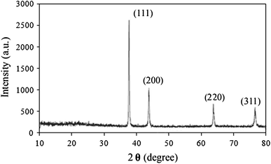
solution were exposed to bright sunlight to prepare the nanoparticles. Characterization of the synthesized nanoparticles by UV–Visible spectrophotometer, X-ray diffraction, Fourier transform infrared spectroscopy, and transmission electron microscopy revealed that the synthesized silver nanoparticles were 10–80 nm in size and polydispersed in nature. Bactericidal effect against common pathogens and cytotoxicity of the synthesized silver nanoparticles was investigated on human promyelocytic leukemic (HL-60) cells. It is concluded that AgNPs synthesized using Naringin as reducing agent showed higher stability and better antibacterial and cytotoxic activities.
Keywords
- Flavonoids,
- Nanoparticles,
- Bio-synthesis,
- Secondary metabolites,
- Reducing agents
References
- Prakash et al. (2006) Polymer memory device based on conjugated polymer and gold nanoparticles (pp. 054309-054313) https://doi.org/10.1063/1.2337252
- LaLonde et al. (2005) Controlled growth of gold nanoparticles on silica nanowires (pp. 3021-3027) https://doi.org/10.1557/JMR.2005.0368
- Manno et al. (2008) Synthesis and characterization of starch-stabilized Ag nanostructures for sensors applications (pp. 5515-5520) https://doi.org/10.1016/j.jnoncrysol.2008.04.059
- Tran et al. (2012) An organoselenium compound inhibits Staphylococcus aureus biofilms on hemodialysis catheters in vivo (pp. 972-978) https://doi.org/10.1128/AAC.05680-11
- WhiteII et al. (2012) Green synthesis of robust, biocompatible silver nanoparticles using garlic extract (pp. 1-12) https://doi.org/10.1155/2012/730746
- Šileikaitė et al. (2006) Analysis of silver nanoparticles produced by chemical reduction of silver salt solution (pp. 287-291)
- Sahu et al. (2012) Synthesis and characterization of silver nanoparticles using Cynodon dactylon leaves and assessment of their antibacterial activity (pp. 999-1004) https://doi.org/10.1007/s00449-012-0841-y
- Prabhu and Poulose (2012) Silver nanoparticles: mechanism of antimicrobial action, synthesis, medical applications, and toxicity effects (pp. 1-10) https://doi.org/10.1186/2228-5326-2-32
- Rico et al. (2011) Interaction of nanoparticles with edible plants and their possible implications in the food chain (pp. 3485-3498) https://doi.org/10.1021/jf104517j
- Sant et al. (2013) Adiantum philippense L. Frond assisted rapid green synthesis of gold and silver nanoparticles (pp. 1-9) https://doi.org/10.1155/2013/182320
- Di Carlo et al. (1999) Flavonoids: old and new aspects of a class of natural therapeutic drugs (pp. 337-353) https://doi.org/10.1016/S0024-3205(99)00120-4
- Jaiswal et al. (2013) Phytochemicals and antioxidative enzymes defence mechanism on occurrence of yellow vein mosaic disease of pumpkin (Cucurbita moschata) (pp. 287-295) https://doi.org/10.1007/s13205-012-0100-6
- Daniel et al. (2012) Rapid biosynthesis of silver nanoparticles using Eichornia crassipes and its antibacterial activity (pp. 125-129) https://doi.org/10.2174/1573413711208010125
- Kar, A: Pharmacognosy and Pharmacobiotechnology (2nd edition) New Age International (P) Ltd (2007)
- Boysen, E, Muir, NC: Nanotechnology for dummies (2nd Edition), Wiley publishing, Inc (2011)
- Gannimani et al. (2014) Synthesis and antibacterial activity of silver and gold nanoparticles produced using aqueous seed extract of Protorhus longifolia as a reducing agent (pp. 1669-1679)
- Rout et al. (2012) Green synthesis of silver nanoparticles using Ocimum sanctum (Tulashi) and study of their antibacterial and antifungal activities (pp. 103-109) https://doi.org/10.5897/JMA11.060
- Umoren et al. (2014) Green synthesis and characterization of silver nanoparticles using red apple (Malus domestica) fruit extract at room temperature (pp. 907-914)
- Hossain and Rahman (2011) Total phenolics, flavonoids and antioxidant activity of tropical fruit pineapple (pp. 672-676) https://doi.org/10.1016/j.foodres.2010.11.036
- Sharma and Janmeda (2014) Extraction, isolation and identification of flavonoid from Euphorbia neriifolia leaves https://doi.org/10.1016/j.arabjc.2014.08.019
- Roy et al. (2014) Green synthesis of silver nanoparticles using fruit extract of Malus domestica and study of its antimicrobial activity (pp. 1137-1147)
- Prathna et al. (2011) Studies on aggregation behavior of silver nanoparticles in aqueous matrices: effects of surface functionalization and matrix composition (pp. 216-224) https://doi.org/10.1016/j.colsurfa.2011.09.047
- Vankar and Shukla (2012) Biosynthesis of silver nanoparticles using lemon leaves extract and its application for antimicrobial finish on fabric (pp. 163-168) https://doi.org/10.1007/s13204-011-0051-y
- Stephen and Seethalakshmi (2013) Phytochemical synthesis and preliminary characterization of silver nanoparticles using hesperidin (pp. 1-6) https://doi.org/10.1155/2013/126564
- Soni et al. (2015) Release transport and toxicity of engineered nanoparticles (pp. 1-47)
- Soni et al. (2014) Stress response of Pseudomonas species to silver nanoparticles at molecular level (pp. 2126-2132) https://doi.org/10.1002/etc.2670
- Kora and Rastogi (2013) Enhancement of antibacterial activity of capped silver nanoparticles in combination with antibiotics, on model gram-negative and gram-positive bacteria (pp. 1-7) https://doi.org/10.1155/2013/871097
- Kim et al. (2010) Cytotoxicity and genotoxicity of nano-silver in mammalian cell lines (pp. 119-125) https://doi.org/10.1007/s13273-010-0018-1
- Kim et al. (2012) Size-dependent cellular toxicity of silver nanoparticles (pp. 1033-1043) https://doi.org/10.1002/jbm.a.34053
- Gliga et al. (2014) Size-dependent cytotoxicity of silver nanoparticles in human lung cells: the role of cellular uptake, agglomeration and Ag release (pp. 1-17) https://doi.org/10.1186/1743-8977-11-11
- Asharani et al. (2009) Cytotoxicity and genotoxicity of silver nanoparticles in human cells (pp. 279-290) https://doi.org/10.1021/nn800596w