 10.1007/s40089-014-0094-7


Published in Issue 2014-02-20
How to Cite
Mirza, A. Z., & Siddiqui, F. A. (2014). Nanomedicine and drug delivery: a mini review. International Nano Letters, 4(1 (March 2014). https://doi.org/10.1007/s40089-014-0094-7


HTML views: 41
PDF views: 150
Share:


Abstract
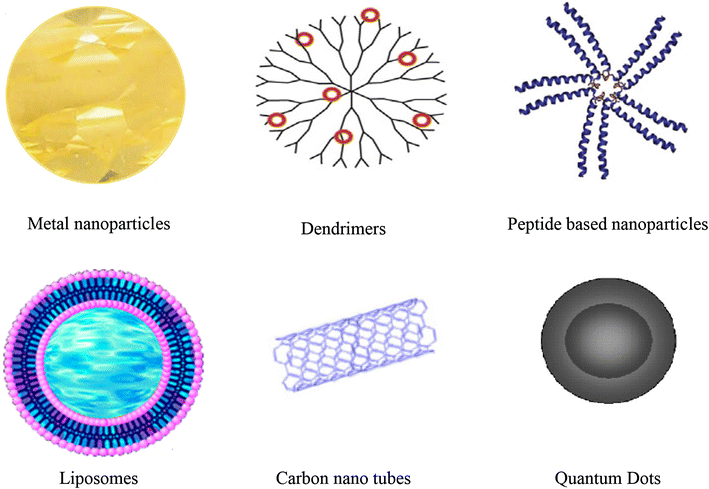
Abstract The field of nanotechnology now has pivotal roles in electronics, biology and medicine. Its application can be appraised, as it involves the materials to be designed at atomic and molecular level. Due to the advantage of their size, nanospheres have been shown to be robust drug delivery systems and may be useful for encapsulating drugs and enabling more precise targeting with a controlled release. In this review specifically, we highlight the recent advances of this technology for medicine and drug delivery systems.Keywords
- Drug delivery,
- Nanotechnology,
- Inorganic nanoparticles,
- Organic nanoparticles
References
- Whitesides (2003) The ‘right’ size in nanobiotechnology (pp. 1161-1165) https://doi.org/10.1038/nbt872
- Lowe (2000) Nanobiotechnology: the fabrication and applications of chemical and biological nanostructures (pp. 428-434)
- Wang et al. (2008) Bioconjugated silica nanoparticles: development and applications (pp. 99-115) https://doi.org/10.1007/s12274-008-8018-3
- John et al. (2002) Recent developments in materials synthesis in surfactant systems https://doi.org/10.1016/S1359-0294(02)00070-5
- Castelvetro and De Vita (2004) Nanostructured hybrid materials from aqueous polymer dispersions (pp. 167-185) https://doi.org/10.1016/j.cis.2003.10.017
- Jang and Shea (2003) Controllable delivery of non-viral DNA from porous scaffolds (pp. 157-168) https://doi.org/10.1016/S0168-3659(02)00369-3
- Shi et al. (2003) Double walled POE/PLGA microspheres: encapsulation of water-soluble and water-insoluble proteins and their release properties (pp. 167-177) https://doi.org/10.1016/S0168-3659(02)00493-5
- Mu and Feng (2001) Fabrication, characterization and in vitro release of paclitaxel (taxol) loaded poly (lactic-co-glycolic acid) microspheres prepared by spray drying technique with lipid/cholesterol emulsifiers (pp. 239-254) https://doi.org/10.1016/S0168-3659(01)00440-0
- Jones et al. (2001) Alumina ultrafiltration membranes derived from carboxylate-alumoxane nanoparticles (pp. 175-184) https://doi.org/10.1016/S0376-7388(01)00490-2
- Gupta and Gupta (2005) Synthesis and surface engineering of iron oxide nanoparticles for biomedical applications (pp. 3995-4201) https://doi.org/10.1016/j.biomaterials.2004.10.012
- Liu et al. (2009) Carbon nanotubes in biology and medicine: in vitro and in vivo detection, imaging and drug delivery (pp. 85-120) https://doi.org/10.1007/s12274-009-9009-8
- Razzacki et al. (2004) Integrated microsystems for controlled drug delivery (pp. 185-198) https://doi.org/10.1016/j.addr.2003.08.012
- Frank et al. (2004) Study of the initial stages of drug release from a degradable matrix of poly(d, l-lactide-co-glycolide) https://doi.org/10.1016/S0142-9612(03)00597-0
- Freitas (2005) What is nanomedicine? (pp. 2-9) https://doi.org/10.1016/j.nano.2004.11.003
- Vinogradov et al. (2002) Nanosized cationic hydrogels for drug delivery: preparation, properties and interactions with cells (pp. 135-147) https://doi.org/10.1016/S0169-409X(01)00245-9
- Orive et al. (2004) Techniques: new approaches to the delivery of biopharmaceuticals (pp. 382-387) https://doi.org/10.1016/j.tips.2004.05.006
- Arayne et al. (2007) Review: nanoparticles in delivery of cardiovascular drugs (pp. 340-348)
- Bala et al. (2004) PLGA nanoparticles in drug delivery: the state of the art (pp. 387-422) https://doi.org/10.1615/CritRevTherDrugCarrierSyst.v21.i5.20
- Karen et al. (2004) Entrapment of bioactive molecules in poly (alkylcyanoacrylate) nanoparticles (pp. 251-259)
- Reddy and Murthy (2004) Pharmacokinetics and biodistribution studies of doxorubicin loaded poly(butyl cyanoacrylate) nanoparticles synthesized by two different techniques (pp. 161-166) https://doi.org/10.5507/bp.2004.029
- Yokoyama et al. (1990) Polymer micelles as novel drug carrier: adriamycin-conjugated poly(ethylene glycol)-poly(aspartic acid) block copolymer https://doi.org/10.1016/0168-3659(90)90139-K
- Yokoyama et al. (1991) Toxicity and antitumor activity against solid tumors of micelle-forming polymeric anticancer drug and its extremely long circulation in blood (pp. 3229-3236)
- Couvreur et al. (1982) Toxicity of polyalkylcyanoacrylate nanoparticles II: doxorubicin-loaded nanoparticles (pp. 790-792) https://doi.org/10.1002/jps.2600710717
- Thrall (2004) Nanotechnology and medicine (pp. 315-318) https://doi.org/10.1148/radiol.2302031698
- Moghimi et al. (2001) Long-circulating and target-specific nanoparticles: theory to practice
- Vauthier et al. (2007) Design aspects of poly(alkylcyanoacrylate) nanoparticles for drug delivery https://doi.org/10.1080/10611860701603372
- Nazarov et al. (2009) Nanosized forms of drugs (a review) (pp. 163-170) https://doi.org/10.1007/s11094-009-0259-2
- Gulyaev et al. (1999) Significant transport of doxorubicin into the brain with polysorbate 80-coated nanoparticles (pp. 1564-1569) https://doi.org/10.1023/A:1018983904537
- Han et al. (2007) Functionalized gold nanoparticles for drug delivery (pp. 113-123) https://doi.org/10.2217/17435889.2.1.113
- Vinagradov et al. (2002) Pluronic block copolymers: novel functional molecules for gene therapy (pp. 223-233) https://doi.org/10.1016/S0169-409X(02)00018-2
- Arayne et al. (2007) Fabrication of solid nanoparticles for drug delivery (pp. 251-259)
- Gelperina et al. (2005) The potential advantages of nanoparticle drug delivery systems in chemotherapy of tuberculosis (pp. 1487-1490) https://doi.org/10.1164/rccm.200504-613PP
- Yamaguchi and Igarashi (2006) Nanotechnology for therapy of type 2 diabetes (pp. 295-300)
- Akamatsu et al. (2000) Preparation and characterization of polymer thin films containing silver and silver sulfide nanoparticles (pp. 55-60) https://doi.org/10.1016/S0040-6090(99)00684-7
- Zeng et al. (2002) Laser ablation of polymer-based silver nanocomposites (pp. 239-247) https://doi.org/10.1016/S0169-4332(01)00991-6
- Hussain et al. (2003) Preparation of acrylate-stabilized gold and silver hydrosols and gold-polymer composite films (pp. 4831-4835) https://doi.org/10.1021/la020710d
- Kumar et al. (2001) Sonochemical preparation and characterization of nanocrystalline copper oxide embedded in poly(vinyl alcohol) and its effect on crystal growth of copper oxide (pp. 1406-1410) https://doi.org/10.1021/la001331s
- Sajinovic et al. (2000) Synthesis and characterization of CdS quantum dots–polystyrene composite (pp. 168-172) https://doi.org/10.1016/S0009-2614(00)00990-8
- Kumar et al. (2000) Preparation of amorphous magnetite nanoparticles embedded in polyvinyl alcohol using ultrasound radiation (pp. 1125-1129) https://doi.org/10.1039/b000440p
- Yu et al. (2001) In situ fabrication and optical properties of a novel polystyrene/semiconductor nanocomposite embedded with CdS nanowires by a soft solution processing route https://doi.org/10.1021/la000941p
- Yang et al. (2000) Doxorubicin-loaded poly(butylcyanoacrylate) nanoparticles produced by emulsifier-free emulsion polymerization (pp. 517-526) https://doi.org/10.1002/1097-4628(20001017)78:3<517::AID-APP60>3.0.CO;2-3
- Huang et al. (2004) Preparation and characterization of metal-chitosan nanocomposites (pp. 31-37) https://doi.org/10.1016/j.colsurfb.2004.08.014
- Egea et al. (1994) Entrapment of cisplatin into biodegradable polyalkylcyanoacrylate nanoparticles (pp. 211-217)
- Santhi et al. (2002) A study on the preparation and anti-tumor efficacy of bovine serum albumin nanospheres containing 5-fluorouracil (pp. 1171-1179) https://doi.org/10.1081/DDC-120014584
- Mu and Feng (2003) A novel controlled release formulation for the anticancer drug paclitaxel (Taxol®): PLGA nanoparticles containing vitamin E TPGS (pp. 33-48) https://doi.org/10.1016/S0168-3659(02)00320-6
- Chawla and Amiji (2002) Biodegradable poly (ε-caprolactone) nanoparticles for tumor-targeted delivery of tamoxifen (pp. 127-138) https://doi.org/10.1016/S0378-5173(02)00483-0
- Jain (2000) The manufacturing techniques of various drug loaded biodegradable poly(lactide-co-glycolide) (PLGA) devices (pp. 2475-2490) https://doi.org/10.1016/S0142-9612(00)00115-0
- Jie et al. (2013) Amphiphilically modified chitosan cationic nanoparticles for drug delivery https://doi.org/10.1007/s11051-013-2123-2
- Shengtang et al. (2013) Folate-conjugated chitosan–polylactide nanoparticles for enhanced intracellular uptake of anticancer drug https://doi.org/10.1007/s11051-013-2096-1
- Murugan et al. (2014) Drug delivery investigations of quaternised poly(propylene imine) dendrimer using nimesulide as a model drug (pp. 121-129) https://doi.org/10.1016/j.colsurfb.2013.10.002
- Abdullah et al. (2013) Carbopol 934, 940 and Ultrez 10 as viscosity modifiers of palm olein esters based nano-scaled emulsion containing ibuprofen, Pak (pp. 75-83)
- Behbehani et al. (2012) The effect of colloidal silica nanoparticles on the activity of α-amylase (pp. 200-202) https://doi.org/10.15228/2012.v02.i04.p08
- Park et al. (2006) Preparation and characterization of self-assembled nanoparticles based on glycol chitosan bearing adriamycin (pp. 763-770) https://doi.org/10.1007/s00396-005-1438-7
- Soppimath et al. (2001) Biodegradable polymeric nanoparticles as drug delivery devices (pp. 1-20) https://doi.org/10.1016/S0168-3659(00)00339-4
- Tan et al. (2004) Bionanotechnology based on silica nanoparticles (pp. 621-638) https://doi.org/10.1002/med.20003
- Stroh et al. (2005) Quantum dots spectrally distinguish multiple species within the tumor milieu in vivo (pp. 678-682) https://doi.org/10.1038/nm1247
- Michalet et al. (2005) Quantum dots for live cells, in vivo imaging, and diagnostics (pp. 538-544) https://doi.org/10.1126/science.1104274
- Daniel and Astruc (2004) Gold nanoparticles: assembly, supramolecular chemistry, quantum-size-related properties, and applications toward biology, catalysis, and nanotechnology (pp. 293-346) https://doi.org/10.1021/cr030698+
- Nichkova et al. (2005) Microarray immunoassay for phenoxybenzoic acid using polymer encapsulated Eu:Gd2O3 nanoparticles as fluorescent labels (pp. 6864-6873) https://doi.org/10.1021/ac050826p
- Chen et al. (2007) Sensitized luminescent terbium nanoparticles: preparation and time-resolved fluorescence assay for DNA (pp. 960-965) https://doi.org/10.1021/ac061477h
- Parveen et al. (2012) Nanoparticles: a boon to drug delivery, therapeutics, diagnostics and imaging, nanomedicine: nanotechnology (pp. 147-166)
- Lee et al. (2010) Uniform mesoporous dye-doped silica nanoparticles decorated with multiple magnetite nanocrystals for simultaneous enhanced magnetic resonance imaging, fluorescence imaging, and drug delivery (pp. 552-557) https://doi.org/10.1021/ja905793q
- Yang et al. (2013) Fluorescent protein capped mesoporous nanoparticles for intracellular drug delivery and imaging (pp. 15378-15383) https://doi.org/10.1002/chem.201302026
- Li et al. (2006) Synthesis, properties, and environmental applications of nanoscale iron-based materials: a review (pp. 405-431) https://doi.org/10.1080/10643380600620387
- Kim et al. (2006) Gold-nanoparticle-based miniaturized laser-induced fluorescence probe for specific DNA hybridization detection: studies on size-dependent optical properties https://doi.org/10.1088/0957-4484/17/13/001
- Dhar et al. (2008) Natural gum reduced/stabilized gold nanoparticles for drug delivery formulations (pp. 10244-10250) https://doi.org/10.1002/chem.200801093
- Selvakannan et al. (2004) Water-dispersible tryptophan-protected gold nanoparticles prepared by the spontaneous reduction of aqueous chloroaurate ions by the amino acid (pp. 97-102) https://doi.org/10.1016/S0021-9797(03)00616-7
- Niemeyer (2003) Functional hybrid devices of proteins and inorganic nanoparticles (pp. 5796-5800) https://doi.org/10.1002/anie.200301703
- Woehrle et al. (2005) Thiol-functionalized, 1.5-nm gold nanoparticles through ligand exchange reactions: scope and mechanism of ligand exchange (pp. 2172-2183) https://doi.org/10.1021/ja0457718
- Liu et al. (2007) Synthesis, stability, and cellular internalization of gold nanoparticles containing mixed peptide—poly(ethylene glycol) monolayers (pp. 2221-2229) https://doi.org/10.1021/ac061578f
- Hurst et al. (2006) Maximizing DNA loading on a range of gold nanoparticle sizes (pp. 8313-8318) https://doi.org/10.1021/ac0613582
- Hwu et al. (2009) Targeted paclitaxel by conjugation to iron oxide and gold nanoparticles (pp. 66-68) https://doi.org/10.1021/ja804947u
- Joshi et al. (2006) Gold nanoparticles as carriers for efficient transmucosal insulin delivery (pp. 300-305) https://doi.org/10.1021/la051982u
- Gu et al. (2003) Presenting vancomycin on nanoparticles to enhance antimicrobial activities (pp. 1261-1263) https://doi.org/10.1021/nl034396z
- Tom et al. (2004) Ciprofloxacin-protected gold nanoparticles (pp. 1909-1914) https://doi.org/10.1021/la0358567
- Gibson et al. (2007) Paclitaxel-functionalized gold nanoparticles (pp. 11653-11661) https://doi.org/10.1021/ja075181k
- Mirza and Shamshad (2011) Preparation and characterization of doxorubicin functionalized gold nanoparticles (pp. 1857-1860)
- Mirza and Shamshad (2013) A versatile approach for the functionalization of gold nanorods and nanoparticles (pp. 1-6)
- Verhaegh and van Blaaderen (1994) Dispersions of rhodamine-labeled silica spheres: synthesis, characterization, and fluorescence confocal scanning laser microscopy (pp. 1427-1438) https://doi.org/10.1021/la00017a019
- Nyffenegger et al. (1993) Synthesis of fluorescent, monodisperse, colloidal silica particles (pp. 150-157) https://doi.org/10.1006/jcis.1993.1306