 10.1007/s40204-019-00123-1
Mechanical properties of highly porous PEEK bionanocomposites incorporated with carbon and hydroxyapatite nanoparticles for scaffold applications
- Md. Nizam Uddin
 1
1 - Puttagounder S. Dhanasekaran1
- Ramazan Asmatulu*1
- Department of Mechanical Engineering, Wichita State University, Wichita, KS, 67260-0133, US

Published in Issue 2019-10-19
How to Cite
Uddin, M. N., Dhanasekaran, P. S., & Asmatulu, R. (2019). Mechanical properties of highly porous PEEK bionanocomposites incorporated with carbon and hydroxyapatite nanoparticles for scaffold applications. Progress in Biomaterials, 8(3 (September 2019). https://doi.org/10.1007/s40204-019-00123-1


Share:


Abstract
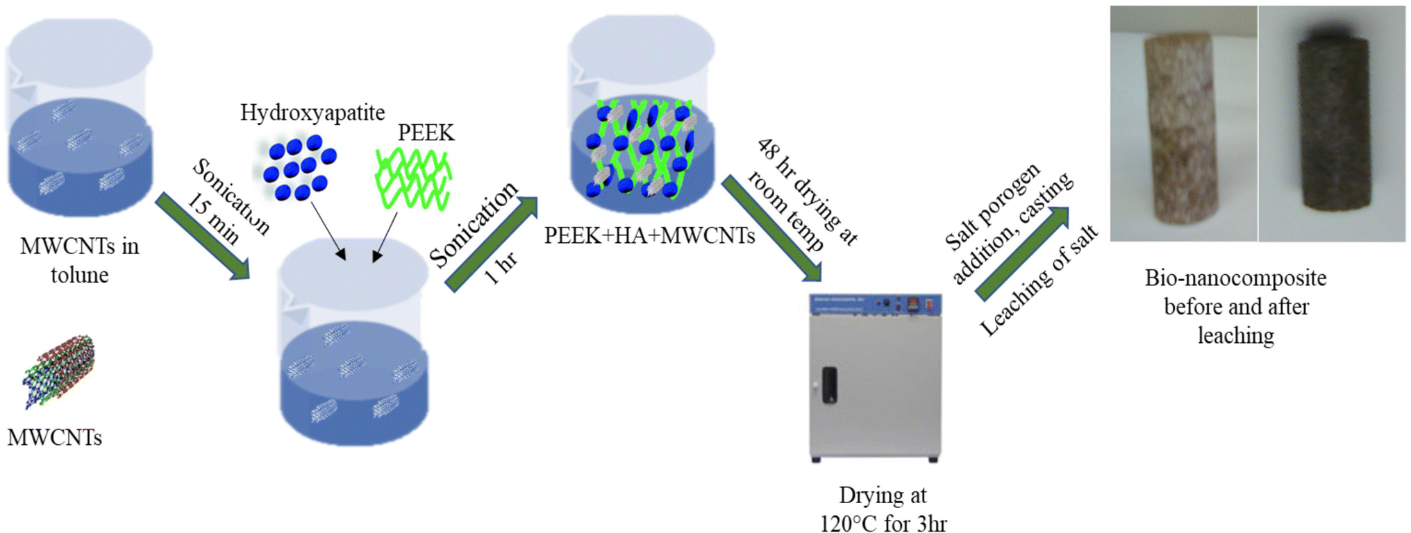
Abstract Bone regeneration is of great importance worldwide, because of various bone diseases, such as infections, tumors, and resultant fracture, birth defects, and bone loss due to trauma, explosion, or accident. Bone regeneration can be achieved by several materials and templates manufactured through various fabrication techniques. Uses of different materials and scaffold fabrication techniques have been explored over the past 20 years. In this research, polyetheretherketone (PEEK) was used to fabricate highly porous bionanocomposite foams for bone scaffolding. Melt casting and salt porogen (200–500 µm size) leaching methods were adapted to create an adequate pore size and the necessary percent of porosity, because pore size plays a vital role in cell implantation and growth. Porosity (75% and 85%) of the prepared scaffolds was adjusted by changing salt concentrations in the PEEK powder. Hydroxyapatite (HA) and carbon particles were used to improve cell attachments and interactions with the porous PEEK and to increase the mechanical properties of the scaffold materials. Carbon fiber (CF) and carbon nanotubes (CNTs) were uniformly dispersed into the PEEK powder before melt casting to enhance the mechanical properties and to observe the influence of the carbon particles on the properties of PEEK bionanocomposite foam. Compression test results of the fabricated bionanocomposites showed that HA and carbon particles are the potential filler materials for the enhancement of bionanocomposite mechanical properties. About 186% enhancement of compression modulus and 43% enhancement of yield strength were observed while incorporating only 0.5 wt% of CNTs into PEEK/HA bionanocomposites having 75% porosity, compared to PEEK/HA 20 wt% bionanocomposites. Micro-computed tomography (micro-CT) test results reveal that pore size and interconnectivity of the nanocomposite foams are in order and within the designed sizes. Mechanical tests proved that PEEK bionanocomposite foam has the potential for use in bone scaffolding and other biomedical applications.Keywords
- Bone regeneration,
- Hydroxyapatite,
- Melt casting,
- Salt leaching,
- PEEK,
- Porous scaffolds
References
- Abedin et al. (2015) Albumin-based micro-composite drug carriers with dual chemo-agents for targeted breast cancer treatment (pp. 38-49) https://doi.org/10.1177/0885328215569614
- Asmatulu et al. (2015) Synthesis and analysis of injection-molded nanocomposites of recycled high-density polyethylene incorporated with graphene nanoflakes (pp. 1565-1573) https://doi.org/10.1002/pc.23063
- Asmatulu et al. (2019) Highly sensitive and reliable electrospun polyaniline nanofiber based biosensor as a robust platform for COX-2 enzyme detections 20(5) (pp. 966-974) https://doi.org/10.1007/s12221-019-1096-x
- Bakar et al. (2003) Tensile properties and microstructural analysis of spheroidized hydroxyapatite-poly (ether ether ketone) biocomposites (pp. 55-63) https://doi.org/10.1016/S0921-5093(02)00289-7
- Bose et al. (2012) Recent advances in bone tissue engineering scaffolds 30(10) (pp. 546-554) https://doi.org/10.1016/j.tibtech.2012.07.005
- Boyan et al. (1996) Role of material surfaces in regulating bone and cartilage cell response (pp. 137-146) https://doi.org/10.1016/0142-9612(96)85758-9
- Converse et al. (2010) Hydroxyapatite whisker-reinforced polyetherketoneketone bone ingrowth scaffolds (pp. 856-863) https://doi.org/10.1016/j.actbio.2009.08.004
- Feng et al. (2016) A nano-sandwich construct built with graphene nanosheets and carbon nanotubes enhances mechanical properties of hydroxyapatite–polyetheretherketone scaffolds (pp. 3487-3500) https://doi.org/10.2147/IJN.S110920
- Huang et al. (2008) Preparation and properties of poly (lactide-co-glycolide) (PLGA)/nano-hydroxyapatite (NHA) scaffolds by thermally induced phase separation and rabbit MSCs cultured on scaffolds (pp. 409-432) https://doi.org/10.1177/0885328207077632
- Karageorgiou and Kaplan (2005) Porosity of 3D biomaterial scaffolds and osteogenesis (pp. 5474-5491) https://doi.org/10.1016/j.biomaterials.2005.02.002
- Keaveny TM, Morgan EF, Yeh OC (2004) Bone mechanics. The McGraw-Hill Companies, pp 1–24
- Mano et al. (2014) Bioinert, biodegradable and injectable polymeric matrix composites for hard tissue replacement: state of the art and recent developments (pp. 789-817) https://doi.org/10.1016/j.compscitech.2003.09.001
- Mozafari et al. (2010) Biomimetic formation of apatite on the surface of porous gelatin/bioactive glass nanocomposite scaffolds 257(5) (pp. 1740-1749) https://doi.org/10.1016/j.apsusc.2010.09.008
- Nga et al. (2015) Biomimetic scaffolds based on hydroxy-apatite nanorod/poly (d, l) lactic acid with their corresponding apatite-forming capability and biocompatibility for bone-tissue engineering (pp. 506-514) https://doi.org/10.1016/j.colsurfb.2015.03.001
- Oryan et al. (2014) Bone regenerative medicine: classic options, novel strategies, and future directions 9(1) https://doi.org/10.1186/1749-799X-9-18
- Pace et al. (2008) Technical and histologic analysis of a retrieved carbon fiber-reinforced poly-etherether-ketone composite alumina-bearing liner 28 months after implantation (pp. 151-155) https://doi.org/10.1016/j.arth.2006.07.012
- Polo-Corrales et al. (2014) Scaffold design for bone regeneration 14(1) (pp. 15-56) https://doi.org/10.1166/jnn.2014.9127
- Saiz et al. (2013) Perspectives on the role of nanotechnology in bone tissue engineering 29(1) (pp. 103-115) https://doi.org/10.1016/j.dental.2012.08.001
- Sarasua and Remiro (1995) The mechanical behavior of PEEK short fiber composites (pp. 3501-3508) https://doi.org/10.1007/BF00349901
- Scolozzi (2012) Maxillofacial reconstruction using polyetheretherketone patient-specific implants by “mirroring” computational planning (pp. 660-665) https://doi.org/10.1007/s00266-011-9853-2
- Seal et al. (2011) Polymeric biomaterials for tissue and organ regeneration (pp. 147-230) https://doi.org/10.1016/S0927-796X(01)00035-3
- Seraz et al. (2017) Investigating electrochemical behavior of antibacterial polyelectrolyte-coated magnesium alloys for biomedical applications 5(1) (pp. 23-33)
- Shuai et al. (2016) Polyetheretherketone/poly (glycolic acid) blend scaffolds with biodegradable properties, journal of biomaterials science 27(14) (pp. 1434-1446)
- Turner et al. (2001) Shear strength and fatigue properties of human cortical bone determined from pure shear tests 69(6) (pp. 373-378) https://doi.org/10.1007/s00223-001-1006-1
- Uddin et al. (2019) Synthesis, characterization, and applications of polymer-based biomaterials (pp. 143-182) Nova Science Publishers Inc
- Uddin et al. (2019) Effects of graphene oxide thin films and nanocomposite coatings on flame retardancy and thermal stability of aircraft composites (pp. 1-7) https://doi.org/10.1115/1.4042663
- Wei and Ma (2008) Nanostructured biomaterials for regeneration (pp. 3568-3582) https://doi.org/10.1002/adfm.200800662
- Williams (2008) Polyetheretherketone for long-term implantable devices 19(1)
- Wong et al. (2009) Mechanical properties and in vitro response of strontium-containing hydroxyapatite/polyetheretherketone composites (pp. 3810-3817) https://doi.org/10.1016/j.biomaterials.2009.04.016
- Zanello et al. (2016) Bone cell proliferation on carbon nanotubes 6(3) (pp. 562-567) https://doi.org/10.1021/nl051861e
- Zheng et al. (2015) Bone-like apatite coating on functionalized poly (etheretherketone) surface via tailored silaniza-tion layers technique (pp. 512-523) https://doi.org/10.1016/j.msec.2015.05.070