Results and discussion
Morphology and apparent porosity
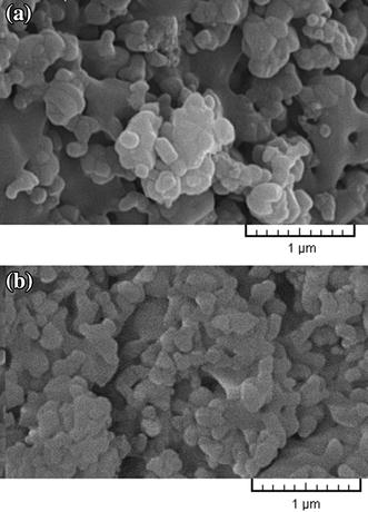
Microstructure and morphology of the surface of samples A and B were assessed by observation of SEM images. The SEM images (Fig.
1
) show that the surface of both types of samples is fully sintered, which is indicative of well-bonded particles and correct density and porosity of the samples. However, the particle size varies after sintering and average of pore size in the surface of samples A and B is between 100 and 300 nm. The difference in thermal expansion coefficient of the composite ingredients during heating and cooling can lead to formation of micro-cracks (Kwok et al.
2009
). In this study, to prevent formation of these cracks, samples were sintered at 1,000 °C for 40 min. As can be seen in Fig.
1
, there are no cracks on the surface of the samples. Table
3
shows the chemical composition of nanobiomaterials.
SEM images
a
sample A,
b
sample B before immersion in SBF EDAX results of nanobiomaterials before immersion in SBF Sample Ca (%) Ti (%) O2 (%) P (%) Al (%) A 7.76 26.53 53.77 4.45 7.49 B 13.43 18.91 53.38 7.1 7.18Fig. 1
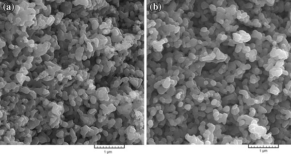
Table 3
The result of samples combination can also be visualized giving a map of elements of particles under the microscope. A homogeneous distribution of nanobiomaterial ingredients (P, Ca, Ti, Al) in structure of all samples is shown in Fig.
2
. The percentage porosity and density of the samples are presented in Table
4
. Sample B had a better particle size distribution, smoother surface, and less porosity (31.3 %) than sample A (41.99 %). An increase in weight percentage of the nano-sized TiO
2
compared to the micron-sized HA in nanobiomaterial A amplifies the likelihood of a porous surface (Harle et al.
2006
). Consequently, with increased percentage of porosity in sample A, vacant space is provided for growth and nutrition of bone cells. But, with reduced HA, the bioactive and osteoconductive properties of this implant were decreased. Samples A and B have densities of 1.994 and 2.016 g/cm
3
, respectively, which are very close to the bone density (1.85 g/cm
3
). Of course, density of sample A compared to sample B has decreased due to the use of nano-sized TiO
2
. Use of HA particles as matrix and addition of titania (30 wt%) and alumina (20 wt%) nanoparticles as reinforcing phase in sample B have improved bioactivity of implant in areas where better osteogenesis and stability of implant and more adhesion of nanobiomaterial to the bone are needed.
Elemental map
a
sample A,
b
sample B Percentage porosity and density of nanobiomaterials Sample Porosity (V %) Density (g/cm3) A 41.99 1.994 B 31.30 2.016Fig. 2
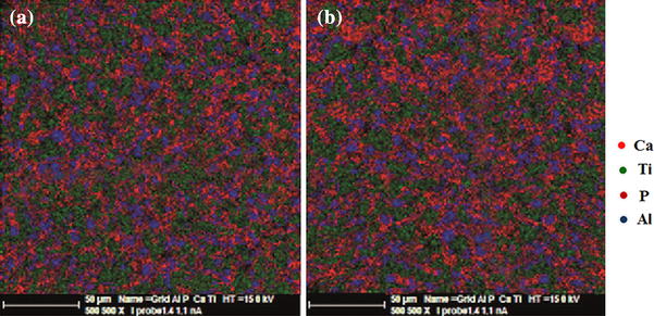
Table 4
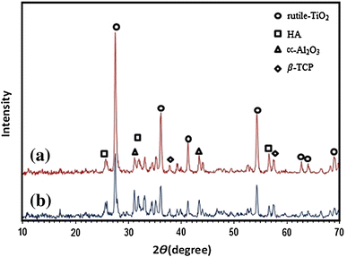
X-ray diffraction pattern
The X-ray diffraction patterns of A and B samples before immersion in SBF solution can be observed in Fig.
3
. Peaks associated with HA are detected with intensity near 2ϴ = 26° and 2ϴ = 32° in (201) and (211) planes, respectively. Also, β-TCP peak can be observed around 2ϴ = 31.5° in (221) plane, which is very close to HA peak. The TiO
2
peak with a high intensity is observed near 2ϴ = 36° in (110) crystal plane. Another reinforcement in nanobiomaterial is alumina that can be seen near 2ϴ = 38° and 2ϴ = 58° in (110) and (116) planes. But Al
2
O
3
has a bigger peak at 2ϴ = 35.5° that is integrated with Titania peak at 2ϴ = 36°. The diffraction pattern peak intensity of the TiO
2
is higher in sample A. It can be seen from the results that after sintering nanobiomaterial for 40 min at 1,000 °C, the nanobiomaterial ingredients maintained their nature, and β-TCP peak with a low intensity is observed in the samples. The intensity of β-TCP peak was higher in sample B due to higher percentage of HA. Also, there was not any peak around 2ϴ = 30° thus, calcium aluminate phase was not observed.
X-ray diffraction pattern of nanobiomaterial
a
sample A,
b
sample B before immersion in SBFFig. 3
Although HA is constant and stable in the body, the presence of secondary phases causes it to dissolve, which consequently leads to degeneration of the implant in body. Thus, increased sintering temperature followed by higher crystallization is necessary for longer life of the implant. Furthermore, sintering temperatures higher than 800 °C lead to decomposition of HA to β-TCP and α-TCP, whose presence in the sample reduces biocompatibility of the implant. Also, when samples are placed in biological environments like blood plasma, β-TCP becomes unstable and gradually degenerates. Thus, the presence of this phase could be considered responsible for the composite strength loss after exposure in SBF solution (Kwok et al. 2009 ). However, in this study, due to sintering temperature (1,000 °C), the intensity of β-TCP peak was low in the structure of both samples, but this weak peak cannot much affect biocompatibility of the samples. During high temperature processes (1,200–1,400 °C) in synthesis of nanomaterial, due to increased surface and reaction area, new phases such as calcium aluminates are created that reduce mechanical properties of the material (Viswanath and Ravishankar 2006 ). In this study, to prevent formation of secondary phases such as calcium aluminate phase, sintering operations were conducted at 1,000 °C.
In vitro bioactivity evaluation
The in vitro bioactive behavior and formation of calcium phosphate phase on the surface of nanobiomaterial in SBF solution were evaluated by SEM images and EDXA analysis. Figure
4
shows SEM images of A and B samples after immersion in SBF solution, and formation of apatite can be observed on both of surfaces. But, there is a considerable difference in ability to form apatite in sample B compared to A. The higher percentage of HA and TiO
2
nanoparticles present in sample B is due to full coverage of its surface by apatite, while in sample A, apatite is scattered in different areas of surface. Researchers have recently reported (Kong et al.
2006
) that the increase in HA in composites encourages the formation of apatite on the surface of samples in SBF solution.
SEM images
a
sample A,
b
sample B after immersion in SBFFig. 4
The biological activity of bioceramics is due to their ability to promote the formation of HA in physiological environments (SBF) (Fujibayashi et al.
2003
; Rámila and Vallet-Regi´
2001
). In this study, formation of apatite and calcium phosphates on ceramic nanobiomaterial was assessed and the relationship between apatite formation and bioactivity of nanobiomaterial was identified. It can be seen from EDAX analysis (Table
5
) that calcium and phosphorus levels significantly increased after 7 days immersion of samples in SBF compared to their levels before immersion (Table
3
). Also, the percentages of Ti, O
2
and Al decreased with formation of an apatite-like material on the nanobiomaterial surface. The ratio of calcium to phosphorus is almost 1.7 and this ratio did not change much in the samples after immersion in SBF. Calcium ion concentration is controlled by the formation of apatite layer in SBF and its release from the samples (Martínez et al.
2000
).
EDXA results of nanobiomaterials after 7 days immersion in SBF Sample Ca (%) Ti (%) O2 (%) P (%) Al (%) A 18.26 20.75 47.61 9.74 3.64 B 25.6 8.33 49.26 14.62 2.19Table 5
Surface hardness and reasons for fabrication of nanobiomaterial with 3 materials
Table
6
presents the results of the surface hardness before and after sintering operation. The surface hardness of both A and B samples nearly doubled after sintering. Obviously, this increase in surface hardness was due to sintering. However, non-homogeneous distribution of powders in nanobiomaterial structures could cause a reduction in hardness and strength of the samples. The surface hardness of sample B before and after sintering was almost half that of sample A, and since the amount of Al
2
O
3
was constant in both of samples; this may have been due to reduction in the amount of TiO
2
in sample B. Another reason for reduced hardness in sample B compared to sample A could be higher formation of beta-tri-calcium phosphate phase in sample B because of increasing in the amount of HA.
The surface hardness of nanobiomaterials before and after sintering operation Sample Hardness before sintering (GPa) Hardness after sintering (GPa) A 3.09 7.01 B 1.54 4.25Table 6
HA is widely used as a biomaterial in medical applications, and alone can amplify bioactivity and tendency to absorb biological materials like protein but, when HA coats on titanium substrate, mechanical properties (fracture toughness) and adhesion strength are weak. Research shows that the presence of TiO 2 alongside HA in composite and HA coating on Ti surface causes increased adhesion strength and corrosion resistance (Wen et al. 2007 ). In addition, it can elevate surface hardness without compromising formation of apatite. The presence of Al 2 O 3 in the sample does not affect calcinations process (Li et al. 1995 ). Besides, addition of 20 % Al 2 O 3 almost doubled fracture toughness and HA strength (Viswanath and Ravishankar 2006 ). As reinforcement in composites, TiO 2 is able to absorb H 2 O and form Ti–OH groups on the surface, which eventually leads to the formation of apatite in SBF solution and enhances bioactivity of samples (Madhan Kumar and Rajendran 2013 ). Therefore, this reinforcement material has an important role in adhesion of implant to the bone (Beherei et al. 2009 ).Particle size and distribution of ingredients in composites affect their strengths. Thus, in this study, 3-phase ceramic HA–TiO 2 –Al 2 O 3 was fabricated.
In vitro behavior of nanobiomaterials
SEM observation revealed the MG-67cell attachment, growth and spreading on different sample surfaces. Figure
5
shows the morphologies of the cells on samples after 3 days of culture. Cells were flattened and well spread across the A and B sample surfaces after 3 days. On both the A and B sample surfaces, cells were seen to adhere to each other with cellular micro-extensions (e.g., filopodia), and were connected to the substrate in addition to neighboring cells. Cells were also found to have migrated into the pores and were observed to bridge them. However, on the A surface, relatively fewer cells were observed (Fig.
6
a), but on the B sample, as is shown in the high-magnification SEM image in Fig.
6
b, cells were observed to grow into the pores. On the B surface cell numbers were increased, and the entire sample surface was covered with cells having numerous filopodia extensions attached to the surface irregularities, as shown in Fig.
5
b.
SEM micrographs illustrating MG-67cell morphology after 3 days of culture on
a
sample A, and
b
sample B high-magnification SEM micrographs illustrating MG-67cell morphology after 3 days of culture on
a
sample A, and
b
sample BFig. 5
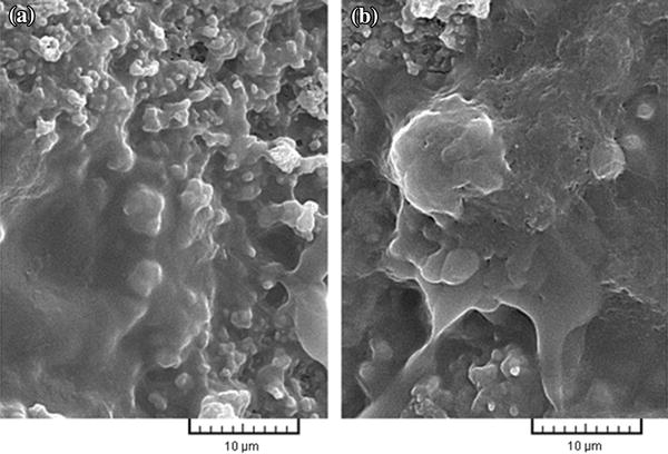
Fig. 6
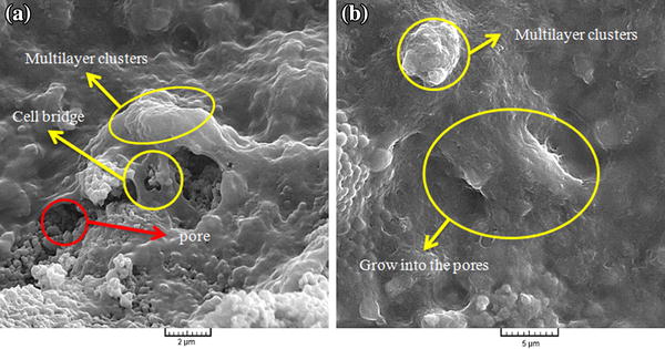
After 3 days of culture, cells appeared to be more elongated and confluent on B, as shown in Fig. 6 b. Evidently, granules were deposited on the cell surfaces because of extracellular matrix (ECM) mineralization. In contrast, on sample A, cells never reached complete confluence over the entire sample surface because of the higher pore volume. As shown in Fig. 6 , on both of samples, cells formed multilayer clusters (i.e., Cells appeared cuboidal and had a three-dimensional morphology with more filopodia) on the smooth surface area. Also, nano-scale grains and high volume fraction of grain boundaries in HA nanomaterial can increase adhesion of osteoblasts, cell proliferation, and mineralization of these composites (Zhang and Kwok 2011 ).
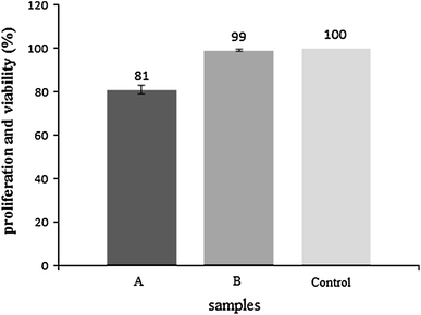
The MTT assay was used to quantitatively determine the proliferation of viable MG-67cells on the A and B sample surfaces. Figure
7
shows a comparison of viable cell densities for samples with negative control after 3 days of culture. For culture durations, cells proliferated in greater numbers on the sample B compared to the sample A. It was observed that, cells proliferated most rapidly on the samples with the highest porosity, i.e., on B in comparison to A sample. Therefore, the results revealed that both nanobiomaterials can be used in different medical applications.
MTT assay of cells on A and B samples after 3 days of incubationFig. 7