Department of Chemistry, Shahre Qods Branch, Islamic Azad University, Tehran, IR
Abstract
In this paper, three kinds of multi-walled carbon nanotubes (MWCNTs) with different diameters (outer diameters: 10 to 20, 30 to 50, and >50 nm) and special surface areas (200, 60, and 40 m
2
/g, respectively) were oxidized in commonly used liquid oxidizers: (1) concentrated nitric acid, (2) a mixture of nitric + sulfuric acids (
V
:
V
, 1:3), (3) hydrogen peroxide, (4) a mixture of hydrogen peroxide + sulfuric acid (
V
:
V
, 1:1), and (5) acidic potassium permanganate. Morphology of the pristine and oxidized MWCNTs was characterized by scanning electron microscopy which provides sufficient resolution for direct visualization of their outer diameter distribution. Full width at half maximum (FWHM) in the X-ray diffraction (XRD) investigation of the MWCNT samples before and after the oxidation process was measured. After treatment with oxidants, a clear decrease in nanotube diameters along the tube walls was observed. Decrease in the degree of crystallites started with the FWHM widening of the XRD diffraction peaks. The particle (crystallite) size (
d002
) calculated by Bragg's law and Scherrer equation increased depending on the kind of oxidants; the procedure can be performed using a mixture of HNO
3
+ H
2
SO
4
on the surface of the MWCNTs with an outer diameter of 10 to 20nm. However, similar are the diffraction patterns of pristine and oxidized MWCNTs. Therefore, the MWCNTs that underwent oxidation process were able to preserve the first features of their structures, even though some narrowing of outer diameters and decreases in crystallites appeared. Nevertheless, the structure of MWCNTs still remains intact to be used as oxidized nanotubes in most applications.
Background
Carbon nanotubes (CNTs) are divided into two types, namely single-walled CNTs (SWCNTs) and multi-walled CNTs (MWCNTs). SWCNTs are a graphite sheet rolled into a cylinder of a few micrometers in length and a few nanometers in diameter. MWCNTs consist of several such cylinders nested inside each other. MWCNTs have attracted great attention as a new kind of nanomaterial since their discovery in 1991. Carbon nanotubes have unique mechanical, electrical, magnetic, optical, and thermal properties. Owing to their inert nature, the nanotubes tend to form bundles with each other and thus do not disperse well in organic matrices in their pristine state. Suitable enhancement of the surface of MWCNTs is thus required in order to optimize their dispersion in the organic matrices. Out of various possible ways to achieve surface functionalization, chemical oxidation means of surface modification are quite common [
1
–
4
]. In this case, in typical experiments, MWCNTs with different diameters were oxidized under reflux condition in different oxidants [
1
–
29
]. The pristine and oxidized MWCNTs were then investigated with regard to their crystallites and morphologies via X-ray diffraction (XRD) and scanning electron microscopy (SEM), respectively. The objective of this research is to study the morphological and crystallographic structure of oxidized MWCNT array that will be used as the first step for other functionalization and applications.
Results and discussion
Morphology and crystallographic structure of MWCNTs
SEM
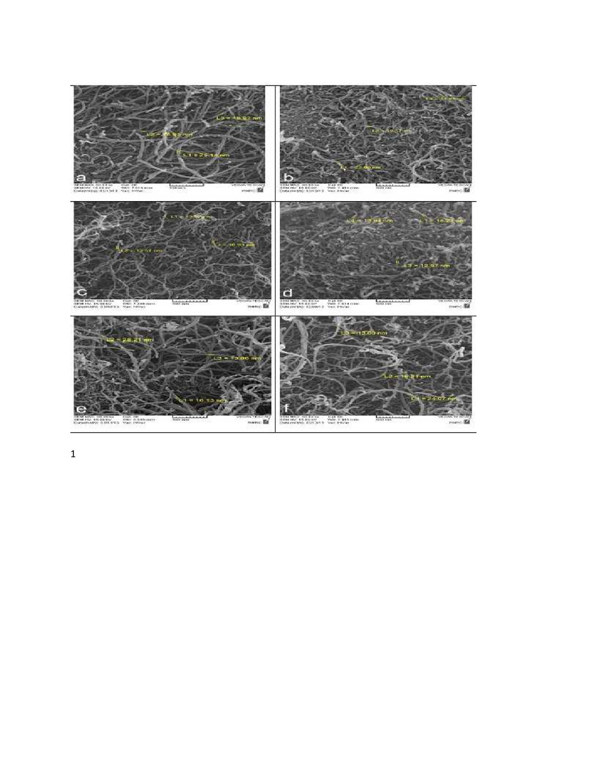
The morphology of MWCNTs before and after oxidation was characterized by SEM. The outer diameter (OD) of the carbon nanotubes varied in the three kinds of MWCNTs (10 to 20, 30 to 50, and >50 nm). The average diameter of MWCNTs before and after oxidation was measured (Table
1
). Figures
1
,
2
, and
3
show the SEM images of MWCNTs. The nanotubes are pure and only carbon nanotubes were observed. After treatment with acids, a clear change in diameter and surface roughness along the tube walls was observed. Through the oxidation process, the diameters of MWCNTs were narrowed down gradually. Table
1
presents the erosion of nanotube surface during oxidation [
1
–
3
].
Table 1
Average diameters of MWCNTs
(
narrowing of nanotube diameters during oxidation
)
Kind of MWCNTs
Average MWCNT diameters (nm)
Outer diameter = 10 to 20 nm
Pristine
20.66
Oxidized with HNO3
16.47
Oxidized with HNO3 + H2SO4
14.25
Oxidized with H2O2
14.90
Oxidized with H2O2 + H2SO4
19.40
Oxidized with KMnO4
18.71
Outer diameter = 30 to 50 nm
Pristine
40.70
Oxidized with HNO3
35.95
Oxidized with HNO3 + H2SO4
34.62
Oxidized with H2O2
38.34
Oxidized with H2O2 + H2SO4
36.42
Oxidized with KMnO4
38.42
Outer diameter > 50 nm
Pristine
74.82
Oxidized with HNO3
47.94
Oxidized with HNO3 + H2SO4
50.25
Oxidized with H2O2
59.44
Oxidized with H2O2 + H2SO4
50.63
Oxidized with KMnO4
55.63
Figure 1
SEM images of MWCNTs with an outer diameter of 10 to 20 nm. (a)
Pristine,
(b)
oxidized with HNO
3
,
(c)
oxidized with HNO
3
+ H
2
SO
4
,
(d)
oxidized with H
2
O
2
,
(e)
oxidized with H
2
O
2
+ H
2
SO
4
, and
(f)
oxidized with KMnO
4
.
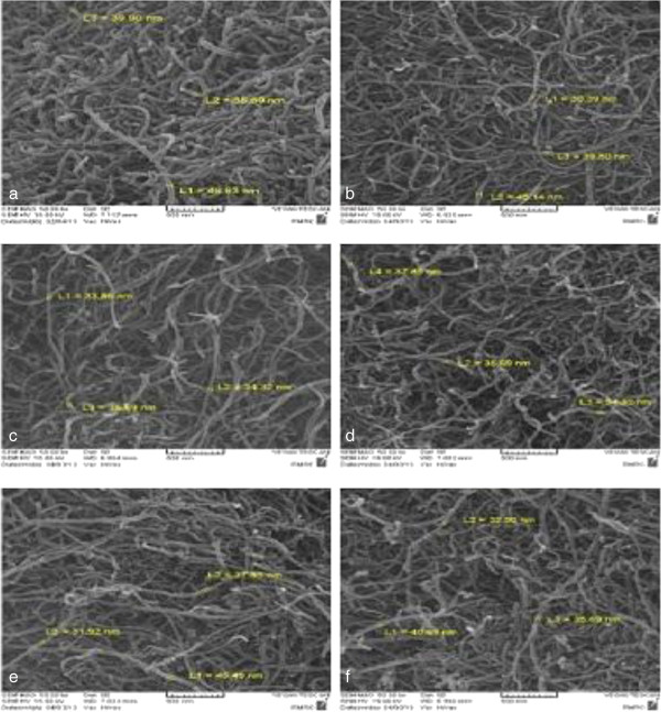
Figure 2
SEM images of MWCNTs with an outer diameter of 30 to 50 nm. (a)
Pristine,
(b)
oxidized with HNO
3
,
(c)
oxidized with HNO
3
+ H
2
SO
4
,
(d)
oxidized with H
2
O
2
,
(e)
oxidized with H
2
O
2
+ H
2
SO
4
, and
(f)
oxidized with KMnO
4
.
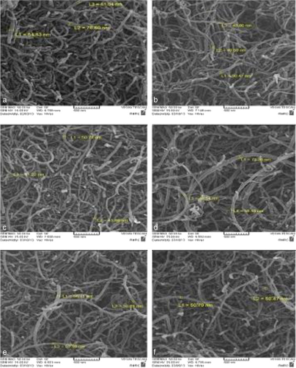
Figure 3
SEM images of MWCNTs with an outer diameter>50 nm. (a)
Pristine,
(b)
oxidized with HNO
3
,
(c)
oxidized with HNO
3
+ H
2
SO
4
,
(d)
oxidized with H
2
O
2
,
(e)
oxidized with H
2
O
2
+ H
2
SO
4
, and
(f)
oxidized with KMnO
4
.
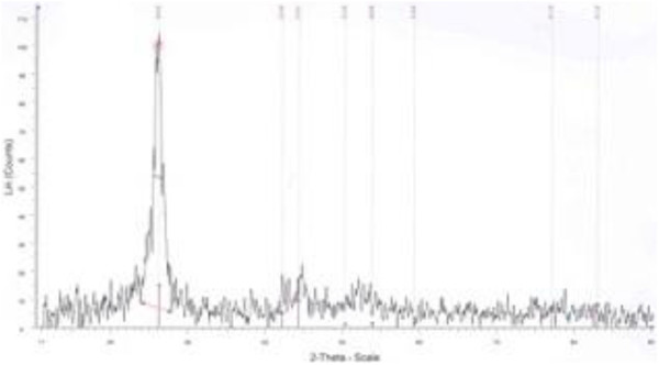
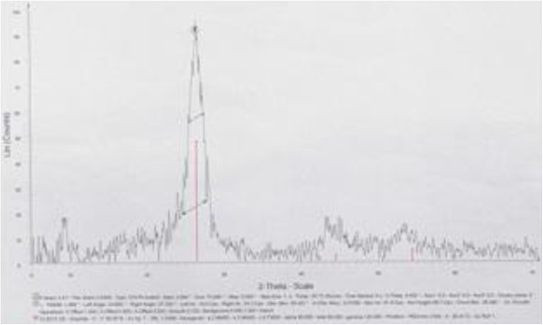
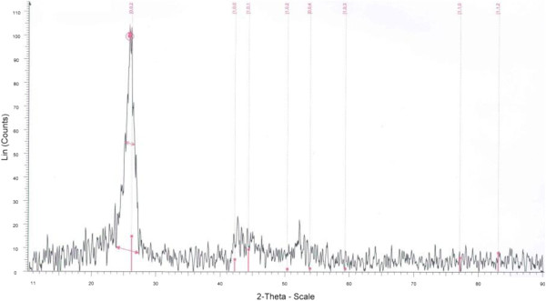
XRD
Figures
4
,
5
,
6
,
7
,
8
,
9
,
10
, and
11
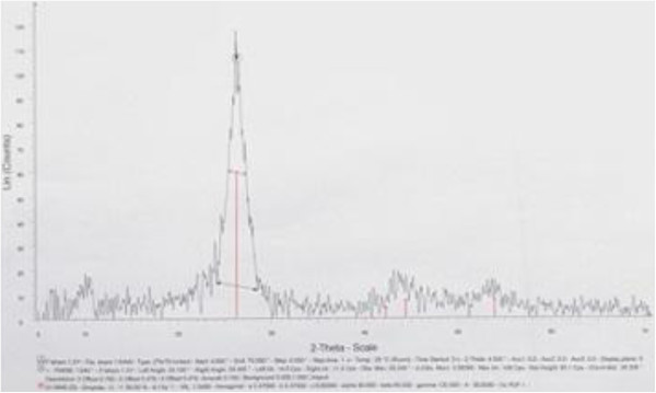
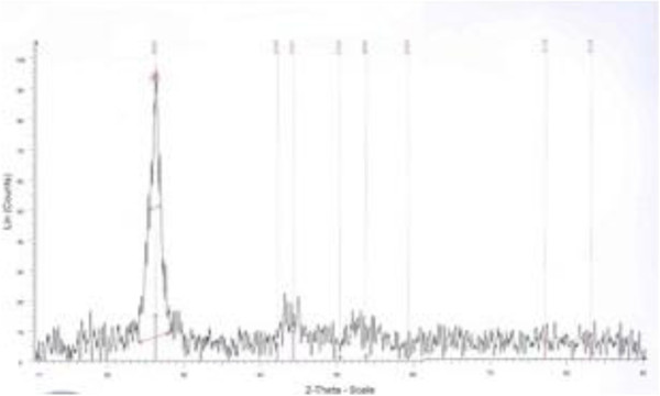
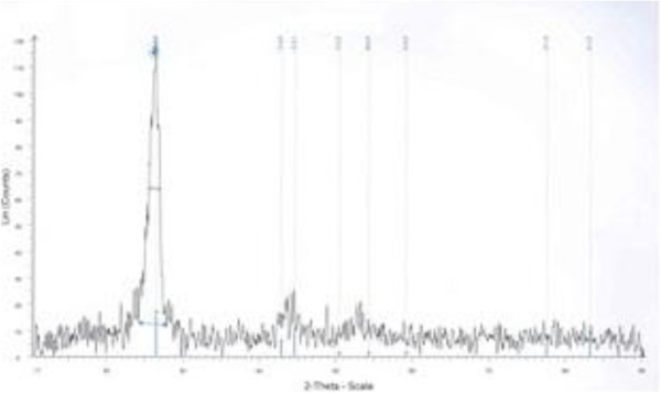
show the XRD profiles of the MWCNTs. It can be found that the pristine and oxidized samples possess a structure similar to that of graphite crystal, which indicates that the functionalization process does not change the bulk structure of the MWCNTs. The strongest and sharpest diffraction peak for all samples at around 2
θ
= 26.3° could be indexed as the C (002) reflection of graphite. The sharpness of the C (002) peak indicates that the graphite structure of MWCNTs was acid-oxidized without significant damage. XRD was used to measure the crystal size and interlayer spacing. Due to the CNT's intrinsic nature, the main features of the X-ray diffraction pattern of CNTs are close to those of graphite, as shown in Figures
4
,
5
,
6
,
7
,
8
,
9
,
10
, and
11
. A comparison between Figure
4
and the others shows that a graphite-like peak (0 0 2) is present at approximately 26° in 2
θ
. Measurements of crystal size (
d
) can be achieved from this peak (Tables
2
and
3
) and the Scherrer equation (Equation 1). As mentioned above, the average crystallite size of the MWCNTs was determined using the XRD patterns, via the well-known Scherrer equation:
d=kλβcosθ=?nm
where
β
is the full width at half maximum (FWHM),
θ
is the diffraction angle,
λ
is the wavelength (1.54 Å),
d
is the particle (crystallite) size, and
k
is the Scherrer constant (0.91).
Figure 4
XRD profiles of the MWCNTs (OD = 10 to 20 nm) before functionalization.
CuKα, graphite.
Figure 5
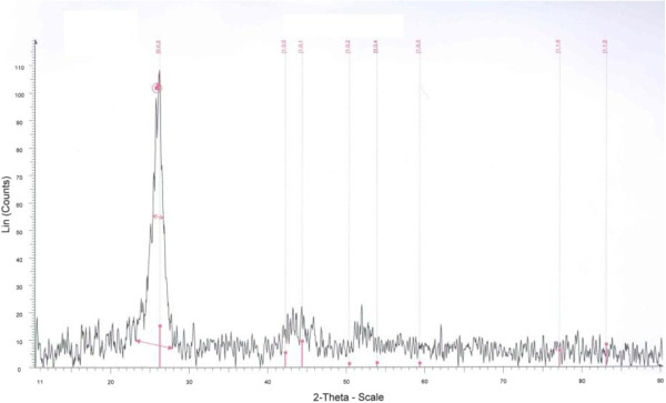
XRD profiles of the MWCNTs (OD = 10 to 20 nm) after functionalization with HNO3+H2SO4.
CuKα, graphite.
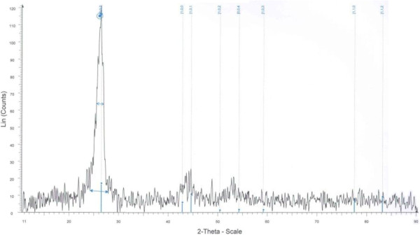
Figure 6
XRD profiles of the MWCNTs (OD = 10 to 20 nm) after functionalization with HNO3.
CuKα, graphite.
Figure 7
XRD profiles of the MWCNTs (OD = 10 to 20 nm) after functionalization with KMnO4.
CuKα, graphite.
Figure 8
XRD profiles of the MWCNTs (OD = 10 to 20 nm) after functionalization with H2O2.
CuKα, graphite.
Figure 9
XRD profiles of the MWCNTs (OD = 10 to 20 nm) after functionalization with H2O2+H2SO4.
CuKα, graphite.
Figure 10
XRD profiles of the MWCNTs (OD = 30 to 50 nm) after functionalization with HNO3.
CuKα, graphite.
Figure 11
XRD profiles of the MWCNTs (OD> 50 nm) after functionalization with HNO3.
CuKα, graphite.
Table 2
Particle
(
crystallite
)
size of MWCNTs with the same diameter and different oxidants
MWCNTs (OD = 10 to 20 nm)
d(C002) (nm)
Pristine
70.07
Oxidized with HNO3 + H2SO4
42.46
Oxidized with HNO3
43.79
Oxidized with KMnO4
51.903
Oxidized with H2O2
58.39
Oxidized with H2O2 + H2SO4
58.39
Table 3
Particle
(
crystallite
)
size of MWCNTs with the same oxidant and different MWCNT diameters
MWCNTs oxidized with HNO3
d(C002) (nm)
OD = 10 to 20 nm
43.79
OD = 30 to 50 nm
63.7
OD > 50 nm
63.7
The Scherrer equation is derived from Bragg's law, and it is limited to nanoscale particles only. It is known that a decrease in the order of crystallinity in carbon materials will make the XRD peaks broader. Accordingly, all treated samples had a wider FWHM, which implies that the oxidation of MWCNTs had actually deteriorated the degree of crystallinity. However, amounts of it are very little (Tables
4
and
5
) [
2
,
27
].
Table 4
The FWHM width with the same MWCNT and different oxidants
MWCNTs (OD = 10 to 20 nm)
FWHM (C002)
2θ
Pristine
1.245°
26.146°
Oxidized with HNO3 + H2SO4
1.958°
26.422°
Oxidized with HNO3
1.940°
26.245°
Oxidized with KMnO4
1.622°
26.208°
Oxidized with H2O2
1.443°
25.808°
Oxidized with H2O2 + H2SO4
1.480°
26.337°
Table 5
The FWHM width with the same oxidant and different MWCNT diameters
MWCNTs oxidized with HNO3
FWHM (C002)
2θ
OD = 10 to 20 nm
1.940°
26.245°
OD = 30 to 50 nm
1.367°
26.170°
OD > 50 nm
1.336°
26.013°
Conclusions
Oxidation of carbon nanotubes was used as a common step in the functionalization process to increase their solubility and compatibility with different materials. However, this procedure should be used with certain caution as it can result in the destruction of the nanotube structure in the case of elevated temperatures and increased oxidation time. In this paper, the structure and morphology of pristine and oxidized MWCNTs were studied using SEM and XRD analyses. SEM examinations on MWCNTs showed that after oxidation process, the diameter of oxidized MWCNTs begin to narrow. However, comparing the SEM images of the pristine MWCNTs and that of the oxidized MWCNTs, there are practically no visual differences between them. XRD and SEM analyses revealed that the chemical treatment did not induce structural damages to the nanotubes. According to the XRD patterns, the least damaging oxidation process for creating hydrophilic sites on the hydrophobic surface of MWCNTs can be achieved using acidic potassium permanganate under reflux and mild oxidation of MWCNTs. The XRD patterns were taken to reveal detailed information about the crystallographic structure of MWCNTs. The 2
θ
ranged from 10° to 90°, where
θ
is the diffraction angle. The strongest and sharpest diffraction peak for all samples at around 2
θ
= 26° could be indexed as the C (0 0 2) reflection of graphite. The sharpness of the C (0 0 2) peak indicates that the graphite structure of the MWCNTs were acid-oxidized without significant damage. The crystallite size particle (
d002
) calculated by Bragg's law changed depending on the kind of oxidants and MWCNTs. It is known that a decrease in the order of crystallinity in carbon materials will make the XRD peaks broader. Accordingly, all treated samples have either a smaller
d002
or a wider FWHM which implies that the oxidation of MWCNTs had actually deteriorated the degree of crystallinity. Moreover, the process seemed to start with widening the FWHM, followed by shifting the C (0 0 2) diffraction towards lower angles. Furthermore, this phenomenon was more significant with oxidation using the mixture HNO
3
+ H
2
SO
4
. These results show that there were more apparent structural changes after acid oxidation with the mixture HNO
3
+ H
2
SO
4
according to the SEM images.
Methods
Material
Multi-walled carbon nanotube features are listed in Table
6
.
Table 6
Multi-walled carbon nanotube features
Specimen 1
Specimen 2
Specimen 3
Purity
>95%
>95%
>95%
Outer diameter
10 to 20 nm
30 to 50 nm
>50 nm
Special surface area
200 m2/g
60 m2/g
40 m2/g
Preparation method
Chemical vapor deposition
Chemical vapor deposition
Chemical vapor deposition
Acid treatment of MWCNTs
Separately, three types of MWCNTs (2.0 g) with different diameters were dispersed in a 200-ml solution of 8 M HNO
3
in a round-bottom flask and refluxed at 60°C for 48 h with continuous stirring (200 rpm) and ultrasonicated in an ultrasonic bath (300 W, 50 kHz) to obtain carboxyl functional groups. Upon cooling, the mixture was thoroughly washed with deionized water to remove traces of untreated acid until the pH value was 7, which signifies zero acidity. Then oxidized MWCNTs were filtered through a centrifuge and a polycarbonate filter (Whatman, pore size 0.2 μm). Then the samples were dried at 120°C in a vacuum oven for 24 h. To compare with the oxidation in HNO
3
, the same process was performed in a mixture of HNO
3
(50 ml) + H
2
SO
4
(150 ml) (
V
:
V
, 1:3), a mixture of H
2
O
2
(100 ml) + H
2
SO
4
(100 ml) and 18% H
2
O
2
(200 ml) at 120°C, and also acidic KMnO
4
(200 ml) in 95°C for 48 h. In the case of functionalization in KMnO
4
, the obtained solid mixture was washed with water, filtered, rewashed with concentrated HCl to remove the produced MnO
2
, then refiltered to remove the produced MnO
2
, and then refiltered again. [
1
–
4
], [
11
], [
18
], [
23
], [
29
]
Characterization methods
Scanning electron microscopy
To investigate the morphologies of MWCNTs before and after oxidation, the SEM model VEGA (TESCAN, Brno, Czech Republic) with a field emission gun was used (without gold-coated samples because the diameter of MWCNTs seems bigger with gold coating).
X-ray diffraction
XRD patterns were taken with an X-ray powder diffractometer (model GNR MPD 300, GNR Analytical Instruments Group, Novara, Italy) to reveal detailed information about the crystallographic structure of the material. The radiation used was CuKα with a wavelength of 1.54 Å. The 2
θ
ranged from 10° to 90°, where
θ
is the diffraction angle. The test condition is shown in Table
7
[
2
,
27
].
Table 7
XRD test condition
Parameters
Reference standard
EN 13925–1:2003
Sample preparation
With crushing
No
Without crushing
Yes
Radiation
Cu voltage
40 kV
Current
30 mA
2θ
10° to 90°
Step size
0.05°
Coating time
0.5 s
λ (wavelength)
1.54 Å
k (Scherrer constant)
0.91
Acknowledgments
The authors wish to acknowledge Professor P. Davami (Razi Metallurgical Research Center, Tehran, Iran) for providing technical assistance provided and also for conducting the SEM, XRD, FT-IR, TGA, and titration equipment and experiment.
Competing interests
Both authors declare that they have no competing interests.
Authors’ contributions
H KH carried out the result analysis, participated to draft the manuscript and in the manuscript elaboration, carried out the experiments, and obtained most of the experimental images. O M coordinated the project, discussed the results, and helped to draft the manuscript. Both authors read and approved the final manuscript.
References
Ilya et al. (2012) Oxidation behavior of multiwall carbon nanotubes with different diameters and morphology (pp. 6272-6280) 10.1016/j.apsusc.2012.03.021
Chen et al. (2012) Influence of surface functionalization via chemical oxidation on the properties of carbon nanotubes (pp. 32-38) 10.1016/j.jcis.2011.12.073
Datsyuk et al. (2008) Chemical oxidation of multiwalled carbon nanotubes (pp. 833-840) 10.1016/j.carbon.2008.02.012
Avilés et al. (2009) Evaluation of mild acid oxidation treatments for MWCNT functionalization (pp. 2970-2975) 10.1016/j.carbon.2009.06.044
Dandekar et al. (1998) Characterization of activated carbon, graphitized carbon fibers and synthetic diamond powder using TPD and DRIFTS (pp. 1821-1831) 10.1016/S0008-6223(98)00154-7
Pereira et al. (2010) Characterization of carbon nanotube 3D-structures infused with low viscosity epoxy resin system (pp. 2252-2257) 10.1016/j.compstruct.2009.08.009
Tsang et al. (1994) A simple chemical method of opening and filling carbon nanotubes 372(6502) (pp. 59-62) 10.1038/372159a0
Moreno-Castilla et al. (1998) Effects of non-oxidant and oxidant acid treatments on the surface properties of an activated carbon with very low ash content (pp. 145-151) 10.1016/S0008-6223(97)00171-1
Zhang et al. (2003) Effect of chemical oxidation on the structure of single-walled carbon nanotubes (pp. 3712-3718) 10.1021/jp027500u
Aksoylu et al. (2001) The effects of different activated carbon supports and support modifications on the properties of Pt/AC catalysts (pp. 175-185) 10.1016/S0008-6223(00)00102-0
Figueiredo et al. (2007) Characterization of active sites on carbon catalysts (pp. 4110-4115) 10.1021/ie061071v
Chen et al. (1998) Solution properties of single-walled carbon nanotubes 282(5386) (pp. 95-98) 10.1126/science.282.5386.95
Cuervo et al. (2008) Modification of the adsorption properties of high surface area graphites by oxygen functional groups (pp. 2096-2106) 10.1016/j.carbon.2008.08.025
Zhao et al. (2004) Synthesis and properties of a water-soluble single-walled carbon nanotube-poly(maminobenzene sulfonic acid) graft copolymer 14(1) (pp. 71-76) 10.1002/adfm.200304440
Osorio et al. (2008) H2SO4/HNO3/HCl—functionalization and its effect on dispersion of carbon nanotubes (pp. 2485-2489) 10.1016/j.apsusc.2008.07.144
Lin et al. (2006) Functionalizing multi-walled carbon nanotubes with poly(oxyalkylene)- amidoamines 17(13) (pp. 3197-3203) 10.1088/0957-4484/17/13/020
Liang et al. (2005) Preparation and characterization of multi-walled carbon nanotubes supported PtRu catalysts for proton exchange membrane fuel cells (pp. 3144-3152) 10.1016/j.carbon.2005.06.017
Nobili et al. (1990) Characterization of electrophoretic fractions of humic substances with different electrofocusing behaviour 150(5) (pp. 763-770) 10.1097/00010694-199011000-00002
Ötvös et al. (2006) Surface oxygen complexes as governors of neopentane sorption in multiwalled carbon nanotubes (pp. 1665-1672) 10.1016/j.carbon.2006.01.005
Figueiredo et al. (1999) Modification of the surface chemistry of activated carbons (pp. 1379-1389) 10.1016/S0008-6223(98)00333-9
Tran et al. (2007) Thermal oxidative cutting of multi-walled carbon nanotubes 45(12) (pp. 2341-2350) 10.1016/j.carbon.2007.07.012
Toebes et al. (2004) The influence of oxidation on the texture and the number of oxygen-containing surface groups of carbon nanofibers (pp. 307-315) 10.1016/j.carbon.2003.10.036
Ajayan et al. (1993) Opening carbon nanotubes with oxygen and implications for filling 362(6420) (pp. 522-525) 10.1038/362522a0
Barroso-Bujans et al. (2007) Degree of functionalization of carbon nanofibers with benzenesulfonic groups in an acid medium (pp. 1669-1678) 10.1016/j.carbon.2007.03.039
Hu et al. (2003) Nitric acid purification of single-walled carbon nanotubes 107(50) (pp. 13838-13842)
Yu-Chun et al. (2011) The influence of treatment duration on multi-walled carbon nanotubes functionalized by H2SO4/HNO3 oxidation (pp. 2401-2410) 10.1016/j.apsusc.2010.09.110
Wang et al. (2007) Adsorption of fulvic acids from aqueous solutions by carbon nanotubes 82(8) (pp. 698-704) 10.1002/jctb.1708
Li et al. (2008) Oxidation of single-walled carbon nanotubes in dilute aqueous solutions by ozone as affected by ultrasound (pp. 466-475) 10.1016/j.carbon.2007.12.012