Department of Medical Biotechnology, School of Advanced Medical Technologies, Tehran University of Medical Sciences, Tehran, 1417614411, IR
Department of Materials Engineering, University of Tarbiat Modares, Tehran, 14115-143, IR
Biotechnology Research Center, Faculty of Pharmacy, Tehran University of Medical Sciences, Tehran, 1417614411, IR
Pharmaceutical Sciences Research Center, School of Pharmacy, Tehran University of Medical Sciences, Tehran, 417614411, IR
Abstract
Some undesired nanoparticles, such as malformed structures or incomplete core/shell Fe
3
O
4
-Au nanocomposites, might be formed when Fe
3
O
4
-Au core/shell nanocomposites are being synthesized. These impurities should be separated before any applications are performed. In this investigation, magnetic cores (Fe
3
O
4
) were synthesized using a conventional fabrication method involving coprecipitation of Fe
2+
and Fe
3+
. Carboxyl-capped magnetite-gold composite nanoparticles, measuring ≤50 nm, were then synthesized using a plant extract (
Eucalyptus camaldulensis
). The prepared carboxyl-capped magnetite-gold composite nanoparticles were further subjected to acid treatment for 18 h and characterized with different instrumentation methods. The results of the investigation showed that acid treatment can be applied successfully to separate defect-free Fe
3
O
4
-Au core/shell nanoparticles from different types of Fe
3
O
4
-Au nanocomposites, without any considerable changes in their physiochemical properties.
Background
Magnetic nanoparticles (MNPs) have been known as one of the nanostructures that provide the widest uses in biomedicine[
1
–
5
]. For example, MNPs that are composed of magnetite (Fe
3
O
4
) or maghemite (
γ
-Fe
2
O
3
) have unique thermal, chemical, and magnetic properties that make these particles particularly well suited for medical applications[
5
,
6
]. However, producing nanoparticles (NPs) with high stability and biocompatibility presents one of the greatest challenges. To address this, MNPs should be covered with an external quite inert shell, in order to protect the magnetic core against chemical changes. In addition, the surfaces of these particles should be functionalized with organic molecules, to allow them to bind chemically to other biomolecules such as DNA, proteins, amino acids, etc.[
7
–
9
]. Gold is a chemically quite inert element, and no single acid dissolves it. This metal is resistant to chemical reactions that take place in biological fluids and is a very useful element for providing a coating layer to protect MNPs[
10
,
11
]. Also, gold metal does not decrease the magnetic properties of Fe
3
O
4
MNPs and, consequently, will not limit the applications in which these particles are used[
12
,
13
].
At present, a number of different chemical and biological methods have been reported for fabricating magnetite Fe
3
O
4
-Au composite NPs[
4
,
6
,
9
]. These nanocomposites contain both defect-free Fe
3
O
4
core@Au shell MNPs and other types of Fe
3
O
4
-Au nanocomposites such as malformed structures or incomplete core/shell Fe
3
O
4
-Au nanocomposites (Figure
1
). As mentioned above, the importance of the external inert shell in protecting the magnetic core is well established, and different reports have been published on the synthesis of Fe
3
O
4
core@Au shell MNPs[
4
,
13
]. However, in these studies, no further treatments such as acid washing have been applied for separating the defect-free Fe
3
O
4
core@Au MNPs from other types of Fe
3
O
4
-Au nanocomposites. An intact gold layer on the Fe
3
O
4
nucleus can protect the core against acid wash treatment. In contrast, Fe
3
O
4
minerals in malformed structures or other structures with incomplete inert shell will be dissolved with exposure to acid. In the current study, carboxyl-capped Fe
3
O
4
-Au composite MNPs were prepared by a combined chemical and biological reducing method[
14
]. Hydrochloric acid (HCl) was used to dissolve all types of Fe
3
O
4
-Au composite MNPs, except for Fe
3
O
4
@Au core/shell MNPs. In the next step, the remaining Fe
3
O
4
MNPs with defect-free gold shell, which has been designated in this report as ‘Fe
3
O
4
core@Au shell MNPs,’ were separated from the reaction mixture with a magnet and fully characterized using different instrumentation methods.
Figure 1
The effect of acid wash treatment on the different structure types of magnetite nanoparticles.
Results and discussion
This study reports a simple acid wash treatment for separating defect-free Fe
3
O
4
core@Au shell MNPs from other types of Fe
3
O
4
-Au composite NPs (i.e., malformed structures and incomplete core/shell MNPs) (Figure
1
). First, crude magnetite Fe
3
O
4
-Au nanocomposites were synthesized using the method described above, and their shapes and sizes were studied by transmission electron microscopy (TEM). Figure
2
shows two representative TEM images recorded from the drop-coated film of the Fe
3
O
4
-Au nanocomposites that were fabricated by a recently described semi-biosynthesis method[
14
]. Two dark and gray particles with sizes of ≤50 nm can be observed in the TEM images that have been illustrated in Figure
2
a,b.
Figure 2
Transmission electron microscopic images.
Of both magnetic-gold composite nanoparticles (dark particles) and bare Fe
3
O
4
magnetite nanoparticles (gray particles), recorded by different magnification (
a
and
b
). Lower illustrations show energy-dispersive spectroscopy patterns of these gray (
c
) or black (
d
) nanoparticles.
It should be noted that Fe
3
O
4
magnetite NPs are seen as a gray color because elemental gold (Au) has a higher electron density than Fe
3
O
4
and, consequently, more electrons are transmitted in bright field imaging[
15
]. The analysis of the gray particles by energy-dispersive spectroscopy (EDS) confirmed the presence of Fe and O element signals (Figure
2
c). The peaks shown in Figure
2
c are attributed to Fe and O elements, and this confirmed the existence of iron-oxide NPs (copper and carbon peaks existed due to the grid used for TEM imaging). The EDS pattern of the dark particles indicated the presence of gold, iron, and oxygen, as illustrated in Figure
2
d. The EDS experiment was repeated to analyze different other dark NPs, and the obtained EDS spectra confirmed that the other dark NPs were also made up of both elemental gold and Fe
3
O
4
magnetite ingredients (Figure
3
a,b). In addition, further analysis of some gray particles (Figure
3
a) by EDS showed a gold element signal with lower density, confirming the presence of other types of Fe
3
O
4
-Au nanocomposites, especially malformed Fe
3
O
4
-Au composite NPs (Figure
3
c).
Figure 3
Another transmission electron micrograph of magnetic-gold composite nanoparticles, prepared by the semi-biosynthetic method (a).
Lower illustration shows energy-dispersive spectroscopy patterns of a core/shell Fe
3
O
4
-Au composite nanoparticle (
b
) and other types of Fe
3
O
4
-Au nanocomposites, especially malformed Fe
3
O
4
-Au composite nanoparticles (
c
).
In the next step, a simple dissolution method using hydrochloric acid (1 N) was performed to separate acid-stable magnetite particles, coated with an intact gold layer, from those containing bare Fe
3
O
4
MNPs, malformed structures or incomplete core/shell nanocomposites (Figure
1
). It should be noted that defect-free Fe
3
O
4
core@Au shell MNPs are stable against acid and can easily be separated after the acid wash process by applying an external magnetic field. In contrast, during acid wash treatment, malformed structures or incomplete Fe
3
O
4
@Au core/shell MNPs are decomposed to gold metal and soluble ferric iron (Figure
1
). In addition, bare Fe
3
O
4
MNPs are also completely dissolved in HCl solution (1 N) (Figure
1
). Figure
4
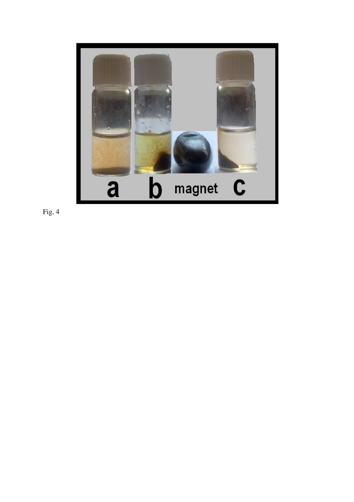
a shows a test tube containing Fe
3
O
4
-Au nanocomposites that has been mixed with HCl and incubated for 18 h. Also, Figure
4
b,c demonstrates two tubes containing acid-stable NPs before and after separation by a magnet.
Figure 4
The images of different test tubes that contain Fe3O4-Au composite MNPs.
In exposure with 1 N hydrochloric acid (
a
) and after separation with external magnetite field (
b
). Tube (
c
) shows the defect-free core/shell fraction of Fe
3
O
4
-Au nanocomposites separated by a magnet and washed three times with distilled water.
The upper illustration in Figure
5
shows different TEM images of the acid-stable NPs after washing with distilled water. As shown in this illustration, dark NPs with sizes of less than 50 nm can be observed in TEM images, indicating different magnetite NPs with higher electron density. The chemical composition of these NPs was studied using the X-ray diffraction (XRD) method and is depicted in the lower illustration in Figure
5
. The Fe
3
O
4
composite MNPs were observed after the acid leaching process and verified the connection between MNPs and elemental gold (also lower illustration in Figure
5
). Some negligible impurities may be remained after acid leaching, so advanced characterization methods such as high resolution TEM or elemental mapping is necessary to differentiate between the core/shell structure and other impurities. The magnetite properties of prepared Fe
3
O
4
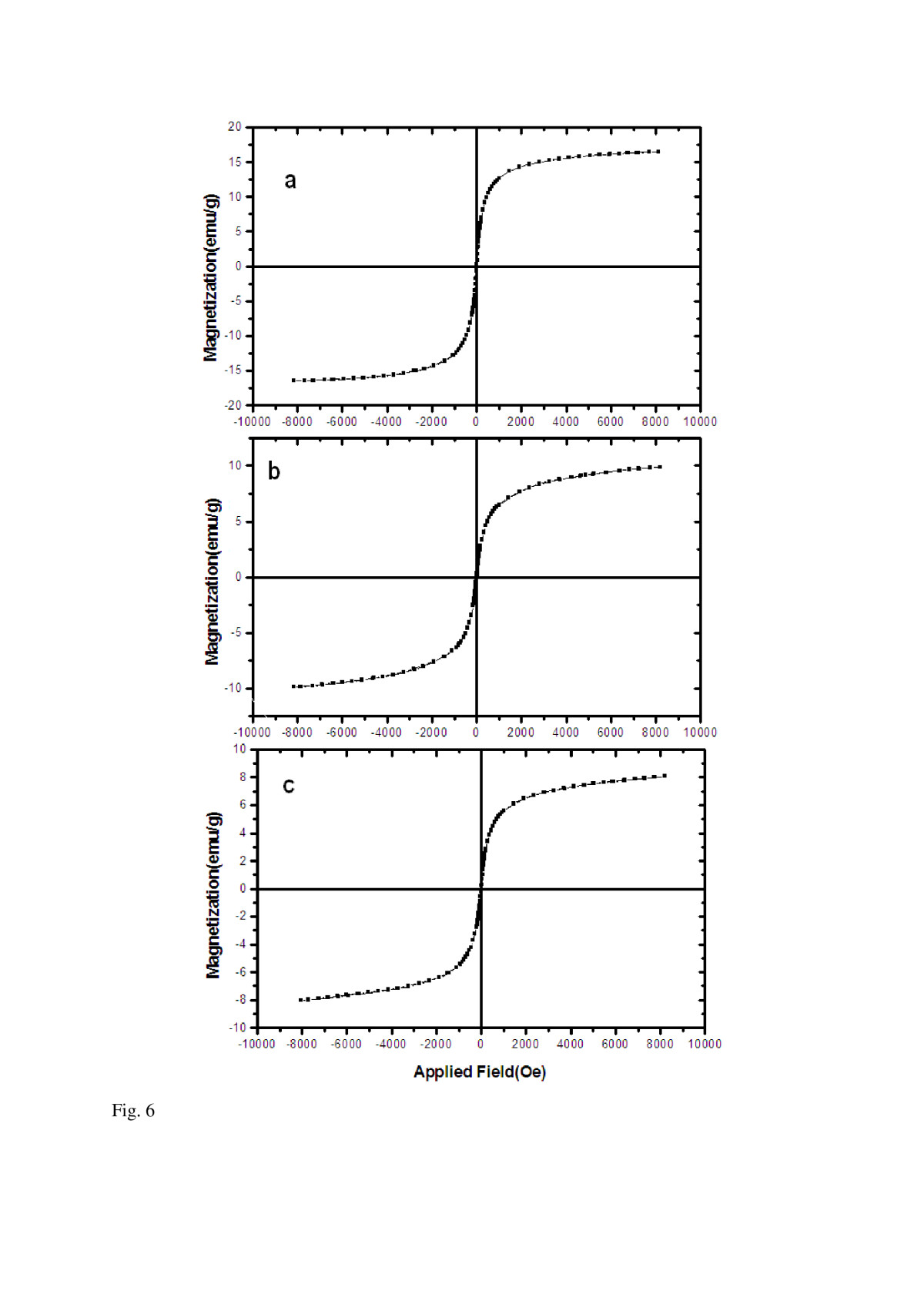
-Au composite MNPs before and after acid wash treatment with HCl (1 N) were also investigated, using the vibrating sample magnetometer (VSM) method. The magnetic hysteresis curves of bare (a) Fe
3
O
4
MNPs, (b) Fe
3
O
4
-Au composites MNPs, and (c) Fe
3
O
4
core@Au shell MNPs are demonstrated in Figure
6
and show magnetite saturation conditions for all magnetite nanomaterials. No considerable change was observed in superparamagnetism properties of this magnetite material (Fe
3
O
4
) either before or after the acid washing process with HCl (1 N) for 18 h (Figure
6
b,c). Furthermore, we have tested HCl solutions with higher normality of 2, 3, 4, and 5 N for the dissolution of non-core/shell composite NPs, but in all experiments, non-magnetic properties were observed from the remaining nanomaterials (data not shown).
Figure 5
Representative transmission electron microscopic images.
Of Fe
3
O
4
-Au composite nanoparticles treated with hydrochloric acid (1 N) for 18 h at room temperature and separated by an external magnetic field (upper illustration). Lower illustration shows X-ray diffraction pattern of these nanoparticles verifying the connection of magnetite nanoparticles with elemental gold.
Figure 6
Magnetite properties of fabricated nanoparticles.
Measured by vibrating sample magnetometer; bare Fe
3
O
4
nanoparticles (
a
), Fe
3
O
4
-Au nanocomposites before (
b
) and after (
c
) acid wash treatment.
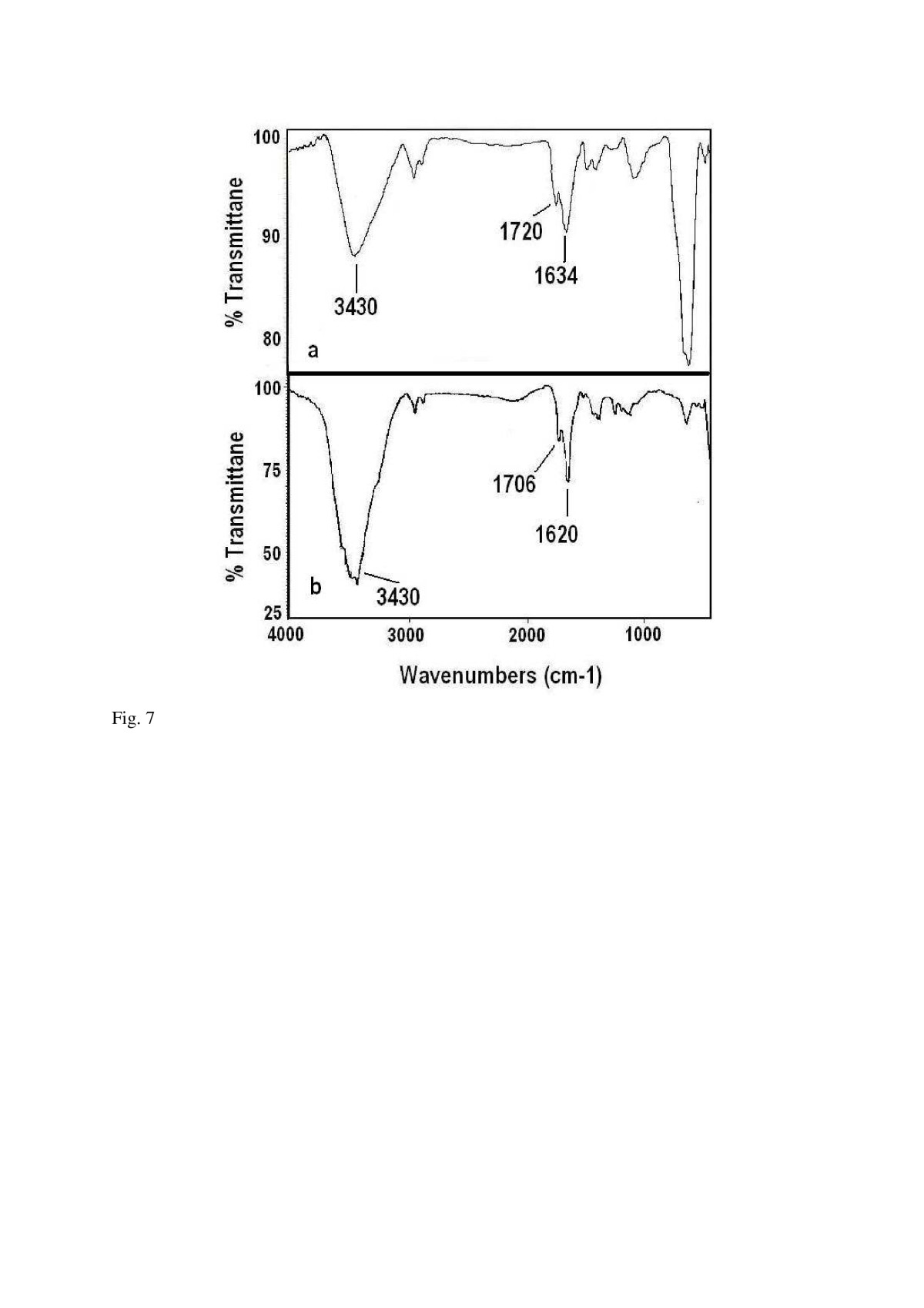
The existence of a carboxyl group on the surface of the prepared magnetite-gold nanocomposites fabricated by a semi-biosynthetic with ethanol extract of
Eucalyptus camaldulensis
has previously been demonstrated by the Fourier transform infrared spectroscopy (FTIR) method[
10
] and confirmed in this investigation (Figure
7
a). IR peaks, centered at 3,430 and 1,720 cm
−1
, confirmed the presence of a carboxyl group on the surface of the semi-biosynthesized Fe
3
O
4
-Au nanocomposites (Figure
7
a). Furthermore, this carboxyl group can also be detected on the surface of Fe
3
O
4
-Au core/shell composites MNPs after acid wash treatment (Figure
7
b) showing that carbonyl groups have not been chemically changed or degraded during acid treatment of Fe
3
O
4
-Au composite MNPs.
Figure 7
Infrared spectra of prepared Fe3O4-Au composite nanoparticles.
Before (
a
) and after (
b
) acid wash treatment by HCL (1 N).
However, it should be noted that the magnetite properties of Fe
3
O
4
-Au composite MNPs was not optimized during this study, and further optimization process should be performed on the magnetization of the Fe
3
O
4
-Au composite MNPs which makes them better candidate for biomedical applications. Also, no assay was carried out to determine the biocompatibility of the above Fe
3
O
4
-Au core/shell composites MNPs and merit further investigation.
Conclusions
Different chemical or biological methods have been reported for synthesizing Fe
3
O
4
MNPs and Fe
3
O
4
core@Au shell MNPs[
16
–
20
]. However, when Fe
3
O
4
-Au core/shell nanocomposites are synthesized, some undesired NPs, such as malformed structures or incomplete core/shell Fe
3
O
4
-Au nanocomposites, may be formed. These types of MNPs are categorized as impurities and need to be removed from the final product. In the current study, an acid wash treatment has been developed to remove all undesired nanomaterials (Figure
1
). The physiochemical properties of isolated Fe
3
O
4
-Au composite MNPs before and after acid wash treatment were investigated by different characterization methods, such as TEM, EDS, XRD, VSM, and FTIR. The results show that the acid wash process did not change the surface chemistry and magnetite properties of Fe3O4 core@Au shell MNPs and can be used to reduce impurities and obtain the defect-free core/shell fraction of Fe
3
O
4
-Au composite MNPS.
Methods
Semi-biosynthesis of magnetite Fe3O4-Au nanocomposites
Carboxyl-capped magnetite Fe
3
O
4
-Au nanocomposites were prepared at room temperature, using the previously described methods[
14
,
21
]. For this purpose, FeCl
3
·6H
2
O (0.487 g) and FeSO
4
·7H
2
O (0.25 g) were precisely weighted and dissolved in 200 ml of deionized water.
The mixture was then treated through a drop-by-drop addition of 10 ml NaOH solution (0.15 M), with mixing (200 rpm), under an argon atmosphere. The final pH of the reaction mixture was adjusted to pH12, with an excess volume of NaOH (0.15 M). The reaction mixture was then further incubated for 30 min at 80°C. The resulting black precipitate was separated by centrifugation (8,000×
g
). The separated MNPs were washed four times with deionized water. The prepared Fe
3
O
4
MNPs were separated from the solution with a magnet. The reduction of Au
3+
onto the surface of Fe
3
O
4
MNPs was carried out using an ethanol extract prepared from
E. camaldulensis
leaves[
14
].
The
E. camaldulensis
leaves were air-dried at room temperature and then pulverized (50 g). The ethanol extract was prepared by macerating the powder (250 g) for 72 h with three changes of the solvent (1,000 ml) at room temperature. The combined solvent extracts were evaporated to yield a brownish or greenish viscous residue. A stock solution (10 mg/ml) of the plant extract was prepared in ethanol for further experiments and reserved in the refrigerator, at 4°C. Chloroauric acid was purchased from Merck, Darmstadt, Germany. MNPs (20 mg) were dispersed in 30 ml of aqueous HAuCl
4
(chloroauric acid) solution (1 mM) and mixed with 1 ml of an ethanol extract of
E. camaldulensis
(10 mg/ml). The resulting mixture was allowed to stand for 45 min at room temperature, and then the prepared magnetite Fe
3
O
4
-Au nanocomposites were separated from the colloid by a magnet. Finally, the NPs were washed three times with distilled water and subjected to the next experiments.
Acid wash treatment
HCl solution (1 N) was used to remove bare Fe
3
O
4
MNPS or other malformed structures from magnetite Fe
3
O
4
-Au nanocomposites (Figure
1
). Prepared composite NPs (100 mg) were mixed with HCl solution (600 ml, 1 N) and incubated for 18 h at room temperature. Then, the remaining composite NPs were separated from the solution by an external magnetic field. These separated NPs were washed three times with deionized water and subjected to further characterization.
Characterizations
All samples were characterized by transmission electron microscopy (model EM 208 Philips, Amsterdam, The Netherlands), EDS, XDS (Philips X
'
Pert Pro), and VSM. The surface chemistry of the composite MNPs before and after acid treatment was also studied by the FTIR(Nicolet Magna 550, Madison, WI, USA).
Acknowledgements
This work has been supported by the Pharmaceutical Sciences Research Center, School of Pharmacy, Tehran University of Medical Sciences, Tehran, Iran. Also, the authors kindly thank the team of Scribendi Company for the language editing and proofreading assistance provided for this article.
Competing interests
The authors declare that they have no competing interests.
Authors’ contributions
ARS is the leader of the group and the main writer of this study's manuscript. HM and AA both participated in synthesis and characterization of nanoparticles. Also, AA and HS commented on EDS and XRD experiments. All authors read and approved the final manuscript.
References
Corchero and Villaverde (2009) Biomedical applications of distally controlled magnetic nanoparticles (pp. 468-476) 10.1016/j.tibtech.2009.04.003
Jain et al. (2008) Magnetic nanoparticles with dual functional properties: drug delivery and magnetic resonance imaging (pp. 4012-4021) 10.1016/j.biomaterials.2008.07.004
Chertoka et al. (2008) Iron oxide nanoparticles as a drug delivery vehicle for MRI monitored magnetic targeting of brain tumors (pp. 487-496) 10.1016/j.biomaterials.2007.08.050
Mahmoudi et al. (2010) Synthesis, surface architecture and biological response of superparamagnetic iron oxide nanoparticles for application in drug delivery: a review (pp. 164-201) 10.1504/IJBNN.2010.034651
Mahmoudi et al. (2010) Recent advances in surface engineering of superparamagnetic iron oxide nanoparticles for biomedical applications (pp. S1-S27) 10.1007/BF03246181
Lu et al. (2006) Synthesis and characterization of composite nanoparticles comprised of gold shell and magnetic core/cores (pp. 44-49) 10.1016/j.jmmm.2005.06.007
Tsuji et al. (2008) Role of chloride ions in the formation of Au@Ag core-shell nanocrystal structures by using a microwave-polyol method10.1016/j.colsurfa.2007.10.023
Rosenholm et al. (2011) Large-pore mesoporous silica-coated magnetite core-shell nanocomposites and their relevance for biomedical applications 145(1–3) (pp. 14-20) 10.1016/j.micromeso.2011.04.022
Maltas et al. (2011) Immobilization of albumin on magnetite nanoparticles 65(23–24) (pp. 3499-3501) 10.1016/j.matlet.2011.07.045
Sun et al. (2007) Theoretical study on gold-coated iron oxide nanostructure: Magnetism and bioselectivity for amino acids (pp. 4159-4163) 10.1021/jp067095x
Cho et al. (2005) Growth mechanisms and oxidation resistance of gold-coated iron nanoparticles (pp. 3181-3186) 10.1021/cm0500713
Tartaj et al. (2003) The preparation of magnetic nanoparticles for applications in biomedicine (pp. 182-197) 10.1088/0022-3727/36/13/202
Haratifar et al. (2009) Semi-biosynthesis of magnetite-gold composite nanoparticles using an ethanol extract of Eucalyptus camaldulensis and study of the surface chemistry10.1155/2009/962021
Yan et al. (2008) Solvothermal, synthesis and characterization of size-controlled Fe3O4 nanoparticles (pp. 487-491) 10.1016/j.jallcom.2007.04.019
Li et al. (2007) SnO2/In2O3 one-dimensional nano-core-shell structures: synthesis, characterization and photoluminescence properties (pp. 441-444) 10.1016/j.ssc.2007.03.031
Lu et al. (2007) Magnetic nanoparticles: synthesis, protection, functionalization, and application (pp. 1222-1244) 10.1002/anie.200602866
Qiua et al. (2010) Facile preparation of magnetic core–shell Fe3O4@Au nanoparticle/myoglobin biofilm for direct Biosensors and Bioelectronics (pp. 1447-1453) 10.1016/j.bios.2009.10.043
Pradhan et al. (2008) Gold–magnetite nanocomposite materials formed via sonochemical methods (pp. 891-897) 10.1016/j.ultsonch.2008.01.004
Lima et al. (2007) Design and synthesis of plasmonic magnetic nanoparticles (pp. 78-83) 10.1016/j.jmmm.2006.10.1169
Martínez-Mera et al. (2007) Synthesis of magnetite (Fe3O4) nanoparticles without surfactants at room temperature (pp. 4447-4451) 10.1016/j.matlet.2007.02.018