Department of Biomolecular Functional Engineering, College of Engineering, Ibaraki University, Hitachi, Ibaraki, 316-8511, JP
Department of Nano-Medical Science, Graduate School of Medicine, Tohoku University, Seiryo-machi, Aoba-ku, Sendai, 980-8575, JP
Department of Surgical Oncology, Graduate School of Medicine, Tohoku University, Seiryo-machi, Aoba-ku, Sendai, 980-8574, JP
Abstract
A preparation method for hollow particles composed of silica shell containing gadolinium compound (GdC) is proposed. GdC nanoparticles with an average size of 40.5 ± 6.2 nm were prepared with a homogeneous precipitation method at 80°C using 1.0 × 10
−3
M Gd(NO
3
)
3
and 0.5 M urea in the presence of 2.0 × 10
−4
M ethylenediaminetetraacetic acid disodium salt dihydrate. Silica-coated GdC (GdC/SiO
2
) nanoparticles with an average size of 100.9 ± 9.9 nm were fabricated with a sol-gel method at 35°C using 5.0 × 10
−3
M tetraethylorthosilicate, 11 M H
2
O, and 1.5 × 10
−3
M NaOH in ethanol in the presence of 1.0 × 10
−3
M GdC nanoparticles. The GdC/SiO
2
particles were aged at 80°C in water after replacement of solvent of the as-prepared GdC/SiO
2
particle colloid solution with water, which provided diffusion of GdC into the silica shell and then formation of a hollow structure. The hollow particle colloid solution revealed good MRI properties. A relaxivity value for longitudinal relaxation time-weighted imaging was as high as 3.38 mM
−1
· s
−1
that attained 80% of that for a commercial Gd complex contrast agent.
Background
Gadolinium compounds (GdC) can function as a contrast agent for magnetic resonance imaging (MRI) in medical diagnosis[
1
–
6
]. Typical commercial GdC-based contrast agents are solutions dissolving homogeneously gadolinium complexes at a molecular level. The GdC molecules are not strongly dragged in fluid because of their small sizes. Consequently, they cannot stay in living bodies for a long period, which provides difficulty taking steady images. Formation of particles of GdC and an increase in their apparent size are promising solutions to the problem because of their projected area which is larger than molecules or nanoparticles. Thus, their residence time will increase, which may realize steady imaging.
Growth of tumor generated in tissue produces a number of slits with a size of several hundred nanometers on the wall of the vessel near the tumor. Polymers or nanoparticles smaller than the slit size may be able to pass through the slit. Their discharge from the tumor is controlled because lymph, which is a way for the discharge, is premature in tumor. This behavior is called an enhanced permeation and retention (EPR) effect[
7
–
9
]. Accordingly, nanoparticle formation has another advantage; nanoparticles that show MRI properties may have the ability to detect the tumor by the EPR effect.
Gadolinium complexes may release free gadolinium ions through dissociation of the ions. The release of gadolinium ions may provoke adverse reactions in some patients[
6
,
10
,
11
]. As one of the methods for reducing the adverse reactions derived from gadolinium complexes, coating of GdC nanoparticles (core) with materials inert to living bodies (shell) can be given because contact of the GdC with living bodies may be controlled with a physical barrier of the shell materials. From this viewpoint, we have also studied on the silica coating of GdC nanoparticles[
12
]. In a preliminary experiment, the silica-coated GdC (GdC/SiO
2
) particles with a core-shell structure became hollow particles after the GdC/SiO
2
particles were washed with a centrifuge and were aged in water. In several previous studies, hollow particles are fabricated by calcining core-shell particles composed of organic compound core and inorganic compound shell to remove the core[
13
–
15
]. The hollow particles in the preliminary experiment were obtained with no calcination. Methods for producing hollow silica particles that do not use calcination process have been proposed by several researchers[
16
–
18
]. They use liquids such as aqueous basic solutions and organic solvents that dissolve core materials. The use of such liquids may damage the silica shell or give environmental load. Care of the problems is not required to be taken for the preliminary experiment since it did not use acid, base, and organic solvent to remove the core. If the components of GdC particles are left in the silica shell after the formation of hollow particles, the hollow particles are expected to function as new MRI contrast agents because many protons contained in the silica shell may interact efficiently with the GdC components compared to the GdC particles localized only at the center of the Gd/SiO
2
particles.
The present work proposes a method for preparing hollow particles composed of GdC and silica. A colloid solution of GdC nanoparticles was prepared with a homogeneous precipitation method, and then the GdC nanoparticles were silica-coated with a modified Stöber method. Elimination of GdC core was performed by replacing the solvent of the reactant with water and aging the aqueous colloid solution. The present work also studied on MRI properties of the hollow particle colloid solution.
Results and discussion
Morphology of particles
GdC nanoparticles
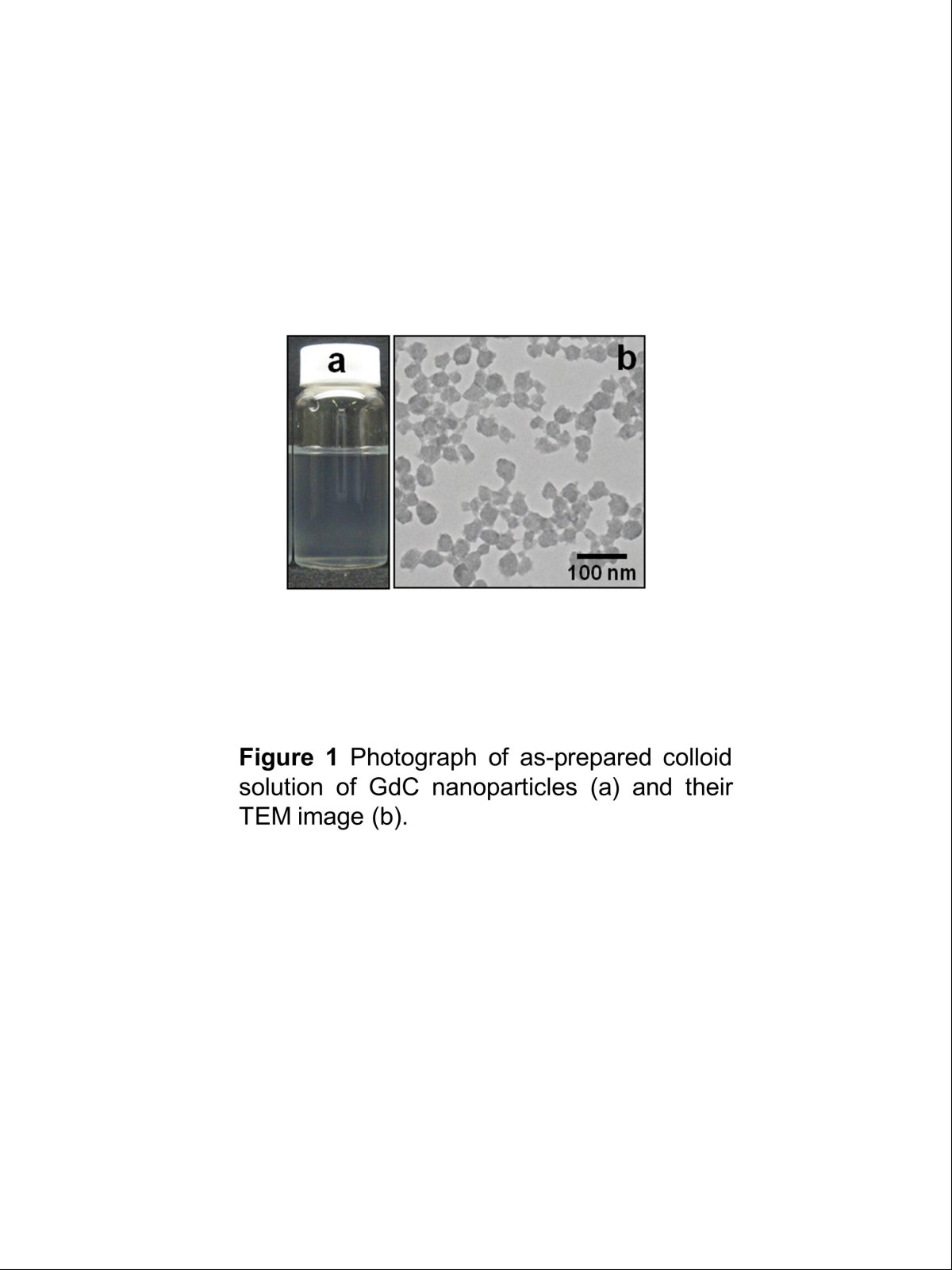
A photograph was taken for the colloid solution of GdC particles, as shown in Figure
1
a. The colloid solution was faintly turbid but colloidally stable. Figure
1
b shows a transmission electron microscopy (TEM) image of the GdC particles. Quasi-spherical particles were obtained, though they partially stuck together. Their particle size was 40.5 ± 6.2 nm. Figure
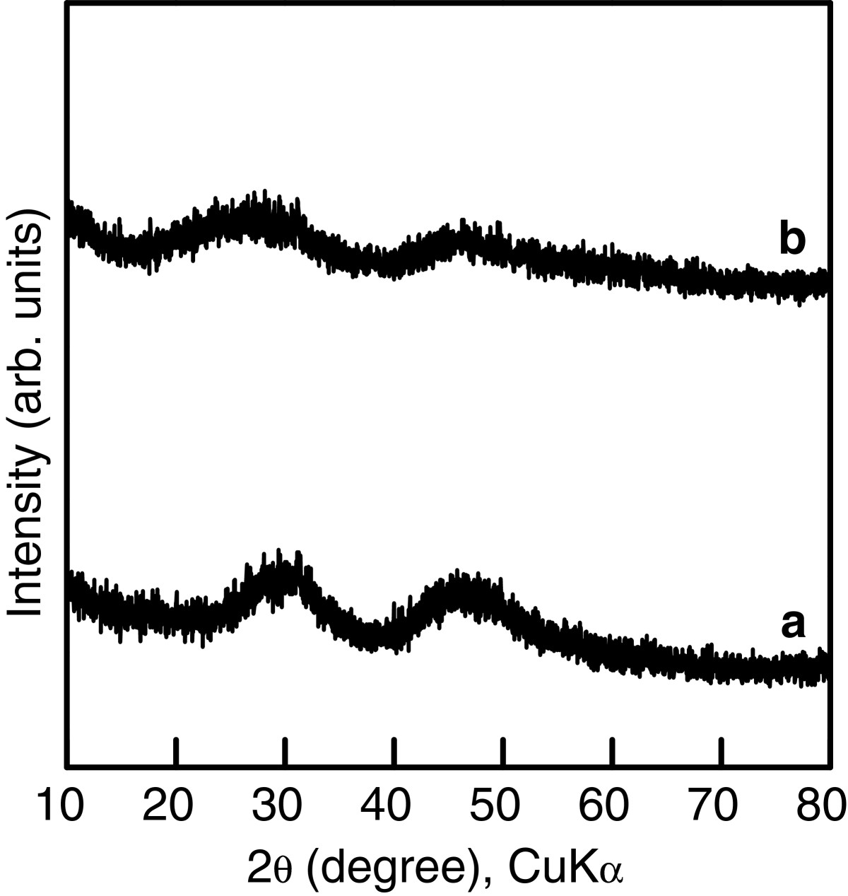
2
(pattern a) shows an X-ray diffractometry (XRD) pattern of the GdC particles. The pattern was structureless, except for broad peaks at
ca.
30° and
ca.
45°. Accordingly, the GdC particles were amorphous or too fine to be detected. The inductively coupled plasma (ICP) measurement showed that Gd concentration in the GdC particle colloid solution was 0.819 mM, which indicated that 81.9% of the Gd(NO
3
)
3
used for production of GdC particles was transformed to GdC particles.
Figure 1
Photograph of as-prepared colloid solution of GdC nanoparticles (a) and their TEM image (b).
Figure 2
XRD patterns of (a) GdC particles and (b) GdC/SiO2particles.
GdC/SiO2 core-shell particles
Figure
3
a shows a TEM image of the GdC/SiO
2
particles, which were as-prepared, i.e., dispersed in H
2
O/ethanol solution with the H
2
O concentration of 11 M. The image was taken several days after preparation. Darker and lighter parts corresponded to GdC and silica, respectively, since the electron density of GdC is significantly higher. The GdC particles were successfully coated with silica shell, though the GdC/SiO
2
particles partially stuck together. No damage was seen for the GdC/SiO
2
particles even several days after preparation. Accordingly, the GdC/SiO
2
particles were chemically stable in the as-prepared solution. The particles had a size of 100.9 ± 9.9 nm, which were composed of core particles with a size of 51.8 ± 8.2 nm and a shell with a thickness of 24.6 nm. The core size was larger than that of the GdC particles prior to the silica coating. Aggregation and particle growth were considered to take place during the silica coating. Hydrolysis of tetraethylorthosilicate (TEOS) provided an increase in ionic strength of the solution. Since an increase in the ionic strength compresses double layer on solid materials such as colloidal particles[
19
–
21
], the double-layer repulsion between the GdC particles probably became small through the hydrolysis. Thus, the GdC particles probably aggregated and grew because of the high ionic strength that would favor the particle aggregation. Figure
2
(pattern b) shows an XRD pattern of the GdC/SiO
2
particles. There was no large difference between the patterns of GdC particles and the GdC/SiO
2
particles. The GdC particles were also amorphous or too fine to be detected. Accordingly, the results of TEM observation and XRD measurement indicated that the silica coating made no chemical effect on the GdC particles. The ICP measurement revealed that an actual Gd concentration in the Gd/SiO
2
particle colloid solution was 0.698 mM, which indicated that 85.2% of Gd contained in the GdC particles was consumed for the formation of GdC/SiO
2
particles.
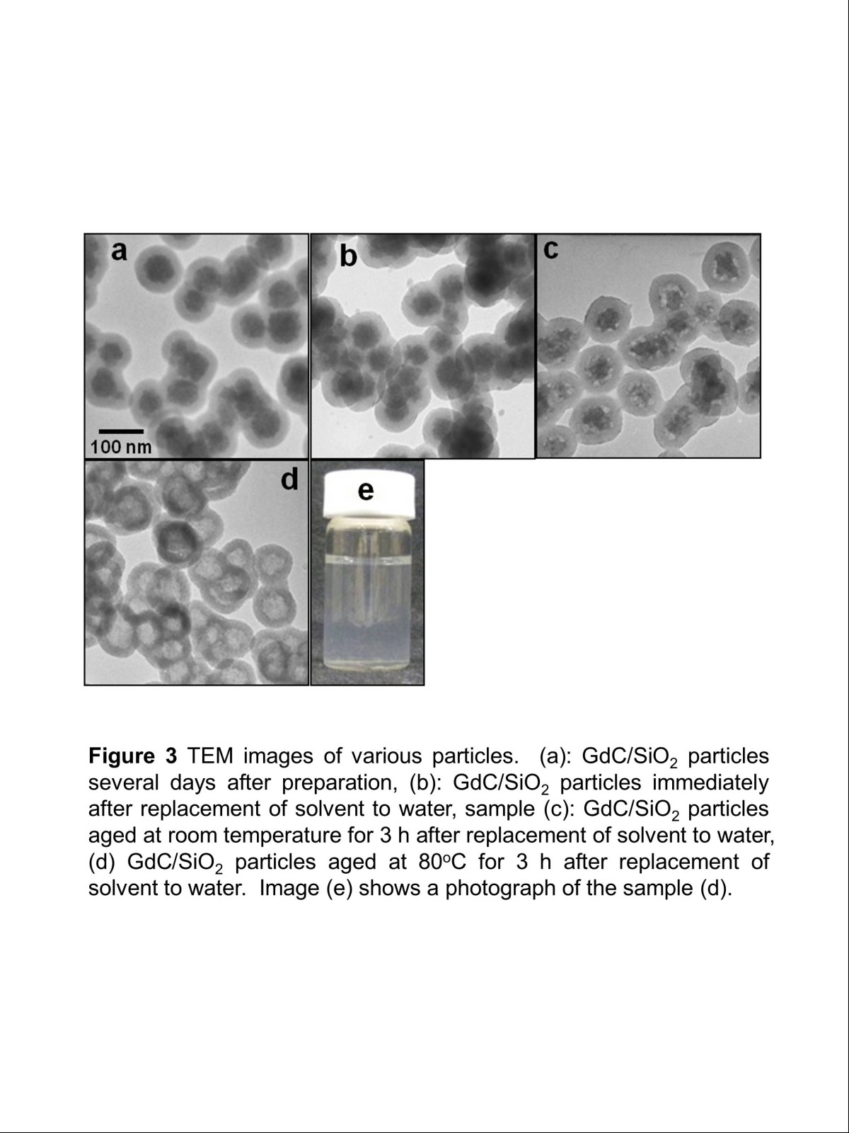
Figure 3
TEM images of various particles.
(
a
) GdC/SiO
2
particles several days after preparation, (
b
) GdC/SiO
2
particles immediately after replacement of solvent to water, (
c
) GdC/SiO
2
particles aged at room temperature for 3 h after replacement of solvent to water, (
d
) GdC/SiO
2
particles aged at 80°C for 3 h after replacement of solvent to water. Image (
e
) shows a photograph of sample (
d
).
Hollow particles
Figure
3
b,c shows TEM images of the GdC/SiO
2
particles, of which solvents were replaced with water. The GdC/SiO
2
particles immediately after the washing process (Figure
3
b) appeared to be similar to those prior to the replacement (Figure
3
a), which indicated that the washing process did not damage their core-shell structure mechanically. For 3 h after the washing (Figure
3
c), the surface of the GdC core appeared to be partially dissolved and then became rough. The silica shell was not damaged in spite of the surface roughing. The surface roughing was promoted by heating at 80°C, as shown in Figure
3
d. Then, hollow particles were produced. The silica shell was still maintained even after the heating. Figure
3
e shows a photograph of the hollow particle colloid solution. The solution was colloidally stable since no sedimentation took place. According to the ICP measurement, an actual Gd concentration in the hollow particle colloid solution was 0.426 mM. This meant that 61.0% of Gd contained in the GdC/SiO
2
particles was considered to still remain in the shell of hollow particles and 39.0% of Gd was removed from the hollow particles.
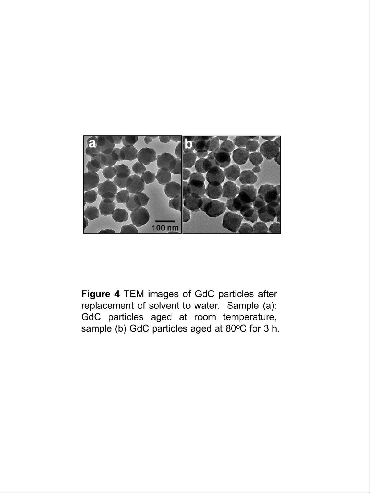
Figure
4
a shows a TEM image of GdC particles after the solvent of their colloid solution was replaced with water. The replacement of solvent with water did not damage the GdC particles mechanically. After the replacement, the colloid solution of GdC particles was aged at 80°C for 3 h. Their TEM image is shown in Figure
4
b. The image appeared to be similar to that prior to the aging (Figure
4
a). Those results indicated that both the replacement and the aging at 80°C did not affect the structure of GdC particles. Accordingly, it could be concluded that the formation of hollow structure required the GdC particles to have contact with the silica shell. The GdC was presumed to diffuse into the silica shell, which provided the production of new compounds such as gadolinium silicate in the shell. Consequently, the hollow particles were produced. However, a precise mechanism of the hollow particle formation is still unclear.
Figure 4
TEM images of GdC particles after replacement of solvent to water.
(
a
) GdC particles aged at room temperature. (
b
) GdC particles aged at 80°C for 3 h.
MRI property
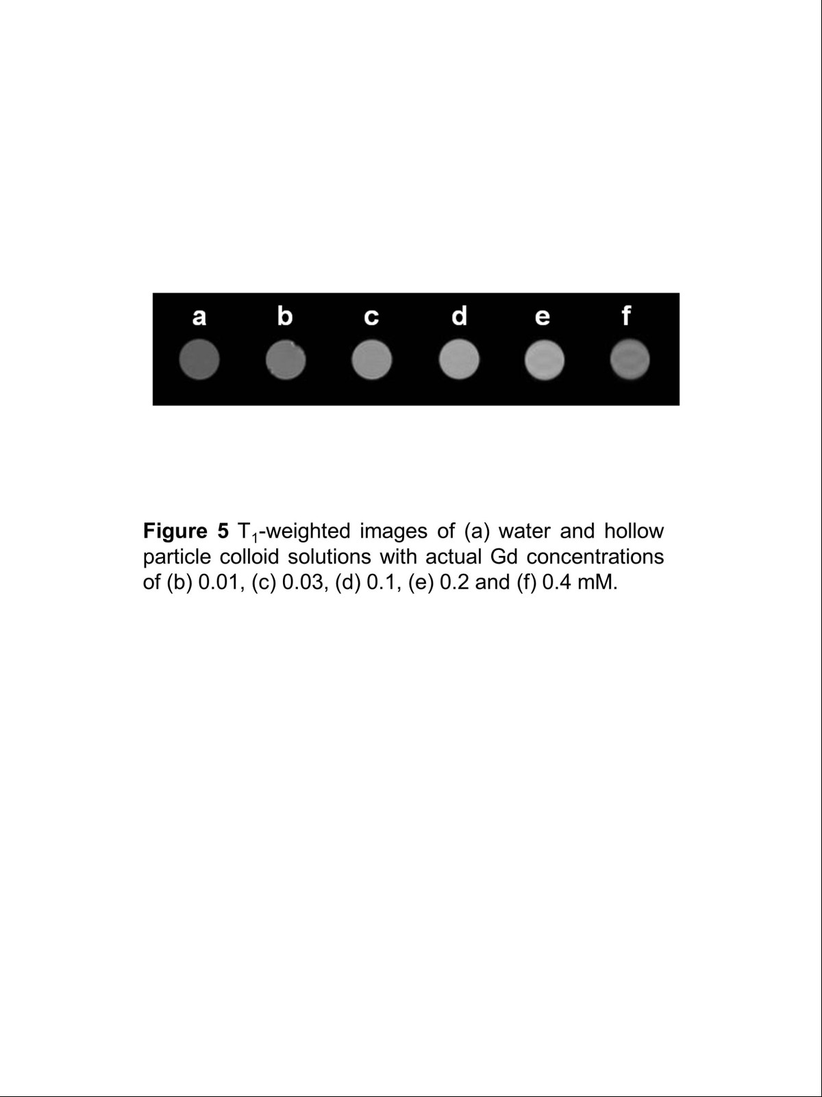
Longitudinal relaxation time (T
1
)-weighted images of the colloid solutions of hollow particles with various actual Gd concentrations are shown in Figure
5
. For T
1
-weighted images, strong magnetic resonance gives positive images with light contrast. All the solutions examined were clearly imaged against a black background, and the light contrast of image increased with increasing actual Gd concentration.
Figure 5
T1-weighted images.
Of (
a
) water and hollow particle colloid solutions with actual Gd concentrations of (
b
) 0.01, (
c
) 0.03, (
d
) 0.1, (
e
) 0.2 and (
f
) 0.4 mM.
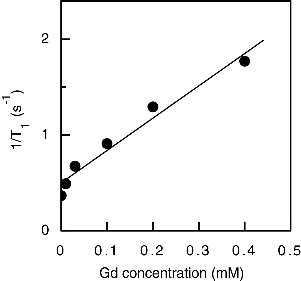
Figure
6
shows a plot of longitudinal relaxation rate (1/T
1
) of the hollow particle colloid solution as a function of the actual Gd concentration. The relaxation rate increased linearly with increasing actual Gd concentration. A value of relaxivity (
r1
) is defined as a slope of relaxation rate with respect to actual Gd concentration and is commonly used as a guideline on the performance of positive contrast agents. The
r1
value, which was calculated by linear fitting, was 3.38 mM
−1
· s
−1
. This value attained 80% of that of Magnevist since an
r1
value of Magnevist, which was measured in our previous work[
22
], was 4.23 mM
−1
· s
−1
. Our previous work also gave that a colloid solution of GdC/SiO
2
particles revealed an
r1
value of 3.11 mM
−1
· s
−1
[
22
], which indicated that the
r1
value of the hollow particle colloid solution was larger than that of the GdC/SiO
2
particle colloid solution. Active exchange of the protons with the contrast agent shortens T
1
, i.e., increases
r1
[
23
,
24
]. Since the GdC present in the shell lay close to the water protons compared to the GdC inside the GdC/SiO
2
particles, the interaction between water protons and gadolinium ions was more significant than that for the GdC/SiO
2
particles. Accordingly, the formation of hollow particles with shell-containing GdC was found to improve imaging ability based on longitudinal relaxation.
Figure 6
Relaxation rates of hollow particle colloid solutions as a function of actual Gd concentration.
Conclusions
A method for producing hollow particles with silica shell containing GdC was proposed. The GdC nanoparticles with the average size of 40.5 nm were prepared by means of the homogeneous precipitation method using aqueous solution of 1.0 × 10
−3
M Gd(NO
3
)
3
, 0.5 M urea, and 2.0 × 10
−4
M ethylenediaminetetraacetic acid disodium salt dihydrate (EDTA). The silica coating of the GdC particles was achieved with the sol-gel method using 5.0 × 10
−3
M TEOS, 11 M H
2
O, and 1.5 × 10
−3
M NaOH in the presence of the GdC particles at the Gd concentration of 1.0 × 10
−3
M, which produced the GdC/SiO
2
particles with the average size of 100.9 nm. The replacement of the solvent of GdC/SiO
2
particle colloid solution with water and the aging of GdC/SiO
2
particles in water at 80°C transformed the GdC/SiO
2
particles to hollow particles due to the diffusion of GdC into the silica shell. The hollow particle colloid solution showed high-contrast T
1
-weighted magnetic resonance images. Its relaxivity value, which was 3.38 mM
−1
· s
−1
, was recorded at the value as high as 80% of that for Magnevist, the commercial Gd complex contrast agent. These results obtained in the present work indicated that the hollow particle colloid solution had an ability of MRI contrast agent.
Methods
Chemicals
Gadolinium nitrate hexahydrate (Gd(NO
3
)
3
· 6H
2
O, 99.5%) and urea (99.0%) were used as a raw chemical for GdC nanoparticles and a precipitation inducer in the preparation of GdC nanoparticles, respectively. The stabilizer used in the preparation of GdC nanoparticles was EDTA (99.0% to 101.0%). A raw chemical for silica shell and a solvent for silica coating were TEOS (95%) and ethanol (99.5%), respectively. A sol-gel reaction of TEOS in the silica coating was catalyzed with sodium hydroxide (NaOH) solution (5 M). The commercial MRI contrast agent used for comparison to the particle colloid solutions in imaging examinations was Magnevist (0.5 M Gd, Bayer Co., Ltd., Leverkusen, Germany). All the chemicals except for the commercial contrast agents were purchased from Kanto Chemical Co., Inc. (Tokyo, Japan) and used as received. The water that was ion-exchanged and distilled with Shimadzu SWAC-500 (Kyoto, Japan) was used in all the preparations.
Preparation of materials
GdC particles
A colloid solution of GdC nanoparticles was prepared in water containing EDTA by a homogeneous precipitation method, according to our previous work[
22
]. Gd(NO
3
)
3
aqueous solution was added to aqueous solution dissolving urea and EDTA in a hermetically sealed flask reactor. The mixture was stirred at 80°C for 3 h. Initial concentrations of Gd(NO
3
)
3
, urea, and EDTA were 1.0 × 10
−3
, 0.5, and 2.0 × 10
−4
M, respectively. The GdC particles in the colloidal solution were washed by repeating centrifugation, removal of supernatant, addition of water, and sonication three times. The solvent was replaced with H
2
O/ethanol solution by adding the H
2
O/ethanol solution at the last washing process.
GdC/SiO2 core-shell particles
GdC/SiO
2
core-shell particles were fabricated by the silica coating of the GdC particles, which was performed in the H
2
O/ethanol solution by a sol-gel method, according to our previous work[
22
]. TEOS and 0.1 M NaOH aqueous solution were successively added to the GdC particle colloid solution at 35°C. The reaction time was 24 h. Initial concentrations of Gd, TEOS, H
2
O, and NaOH were 1.0 × 10
−3
, 5.0 × 10
−3
, 11, and 1.5 × 10
−3
M, respectively.
Hollow particles
Hollow particles were fabricated from the Gd/SiO
2
core-shell particles. The colloidal suspensions of GdC/SiO
2
particles were washed by repeating centrifugation, removal of supernatant, addition of water, and sonication three times, which resulted in the replacement of solvent with water. Then, the washed colloid suspensions were aged in water.
Characterization
The samples were characterized by TEM and XRD. TEM was performed with a JEOL JEM-2000FX II microscope (Akishima, Japan) operating at 200 kV. Samples for TEM were prepared by dropping and evaporating the nanoparticle suspensions on a collodion-coated copper grid. Dozens of particle diameters in TEM images were measured to determine volume-averaged particle size,
dv
, and standard deviation of particle size distribution,
σ
, defined by the following equations:
dv=∑inidi3/∑ini13,σ=∑idi−dv2/∑ini12,
where
ni
is the number of particles with a size of
di
. XRD measurements were carried out with a Rigaku RAD-B (Tokyo, Japan) operating at 7.5 kW Cu K
α
radiation using a wide-angle goniometer. Samples for XRD were obtained with centrifugation of the solution containing the particles, removal of the supernatant, and drying of the residue at room temperature for 24 h in vacuum.
Actual Gd concentrations in particle colloid solutions were measured by ICP emission spectroscopy. ICP measurement was performed with a Shimadzu ICPS-7510 atom emission spectrometer (Kyoto, Japan). The emission was detected at a wavelength of 342.247 nm. Samples for ICP were prepared by dissolving the particles with aqua regia completely and then diluting the obtained solution with water to adjust its concentrations to concentrations suitable for the measurements.
For the estimation of MR signal intensity, T
1
-weighted images of colloid solutions with different Gd concentrations were taken with a Bruker AVANCE III 400WB magnetic resonance imaging system (Madison, WI, USA) at a static magnetic field of 9.4 T. Times of echo and repetition were 8.5 and 1,500 ms, respectively. The colloid solutions were prepared by diluting the as-prepared colloid solutions with water.
Acknowledgements
We express our thanks to Prof. T. Noguchi in the College of Science of Ibaraki University, Japan, for his help in TEM observation.
Competing interests
The authors declare that they have no competing interests.
Authors’ contributions
YK (Kobayashi) drafted the manuscript. KG and NO modified it. HM, TN, and YK (Kubota) participated in some practical work. All authors read and approved the final manuscript.
References
Yim et al. (2011) The performance of gadolinium diethylene triamine pentaacetate-pullulan hepatocyte-specific T1 contrast agent for MRI (pp. 5187-5194) 10.1016/j.biomaterials.2011.03.069
Wang et al. (2011) Effect of gadolinium chelate contrast agents on diffusion weighted MR imaging of the liver, spleen, pancreas and kidney at 3 T (pp. e1-e7) 10.1016/j.ejrad.2010.05.019
Chandrasekharan et al. (2012) Gadolinium chelate with DO3A conjugated 2-(diphenylphosphoryl)-ethyldiphenylphosphonium cation as potential tumor-selective MRI contrast agent (pp. 9225-9231) 10.1016/j.biomaterials.2012.08.071
Shen and New (2012) Promising strategies for Gd-based responsive magnetic resonance imaging contrast agents (pp. 1-9)
Akai et al. (2012) Fate of hypointense lesions on Gd-EOB-DTPA-enhanced magnetic resonance imaging (pp. 2973-2977) 10.1016/j.ejrad.2012.01.007
Telgmann et al. (2013) Determination of gadolinium-based MRI contrast agents in biological and environmental samples: a review: Anal (pp. 1-16) 10.1016/j.aca.2012.12.007
Shalviri et al. (2013) Multifunctional terpolymeric MRI contrast agent with superior signal enhancement in blood and tumor (pp. 11-20) 10.1016/j.jconrel.2013.01.014
Yamashita and Hashida (2013) Pharmacokinetic considerations for targeted drug delivery (pp. 139-147) 10.1016/j.addr.2012.11.006
Doane and Burda (2012) Nanoparticle mediated non-covalent drug delivery
Marshall and Kasap (2012) Adverse events caused by MRI contrast agents: implications for radiographers who inject (pp. 132-136) 10.1016/j.radi.2010.09.007
Mi et al. (2013) Gd-DTPA-loaded polymer-metal complex micelles with high relaxivity for MR cancer imaging (pp. 492-500) 10.1016/j.biomaterials.2012.09.030
Morimoto et al. (2011) X-ray imaging of newly-developed gadolinium compound/silica core-shell particles (pp. 650-657) 10.1007/s10971-011-2540-6
Pi et al. (2010) Biomimetic synthesis of raspberry-like hybrid polymer–silica core–shell nanoparticles by templating colloidal particles with hairy polyamine shell (pp. 193-199) 10.1016/j.colsurfb.2010.02.031
Liu et al. (2011) Micelles of poly(styrene-b-2-vinylpyridine-b-ethylene oxide) with blended polystyrene core and their application to the synthesis of hollow silica nanospheres (pp. 354-359) 10.1016/j.jcis.2011.03.004
Takai et al. (2012) Determine apparent shell density for evaluation of hollow silica nanoparticle (pp. 101-105) 10.1016/j.colsurfa.2012.04.019
Fuji et al. (2012) Shape-controlled hollow silica nanoparticles synthesized by an inorganic particle template method (pp. 562-565) 10.1016/j.apt.2011.06.002
Yang et al. (2013) Preparation of Cu2O@SiO2 particles and their evolution to hollow SiO2 particles (pp. 30-36) 10.1016/j.colsurfa.2012.12.016
Kim et al. (2012) Synthesis of hollow silica particles with tunable size, shell thickness, and morphology
Singh and Song (2007) Experimental correlations of pH and ionic strength effects on the colloidal fouling potential of silica nanoparticles in crossflow ultrafiltration (pp. 112-118) 10.1016/j.memsci.2007.06.072
Yilmaz et al. (2007) AFM interaction study of α-alumina particle and c-sapphire surfaces at high-ionic-strength electrolyte solutions (pp. 116-123) 10.1016/j.jcis.2006.11.010
Li and Xu (2008) Electrical double layers’ interaction between oppositely charged particles as related to surface charge density and ionic strength (pp. 157-161) 10.1016/j.colsurfa.2008.05.023
Kobayashi Y, Morimoto H, Nakagawa T, Gonda K, Ohuchi N:
Preparation of silica-coated gadolinium compound particle colloid solution and its application in imaging.Adv Nano Res
under review under review
Bagher-Ebadian et al. (2011) MRI estimation of gadolinium and albumin effects on water proton (pp. S176-S179) 10.1016/j.neuroimage.2010.05.032
Liu et al. (2011) Gadolinium-loaded polymeric nanoparticles modified with anti-VEGF as multifunctional MRI contrast agents for the diagnosis of liver cancer (pp. 5167-5176) 10.1016/j.biomaterials.2011.03.077