Green synthesis of gold nanoparticles from fruit extract of Terminalia arjuna, for the enhanced seed germination activity of Gloriosa superba

Abstract

This study reveals the synthesis of spherical gold nanoparticles (Au NPs) using aqueous fruit extract of Terminalia arjuna , which contains tannin, terpenoid, saponins, flavonoids, glycosides and polyphenolic compounds. The synthesized Au NPs were characterized by UV–visible spectroscopy (UV–vis), Fourier transform infrared (FTIR), X-ray diffraction (XRD), atomic force microscopy (AFM), energy-dispersive X-ray spectroscopy (EDX), transmission electron microscopy (TEM), dynamic light scattering (DLS) and zeta potential (ZP) analyses. UV–visible spectra of the fruit extract containing Au NPs showed a surface plasmon resonance peak at 523 nm. FTIR analysis was performed to analyze the biomolecules responsible for the reduction of Au NPs. FTIR analysis clearly showed that Au NPs were capped with plant compounds. The EDX analysis was used to identify the elemental composition of the synthesized Au NPs. The high crystallinity of Au NPs with a face-centered cubic phase is evident to XRD patterns. AFM and TEM observations revealed that synthesized Au NPs were spherical shape with the range 20–50 nm. DLS measurement revealed that Au NPs were obtained in the average size of 25 nm and it is found to be stable at 21.9 mV through ZP analysis. The synthesized Au NPs were investigated for its antibacterial activity. By contrast, Au NPs did not show any antibacterial activity against Gram-positive and Gram-negative bacteria. The Au NPs were treated with two different concentrations (500 and 1,000 μM) of Gloriosa superba seeds. Au NPs exposure at 1,000 μM concentration has most significant effect on seed germination rate and vegetative growth of G. superba . This is the first report on Au NPs as a biocompatibility material to enhance the seed yield of this endangered medicinal plant.


Background

Nanotechnology has varied applications in lifestyle. Surface to the volume ratio is the high impact of nanoparticles, and it contains a lot of physical, chemical and optical properties. In the present scenario, the gold nanoparticles (Au NPs) were used for wide range of applications such as biomedical [ 1 ], drug delivery [ 2 ], bio-sensor [ 3 ], catalytic [ 4 ], agriculture [ 5 ], antioxidant [ 6 ], anticancer [ 7 ], antibacterial [ 8 ], antifungal [ 9 ], antibiofilm [ 10 ] and larvicidal activity [ 11 ]. Generally, Au NPs can be synthesized by physical and chemical methods such as physical vapor deposition [ 12 ], chemical vapor deposition [ 13 ], Microwave irradiation [ 14 ], UV irradiation [ 15 ], sol–gel technique [ 16 ], aerosol technology [ 17 ], sono-chemical method [ 18 ] and photochemical reduction [ 19 ]. However, most of these techniques are expensive, hazardous and employed by toxic chemicals. Hence, there is an ever-growing need to develop the cost-effective, simplicity and mainly environmentally benign synthesis protocol. Recently, plant extracts have been reported to show high efficacy in Au NPs synthesis such as Anacardium occidentale [ 20 ], Centella asiatica [ 21 ], Chenopodium album [ 22 ], Coleus amboinicus [ 23 ], Crocus sativus [ 24 ], Euphorbia hirta [ 8 ], Mangifera indica [ 25 ], Macrotyloma uniflorum [ 26 ], Trianthema decandra [ 27 ], Murraya koenigii [ 28 ], Piper betle [ 29 ], and seed extracts used for Terminalia chebula [ 30 ] and Cuminum cyminm [ 31 ]. Very few works had been done in the Au NPs synthesis using fruit extract such as Abelmoschus esculentus [ 9 ], Citrus limon , Citrus reticulata and Citrus sinensis [ 32 ]. So, our attempt was carried out to synthesize Au NPs using Terminalia arjuna fruit extract. Synthesis of 40 nm citrate stabilized Au NPs (20 ml) cost value of $50.00 [ 33 ]. However, our green synthesis method is very useful compared to the chemical method to minimize the cost of value. This methodology offers huge advantages of cost effectiveness, biocompatibility for drug delivery application and large-scale production.

Gloriosa superba L., (family Colchicaceae). It is a climbing herb and native of South Africa. Its flower is a national flower of Tamil Eelam, Zimbabwe, and state flower of Tamil Nadu. It is an endangered medicinal plant in southern India. Since 2000 B.C., it is being used as a traditional medicine by the tribes. The entire plant has been used in Siddha, Ayurveda and Unani medicine. G. superba is a tuberous plant with plow- (or) finger-shaped cylindrical tubers that are pure white when it is young [ 34 ]. It contains two major alkaloids namely Colchicines and Colchicosides. The seeds consist of colchicines, which are 2–5 times higher than in the tubers. Due to the medicinal value, this plant is collected from the wild and it belongs to rare plant species in India. It has been included in the Red data book. The major problems in commercial cultivation of this plant are because of low propagation rate of seeds and the viability of seeds being only 50 % [ 35 , 36 ]. It takes four or five vegetative cycles to finish the reproductive phase [ 37 ]. These conventional propagation methods are less efficient. Hence, there is a need to improve the mass propagation of medicinal plant through nanotechnology applications. Nowadays, metal and metal oxide nanoparticles are used in crop production [ 5 , 38 , 39 ]. Arora et al. [ 40 ] have reported that 10 ppm of Au NP-treated Braceca juncea seedlings have enhanced the net productivity of seed yield.

Terminalia arjuna belongs to Combretaceae family. It is a large evergreen tree with spreading crown and drooping branches. It is a 20- to 30-m tall tree distributed in Burma, India and Sri Lanka [ 41 ]. T. arjuna bark stem powder has been used for treatment of coronary artery disease, heart failure and hypercholesterolemia [ 4244 ]. The phytochemical investigations showed that arjunic acid, arjugenin, arjunetin, arjunoglucoside, flavonoids, triterpenoids and tannins were separated from the bark of T. arjuna [ 45 , 46 ]. Its leaf extract contains leucoanthocyanidins and hydrolysable tannins [ 47 ] and fruit extract contains saponins, flavonoids, glycosides and polyphenolic compounds [ 48 ].

In this paper, the green synthesis and characterization of Au NPs using T. arjuna fruit extract and their potential application for antibacterial activity, in vivo seed germination activity and the vegetative growth of G. superba seedling. To the best of our knowledge, this work is the first example on the synthesis of Au NPs and can be used for improving the mass propagation of endangered medicinal plant.

Results and discussion

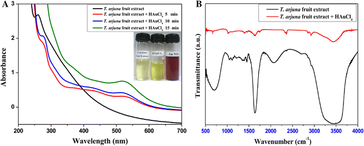
The synthesized Au NPs were clearly observed, when T. arjuna fruit extract added with HAuCl 4 solution within 15 min. The solution has been modified from yellow to reddish wine color, which indicates the formation of Au NPs.

UV–vis spectroscopy and Fourier transform infrared spectroscopy analysis

The mixture of fruit extract and HAuCl 4 solution was subjected to UV–vis spectroscopy analysis. The UV–vis spectra showed an absorption peak at 523 nm, which is due to the surface plasmon resonance for the formation of Au NPs (Fig.  1 a). The similar results were reported earlier [ 20 , 26 , 30 , 4951 ]. The Fourier transform infrared (FTIR) analysis of T. arjuna fruit extract before and after bioreduction is shown in Fig.  1 b. FTIR spectra are carried out to identify the possible biomolecules responsible for the reduction of the Au 3+ ions and capping of the reduced Au 0 NPs synthesized using T. arjuna fruit extract. The strong IR bands were observed at 3,464, 2,921, 2,849, 2,066, 1,629, 1,372, 1,109 and 667 cm −1 . The bands appeared at 3,464 cm −1 correspond to N–H stretching vibration of primary amines, thereafter the band at 2,921 and 2,849 cm −1 corresponds to C–H stretching of asymmetry and symmetry vibration, respectively. The band at 2,066 cm −1 corresponds to C–N stretching of any R–N=C=S, the medium band at 1,629 cm −1 corresponds to similar conjugation effects of N–H stretching. The IR bands observed at 1,372 and 1,109 cm −1 may be ascribed to C–N and –C–O–C stretching modes, respectively. The band observed at 667 cm −1 is due to the bending vibrations of N–H groups in proteins. Hence, tannin, terpenoid, saponins, flavonoids, glycosides and polyphenolic compounds present in the fruit extract of T. arjuna. They act as reducing and capping agent during the synthesis of Au NPs. The three new strong bands recorded at 2,334, 1,730 and 533 cm −1 in the spectra of the synthesized material were assigned to CO 2 -stretching, C=O carbonyl stretching and metal, respectively. The formation of carbonyl and metal peaks is due to the reduction of gold chloride to Au NPs. So that flavonoids and polyphenolic compounds act as a capping layer of the metal nanoparticles to prevent the agglomeration [ 52 ]. Water soluble polyols of plant origin are reported to the potent reducing agent in green synthesis of metal nanoparticles [ 53 ].

Fig. 1

a UV–vis spectra of Au NPs synthesized by reacting 1 mM HAuCl 4 aqueous solution with T. arjuna fruit extract, b FTIR spectra of Au NPs synthesized using T. arjuna fruit extract

X-ray diffraction and energy-dispersive X-ray spectroscopy analysis

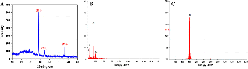
X-ray diffraction (XRD) pattern was recorded for the synthesized Au NPs (Fig.  2 a). The XRD peaks are located at angles (2 θ ) 38.24°, 44.45° and 66.30° corresponding to (1 1 1), (2 0 0) and (2 2 0) planes of the Au NPs. The standard diffraction peaks show the face-centered cubic phase of gold as per the JCPDS card no. 04-0784. The well-resolved and intense XRD pattern clearly showed that the Au NPs formed by the reduction of Au 3+ ions using T. arjuna fruit extract are crystalline in nature. Similar results were reported for Au NPs in the literature [ 20 , 26 ]. Energy-dispersive X-ray spectroscopy (EDX) (Fig.  2 b) illustrated the chemical nature of synthesized Au NPs using T. arjuna fruit extract. The peak was obtained at the energy of 2.12 keV for Au NPs. In addition, some of the weak peaks such as C and O are also found. The major peak of Al was found in the sample, which is due to the aluminum foil substrate (Fig.  2 c). The emission energy at 2.12 keV indicates the reduction of gold ions. Similarly, T. chebula seed extract-derived Au NPs showed an EDX spectrum with emission energy at 2.12 keV for gold and some of the weak signals from C and Cu observed [ 30 ].

Fig. 2

a XRD pattern of Au NPs synthesized by treating the fruit extract of T. arjuna with HAuCl 4 aqueous solution, b EDX analysis of Au NPs, c EDX analysis of aluminum foil substrate

Atomic force microscopy and transmission electron microscopy

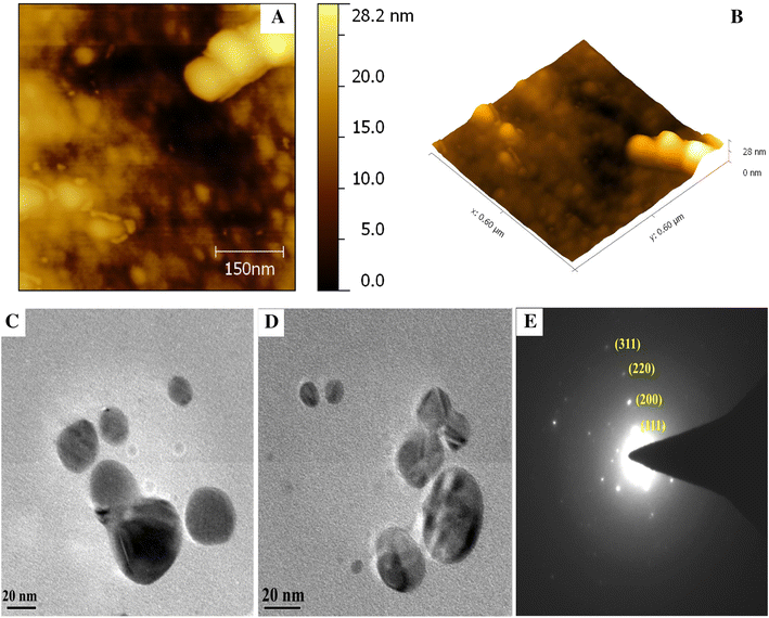
Surface morphology of the synthesized Au NPs was studied by atomic force microscopy (AFM) analysis (Fig.  3 a, b). The micrographs clearly showed that the formulated Au NPs possess spherical shape and calculated size in the range 20–50 nm. The transmission electron microscopy (TEM) images (Fig.  3 c, d) further ascertain that the Au NPs predominantly spherical in morphology with their sizes ranging from 5 to 50 nm have an average size of about 25 nm. The selected area electron diffraction (SAED) pattern (Fig.  3 e) of Au NPs resulted in the characteristics ring pattern of face-centered cubic (fcc) and it manifest the higher degree of crystallinity of Au NPs.

Fig. 3

AFM image of synthesized Au NPs: a 2D image, b 3D image, c , d TEM image of Au NPs formed by reduction of Au 3+ ions using the fruit extract of T. arjuna and e selected area electron diffraction (SAED) pattern of Au NPs

Dynamic light scattering and zeta potential analysis

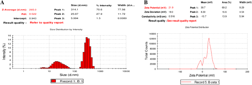
Dynamic light scattering (DLS) analysis determined the average particles size distribution profile of synthesized nanoparticles and capping agent enveloped the metallic particles along with the particular size of the metallic core [ 32 ]. In our result, the size of Au NPs is in the range 5–60 nm and average size is 25 nm (Fig.  4 a). In addition, some of the large sized particles appeared in DLS result, which is due to the agglomeration Au NPs in the solution. Zeta potential (ZP) analysis provided a clear information on the surface charge as well as stability of the synthesized nanoparticles. Even though the ZP value is higher than 30 mV or less than −30 eV, the dispersion is found to be stable. In our study, ZP value of the synthesized Au NPs is 21.9 mV (Fig.  4 b).

Fig. 4

a DLS measurement to determine the size distribution of Au NPs, b zeta potential analysis to determine the T. arjuna fruit extract on Au NPs stability

Antibacterial activity

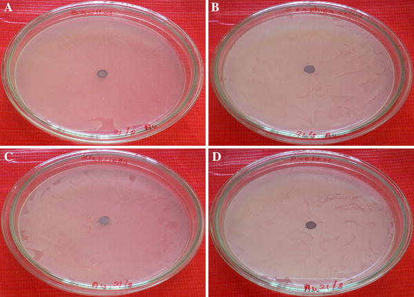
The antibacterial activity was investigated against Gram-positive and Gram-negative bacterial pathogens using green synthesized Au NPs and no significant growth inhibition was observed (Fig.  5 a–d). Even though the Au NPs bind with cytoplasmic membrane, they may not kill the bacterial cell due to its non-toxicity. Similarly, Au NPs induced the cell division without any endocytosis and cytotoxicity effect of HeLa and E. coil cells [ 54 ]. Recently, synthesized Au NPs using Turbinaria conoides aqueous extract and Au NPs alone did not showed any antibacterial and antimicrofouling activity [ 55 ].

Fig. 5

Antibacterial study of Au NPs: a activity of Au NPs loaded with 100 μl/6 mm disc against B. subtilis , b S. aureus gram-positive bacteria and activity of c K. pneumoniae , d P. vulgaris gram-negative bacteria

Effect of Au NPs on seed germination and seedling vegetative growth

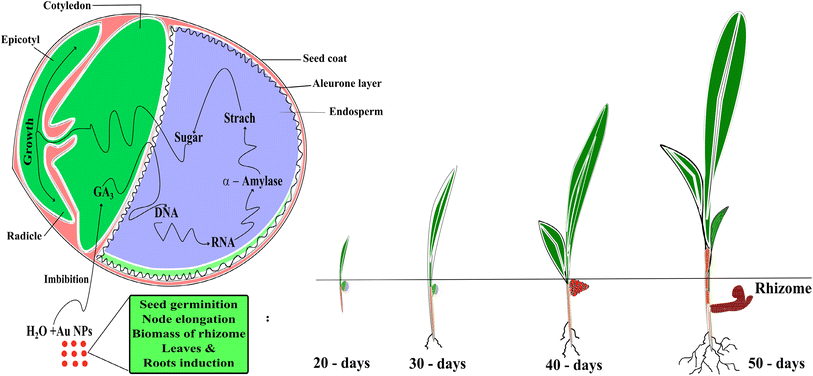
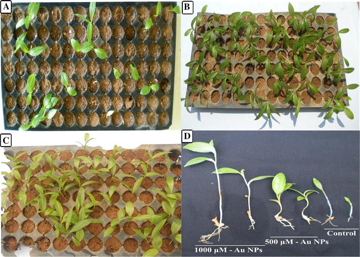
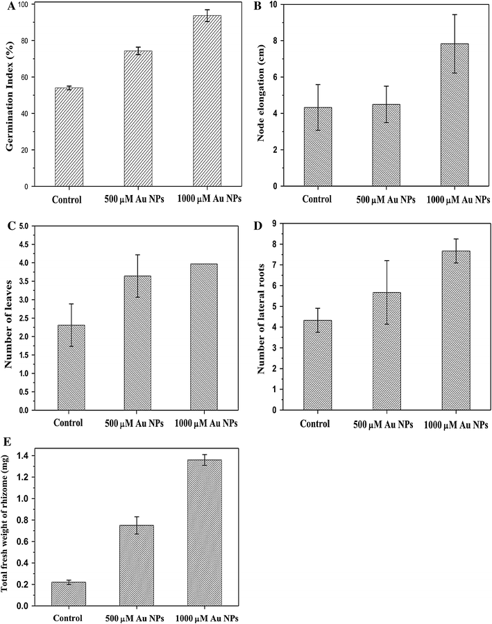
Germination is a physiological process beginning with water imbibition by seed. It initiated the metabolic activity of the emerging seedling [ 56 ]. Seed germination is a rapid growing process and widely used for phytotoxicity analysis, and also more advantages like sensitivity, simplicity, cost-effective and suitability for tested chemical sample [ 57 ]. To evaluate the effect of Au NPs on the seed germination, node elongation, increase in biomass of rhizome, leaf and root initiation were calculated with the tested samples of G. superba seeds. Au NPs were treated with two different concentrations at 500 and 1,000 μM exhibiting an increment in the frequency of germination index (Supplementary files, Table. S1) of 74.33 ± 2.08 and 93.6 ± 3.21, respectively. Finally, empirical values are observed at 1,000 μM Au NPs, which exhibit significant effect on germination activity. In control samples, seeds soaked in the distilled water have up taken the water molecules and passed through the embryo cells. From gibberellic acid (GA 3 ) the embryo cells were collected in germination signal and have activated the DNA coding for the synthesis of alpha amylase enzyme in the aleurone cell layer. The alpha amylase is an enzyme, which breaks down the starch into simple sugar that provides energy for the growing seedling [ 58 ] of G. superba seeds. Au NP-treated seeds were up taken by the imbibition process and breaking the seed coat of G. superba seeds, which enters the high affinity uptake system of gold ions and interact with embryo cells. This interference with the intercellular signaling stimulates the GA 3 activity and promotes the DNA coding for the synthesis of alpha amylase enzyme in the aleurone cell layer. This enzyme rapidly breaks down starch into simple sugar, which offers the energy for growing seedlings (Fig.  6 ). The dynamic growth of seed germination activity was evaluated at two different concentrations of Au NPs treatment. Au NPs exhibited considerable variation in the growth of node elongation, increase in biomass of rhizome, leaf and root initiation at different day intervals (Fig.  7 ) when compared to the control. The seed germination index showed that the 500 and 1,000 μM Au NPs have been enhanced by 20.33 and 39.67 % of seed germination when compared to the control. Au NPs can increase permeability of the seed coat facilitating the admission of H 2 O and O 2 into the cells, which accelerates the metabolic activity and germination process [ 59 ]. Node elongation was also found to follow the same treatment. In 500 μM Au NPs treatment of the node elongation was relevant to the control. By contrast, more significant effect on node elongation was observed in 1,000 μM of Au NPs (Supplementary files, Table. S2) (Fig.  8 ). The observed phenomenon might be attributed to an increased level of GA 3 and it is due to the responsible of shoot elongation [ 60 ]. The Au NPs treatment interferes with the action of endogenous plant hormones and induced the phenotype in the treated seedlings. Similarly, 10 ppm of Au NPs treated in B. juncea seedling increased the plants height 8–9 and 35 % stem diameter when compared to the untreated seedling [ 40 ]. Moreover, there was an increase in the number of leaves and lateral roots in the case of Au NP-treated seedlings (Supplementary files, Table. S3, S4) (Fig.  8 ). Ethylene is a gaseous plant hormone and it controls the leaf number by regulating leaf abscission. It has been observed that inhibition of ethylene action reduces the function of abscission, which is due to the increase in the number of leaves in treated seedling. However, the antagonistic impact of Au NPs reduced the function of ethylene and increased the leaf number of G. superba seedling. Recently, 25 % of leaves initiation was increased in the 10 ppm of Au NPs treated in B. juncea seedling, as compared to the control [ 40 ]. In our previous studies, Au NP-treated Allium cepa root cells interact with intracellular chromatin (DNA + Histone protein), mitotic interphase and induced the protein synthesis. Consequently, increase in the dose of Au NPs have enhanced the mitotic index without any chromosomal aberration of the A. cepa root tip cells as well as Au NPs involved intercellular communication and intracellular signaling with cells [ 61 ]. In addition, the total fresh weight of G. superba seedling rhizome was also increased by 2.40 and 5.18 times exposure to 500 and 1,000 μM of Au NPs and they are compared with the control (Supplementary files, Table. S5). In the actual mechanism, the Au NPs increased the chlorophyll content for the treated seedling and induced the gross light absorbance by accelerating the photochemical reaction, and consequently, higher availability of reducing power (NADPH + H + ) and energy (ATP) to carry out CO 2 fixation [ 62 ]. It improves the CO 2 fixation and increases total sugar content of the rhizome of all treated seedling. Previously, 43 % of sugar contents were increased by 25 ppm Au NP-treated B. juncea seedling [ 40 ]. Therefore, seed germination activity and plant vegetative growth showed dose-dependent response to Au NPs. Recently, Lin and Xing have investigated the seed germination activity of different metal and metal oxide nanoparticles such as MWCNT, Al, Al 2 O 3 , Zn, and ZnO NPs treated with six various plant species like corn, cucumber, radish, rape and ryegrass. The Zn and ZnO NPs have significantly inhibited the seed germination and root growth of all treated plant species [ 57 ]. Similarly, lettuce and cucumber seeds were treated with different concentrations of Au NPs and the results showed significant effect in lettuce seed than that of cucumber seeds [ 63 ]. Arabidopsis thaliana seeds treated with 10 μg/ml of Au NPs (24 nm) enhance the seed yield and consequently, 80 μg/ml dose of Au NPs was recommended for fodder crops productivity [ 5 ]. It is suggested that 1,000 μM Au NPs treatment was able to enhance the seed germination of other rare species.

Fig. 6

Schematic representation of effect of Au NPs induced the G. superba seed germination, node elongation, biomass of rhizome, leaf and root initiation

Fig. 7

Effect of Au NPs on G. superba seed germination: a Control, b 500 μM Au NPs, c 1,000 μM Au NPs for a duration of 30 day, d Induction of node elongation, biomass of rhizome leaf and root initiation of Au NP-treated samples for a duration of 40 day

Fig. 8

Effect of Au NPs on G. superba seed germination and vegetative growth: a Germination rate of seed in the presence and absence of Au NPs for a duration of 50 day, b node elongation of G. superba seedling growth on with and without treatment of Au NPs, c bar diagram representing number of leaves initiated at presence and absence of Au NPs, d image shows lateral roots produced in with and without treatment of Au NPs, e total fresh weight of rhizome in G. superba seedling for a duration of 50 day. Results are presented as average ± SD of triplicate

Conclusion

In this paper, we have developed the green synthesis of Au NPs using T. arjuna fruit extract. The TEM image clearly showed the spherical shape with an average size of 25 nm. The antibacterial activity was tested for the Au NPs and has no significant inhibition effect. Hence, biocompatibility of Au NPs was treated with G. superba seed germination activity. The results confirmed that the Au NPs have great potential in inducing the seed germination as well as have positive impact on the plant vegetative growth of G. superba seedling. It is suggested that green synthesis of nanoparticles has beneficial application in the field of agriculture crops and seed germination of endangered plant species.

Methods

Collection of plant materials

T. arjuna fruits and G. superba seeds were collected from Endangered Medicinal Plant Conservation Centre, Science Campus, Alagappa University, Karaikudi, Tamil Nadu, India. Taxonomic identification was made by Dr. S. John Britto, The Rapinat Herbarium and Centre for Molecular Systematics, St. Joseph’s College, Tiruchirappalli, Tamil Nadu, India. The voucher specimens were numbered (KG-002 and KG-001) and preserved in the Department of Nanoscience and Technology, Alagappa University, Karaikudi.

Synthesis of Au NPs using T. arjuna fruit extract

Fresh T. arjuna fruits were cleaned in running tap water and then by distilled water. 10 g of fruits were added with 100 ml of double-distilled water and boiled at 50–60 °C for 5 min. The obtained extraction was filtered using Whatman No. 1 filter paper and the filtrate was collected in 250 ml Erlenmeyer flask and stored at room temperature for further usage. Thereafter, 1 ml of T. arjuna fruit extract was added to 100 ml of 1 mM HAuCl 4 solution at room temperature and reduction of Au NPs was clearly observed within next 15 min.

Characterization

UV–visible spectra of the synthesized Au NPs were recorded in the wavelength range of 200–700 nm using Shimadzu spectrophotometer (Model UV-1800) operating at a resolution of 1 nm. Moreover, Fourier transform infrared spectroscopy (FTIR) analysis was carried out in the range 500–4,000 cm −1 . XRD pattern was recorded using Cu Kα radiation ( λ  = 1.54060 Å) with nickel monochromator in the range of 2 θ from 10° to 80°. The average crystalline size of the synthesized Au NPs was calculated using Scherrer’s formula ( D  = 0.9 λ / β cos θ ). EDX analysis was performed for a thin-film sample prepared using the Au NPs by spin coating method (1,500 rpm) on a aluminum foil (1 × 1 cm) by dropping 100 μl of the sample on the foil and is allowed to dry for 30 min at room temperature for further use. For AFM analysis, sample was prepared on a glass slide (1 × 1 cm) by dropping 100 μl of the sample on the slide and allowed to dry for 30 min. Then the slides were scanned with AFM (APE Research model no: A100-SGS). The AFM characterization was carried out in ambient temperature in non-contact mode using silicon nitrite tips with varying resonance frequencies. The morphology of the synthesized Au NPs was examined using TEM. Samples for TEM analysis were prepared by drop coating the Au NPs solutions on carbon-coated copper grids at room temperature. The excess Au NPs solution was removed with filter paper. The copper grid was finally dried at room temperature and was subjected to TEM analysis by the instrument Tecnai F20 model operated at an accelerating voltage of 200 kV. The particles size distribution of synthesized Au NPs was evaluated by DLS measurement and stability was also determined by ZP analysis using a Zetasizer, version 6.32. The measurement of zeta potential is due to the direction and velocity of particles under the influence of known electric field.

Antibacterial activity of Au NPs

The antibacterial activity of the green synthesized Au NPs was examined using two Gram-positive ( Staphylococcus aureus and Bacillus subtilis ) and two Gram-negative bacteria ( Proteus vulgaris and Klebsiella pneumoniae ) by disc diffusion method. These four bacterial strains were grown in nutrient broth at 37 °C until the bacterial suspension has reached 1.5 × 10 8 CFU/ml. Approximately, 20 ml of molten nutrient agar was poured into the Petri dishes. All the bacterial suspensions were swapped over the medium and the discs loaded with 100 μl of Au NPs were placed over the medium using sterile forceps. The plates were incubated for 24 h at 37 °C and inhibition zone of each disc was measured. Each experiments were conducted in triplicate.

In vivo seed germination of G. superba treated with Au NPs

Collected G. superba seeds were washed in running tap water for about 15–20 min to remove the soil particles, cleaned with liquid detergent Tween-20 (1 % v/v) for 5–10 min and then rinsed with sterile double-distilled water. They were surface sterilized with 0.01 % (HgCl 2 w/v) solution for 2 min and again washed well in distilled water for about 3–4 times to remove all the traces of HgCl 2 (Mercuric chloride). After the surface sterilization to remove the microbes, the synthesized Au NPs were suspended directly in deionized water and dispersed by ultrasonic vibration (100 W, 30 kHz) for 30 min to construct 100 ml of two different concentrations (500 and 1,000 μM) of Au NPs and they were autoclaved at 121 °C and 15 lb for 20 min. Surface sterilized G. superba ( n  = 100) seeds were soaked in 100 ml of 500 μM and 1,000 μM Au NPs and kept in dark atmosphere at room temperature (25 ± 1 °C) for one day. Au NP-treated seeds were transferred into the polystyrene seedling trays, which contain sterile sand and were kept in a mist house. The seed germination percentage, node elongation, increase in biomass of rhizome, leaf and root initiation were calculated at 20, 30, 40 and 50 days intervals, respectively. All the experiments were conducted in triplicate. The data were statistically analyzed using the one-way analysis of variance (ANOVA) and significant differences between the mean values. The germination index was calculated using the following formula:


Acknowledgments

The authors gratefully thank School of Physics, Alagappa University for extending the XRD facility and also the Department of Industrial Chemistry, Alagappa University for providing the EDX facility.

Conflict of interests

The authors declare that they have no competing interests.

Authors’ contributions

KG, SG and VK carried out the gold nanoparticles synthesis and characterization. Synthesized gold nanoparticles were investigated in antibacterial and seed germination activity. AA carried out the manuscript preparation. All authors read approved the final manuscript.


References

  1. Castaneda et al. (2007) Electrochemical genosensors for biomedical applications based on gold nanoparticles (pp. 1961-1967) 10.1016/j.bios.2006.08.031
  2. Ganeshkumar et al. (2013) Spontaneous ultra fast synthesis of gold nanoparticles using Punica granatum for cancer targeted drug delivery (pp. 208-216) 10.1016/j.colsurfb.2013.01.035
  3. Kesik et al. (2013) A functional immobilization matrix based on a conducting polymer and functionalized gold nanoparticles: Synthesis and its application as an amperometric glucose biosensor (pp. 4463-4471) 10.1016/j.polymer.2013.06.050
  4. Maity et al. (2012) Green synthesis of gold nanoparticles using gum polysaccharide of Cochlospermum religiosum (katira gum) and study of catalytic activity (pp. 130-134) 10.1016/j.physe.2012.07.020
  5. Kumar et al. (2013) Gold nanoparticle exposure induces growth and yield enhancement in Arabidopsis thaliana (pp. 462-468) 10.1016/j.scitotenv.2013.05.018
  6. Liu et al. (2012) Evaluation of antioxidant activity of chrysanthemum extracts and tea beverages by gold nanoparticles-based assay (pp. 348-352) 10.1016/j.colsurfb.2011.12.007
  7. Bhat et al. (2013) Photo-bio-synthesis of irregular shaped functionalized gold nanoparticles using edible mushroom Pleurotus florida and its anticancer evaluation (pp. 63-69) 10.1016/j.jphotobiol.2013.05.002
  8. Annamalai et al. (2013) Green synthesis, characterization and antimicrobial activity of Au NPs using Euphorbia hirta L. leaf extract (pp. 60-65) 10.1016/j.colsurfb.2013.02.012
  9. Jayaseelan et al. (2013) Green synthesis of gold nanoparticles using seed aqueous extract of Abelmoschus esculentus and its antifungal activity (pp. 423-429) 10.1016/j.indcrop.2012.12.019
  10. Apte et al. (2013) Melanin mediated synthesis of gold nanoparticles by Yarrowia lipolytica (pp. 149-152) 10.1016/j.matlet.2012.12.087
  11. Kumar et al. (2013) Bioefficacy of plant mediated gold nanoparticles and Anthocepholus cadamba on filarial vector, Culex quinquefasciatus (Insecta: Diptera: Culicidae) (pp. 1053-1063) 10.1007/s00436-012-3232-z
  12. Moreau et al. (2009) PVD synthesis and transfer into water-based solutions of functionalized gold nanoparticles (pp. S888-S892) 10.1002/ppap.200932210
  13. Dillon et al. (2006) Hot-wire chemical vapor synthesis for a variety of nano-materials with novel applications (pp. 216-220) 10.1016/j.tsf.2005.07.182
  14. Gutierrez-Wing et al. (2012) Microwave-assisted synthesis of gold nanoparticles self-assembled into self-supported superstructures (pp. 2281-2287) 10.1039/c2nr12053d
  15. Yang et al. (2007) Continuous synthesis of gold nanoparticles and nanoplates with controlled size and shape under UV irradiation (pp. 37-44) 10.1016/j.colsurfa.2006.09.017
  16. Sobhani et al. (2008) Sol-gel synthesis of aluminum titanate (Al2 TiO5) nano-particles (pp. 282-285) 10.1016/j.jmatprotec.2007.12.023
  17. Magnusson et al. (1999) Size-selected gold nanoparticles by aerosol technology (pp. 45-48) 10.1016/S0965-9773(99)00063-X
  18. Kenji et al. (2005) Sonochemical synthesis of gold nanoparticles: effects of ultrasound frequency (pp. 20673-20675) 10.1021/jp0549374
  19. McGilvray et al. (2006) Facile photochemical synthesis of unprotected aqueous gold nanoparticles (pp. 15980-15981) 10.1021/ja066522h
  20. Sheny et al. (2011) Phytosynthesis of Au, Ag and Au-Ag bimetallic nanoparticles using aqueous extract and dried leaf of Anacardium occidentale (pp. 254-262) 10.1016/j.saa.2011.02.051
  21. Kumar Das et al. (2010) Green synthesis of gold nanoparticles using ethanolic leaf extract of Centella asiatica (pp. 1445-1447) 10.1016/j.matlet.2010.03.051
  22. Dwivedi and Gopal (2010) Biosynthesis of silver and gold nanoparticles using Chenopodium album leaf extract (pp. 27-33) 10.1016/j.colsurfa.2010.07.020
  23. Narayanan and Sakthivel (2010) Phytosynthesis of gold nanoparticles using leaf extract of Coleus amboinicus Lour (pp. 1232-1238) 10.1016/j.matchar.2010.08.003
  24. Vijayakumar et al. (2011) Green synthesis and characterization of gold nanoparticles using extract of anti-tumor potent Crocus sativus (pp. 665-671) 10.1016/j.physe.2011.11.002
  25. Philip (2010) Rapid green synthesis of spherical gold nanoparticles using Mangifera indica leaf (pp. 807-810) 10.1016/j.saa.2010.08.008
  26. Aromal et al. (2012) Green synthesis of well-dispersed gold nanoparticles using Macrotyloma uniflorum (pp. 99-104) 10.1016/j.saa.2011.09.035
  27. Geethalakshmi and Sarada (2013) Characterization and antimicrobial activity of gold and silver nanoparticles synthesized using saponin isolated from Trianthema decandra L (pp. 107-115) 10.1016/j.indcrop.2013.08.055
  28. Philip et al. (2011) Murraya koenigii leaf-assisted rapid green synthesis of silver and gold nanoparticles (pp. 899-904) 10.1016/j.saa.2010.12.060
  29. Sneha et al. (2010) Counter ions and temperature incorporated tailoring of biogenic gold nanoparticles (pp. 1450-1458) 10.1016/j.procbio.2010.05.019
  30. Kumar et al. (2012) Terminalia chebula mediated green and rapid synthesis of gold nanoparticles (pp. 490-494) 10.1016/j.saa.2011.11.001
  31. Sneha et al. (2011) Biosynthesis of Au nanoparticles using cumin seed powder extract (pp. 1811-1814) 10.1166/jnn.2011.3414
  32. Sujitha and Kannan (2013) Green synthesis of gold nanoparticles using Citrus fruits (Citrus limon, Citrus reticulata and Citrus sinensis) aqueous extract and its characterization (pp. 15-23) 10.1016/j.saa.2012.09.042
  33. http://www.cytodiagnostics.com/store/pc/40nm-Stabilized-Gold-Nanoparticles-20ml-141p108.htm
  34. Jana and Shekhawat (2011) Critical review on medicinally potent plant species: Gloriosa superba (pp. 293-301) 10.1016/j.fitote.2010.11.008
  35. Sumy et al. (2000) Foundation for revitalization of local health traditions
  36. Arumugam and Gopinath (2012) In vitro micropropagation using corm bud explants: an endangered medicinal plant of Gloriosa superba L (pp. 120-128) 10.3923/ajbkr.2012.120.128
  37. Samarajeewa et al. (1993) Clonal propagation of Gloriosa superba (pp. 719-720)
  38. An et al. (2008) Physical, chemical and microbiological changes in stored green asparagus spears as affected by coating of silver nanoparticles-PVP (pp. 1100-1107) 10.1016/j.lwt.2007.06.019
  39. Hong et al. (2005) Effect of Nano-TiO2 on photochemical reaction of chloroplasts of spinach (pp. 269-279) 10.1385/BTER:105:1-3:269
  40. Arora et al. (2012) Gold nanoparticle induced enhancement in growth and seed yield of Brassica juncea (pp. 303-310) 10.1007/s10725-011-9649-z
  41. Dhingra et al. (2013) Forensic and pharmacognostic studies of the Terminalia arjuna Bark (pp. 15-19)
  42. Dwivedi and Jauhari (1997) Beneficial effects of Terminalia arjuna in coronary artery disease (pp. 507-510)
  43. Antani et al. (1991) Terminalia arjuna in congestive heart failure (Abstract)
  44. Tiwari et al. (1990) Effect of Terminalia arjuna on lipid profiles of rabbit fed hypercholesterolemic diet (pp. 43-47)
  45. Saxena et al. (2007) Cytotoxic agents from Terminalia arjuna (pp. 1486-1490) 10.1055/s-2007-990258
  46. Dwivedi (2007) Terminalia arjuna Wight & Arn. A useful drug for cardiovascular disorders (pp. 114-129) 10.1016/j.jep.2007.08.003
  47. Arumugam and Gopinath (2011) In-vitro callus development of different explants used for different medium of Terminalia arjuna (pp. 564-572) 10.3923/ajbkr.2011.564.572
  48. Ghosh et al. (2010) Protective effect of the fruits of Terminalia arjuna against cadmium-induced oxidant stress and hepatic cell injury via MAPK activation and mitochondria dependent pathway (pp. 1062-1075) 10.1016/j.foodchem.2010.05.062
  49. Sathishkumar et al. (2010) Green recovery of gold through biosorption, biocrystallization, and pyro-crystallization (pp. 7129-7135) 10.1021/ie100104j
  50. Binupriya et al. (2010) Biocrystallization of silver and gold ions by inactive cell filtrate of Rhizopus stolonifer (pp. 531-534) 10.1016/j.colsurfb.2010.05.021
  51. Binupriya et al. (2010) Bioreduction of trivalent aurum to nano-crystalline gold particles by active and inactive cells and cell-free extract of Aspergillus oryzae var. viridis (pp. 539-545) 10.1016/j.jhazmat.2009.12.066
  52. Ghoreishi et al. (2011) Green synthesis of silver and gold nanoparticles using Rosa damascena and its primary application in electrochemistry (pp. 97-104) 10.1016/j.physe.2011.07.008
  53. Sathishkumar et al. (2009) Cinnamon zeylanicum bark extract and powder mediated green synthesis of nano-crystalline silver particles and its bactericidal activity (pp. 332-338) 10.1016/j.colsurfb.2009.06.005
  54. Cui et al. (2012) Effects of aggregation and the surface properties of gold nanoparticles on cytotoxicity and cell growth (pp. 46-53) 10.1016/j.nano.2011.05.005
  55. Sri Ramkumar et al. (2014) Synthesis and characterization of silver and gold nanoparticles using aqueous extract of seaweed, Turbinaria conoides and their anti-microfouling activity 10.1155/2014/938272
  56. Kordan (1992) Seed viability and germination: a multi-purpose experimental system (pp. 247-251) 10.1080/00219266.1992.9655281
  57. Lin and Xing (2007) Phytotoxicity of nanoparticles; inhibition of seed germination and root growth (pp. 243-250) 10.1016/j.envpol.2007.01.016
  58. Sun and Henson (1991) A quantitative assessment of the importance of barley seed α-amylase, β-amylase, debranching enzyme, and α-glucosidase in starch degradation (pp. 298-305) 10.1016/0003-9861(91)90299-X
  59. Zheng et al. (2005) Effect of nano-TiO2 on strength of naturally aged seeds and growth of spinach (pp. 83-91) 10.1385/BTER:104:1:083
  60. Stepanova et al. (2007) Multilevel interactions between ethylene and auxin in Arabidopsis roots (pp. 2169-2185) 10.1105/tpc.107.052068
  61. Gopinath et al. (2013) Green synthesis of gold nanoparticles from leaf extract of Terminalia arjuna for the enhanced mitotic cell division and pollen germination activity (pp. 737-742) 10.1016/j.indcrop.2013.08.060
  62. Krause et al. (1998) Sucrose metabolism in cold-stored potato tubers with decreased expression of sucrose phosphate synthase (pp. 285-299) 10.1046/j.1365-3040.1998.00271.x
  63. Barrena et al. (2009) Evaluation of the ecotoxicity of model nanoparticles (pp. 850-857) 10.1016/j.chemosphere.2009.01.078