A new procedure for preparation of polyethylene glycol-grafted magnetic iron oxide nanoparticles

Abstract

Magnetic iron oxide nanoparticles (MNPs) have been widely explored for use in biomedical applications. In the present study, iron oxide nanoparticles were connected to methoxy poly(ethylene glycol) (mPEG) via a new method. The mPEG was acrylated at first and Michael reaction was carried out between acrylated mPEG and 3-aminopropyl triethoxysilane as a coupling agent. The chemical structures of modified mPEG were characterized by Fourier transform-infrared spectroscopy (FT-IR) and nuclear magnetic resonance. In the next step, iron oxide nanoparticle was coupled with the above-mentioned adduct. Preparation of magnetic nanoparticles with average particle size of 20–30 nm was proved by scanning electron microscopy. The structure of mPEG grafted on the surface of MNPs was confirmed by FT-IR spectroscopy and thermal gravimetric analysis. The synthesized nanoparticles have the potential to be used in different biomedical applications.


Background

Magnetic iron oxide nanoparticles with tailored surface chemistry have been widely used experimentally for numerous in vivo applications such as magnetic resonance imaging, contrast enhancement, tissue repair, immunoassay, detoxification of biological fluids, hyperthermia, drug delivery and in cell separation [ 15 ]. Coating or modifying these nanoparticles with hydrophilic molecules, especially biomolecules, is a crucial step for the preparation of water-dispersible magnetite nanoparticles (MNPs) for use in biological applications [ 6 ]. Several synthetic and natural polymers have been employed to modify the surface of the MNPs. These polymers include dextran [ 7 ] poly(ethylene glycol)s (PEG) [ 8 ], poly(vinylpyrrolidone) (PVP) [ 9 ] and chitosan [ 10 ] all of which are known to be biocompatible and result in a long blood-circulating MNPs. The PEG, because of its hydrophilicity, nontoxicity and absence of antigenicity and immunogenicity, can be selected to be attached to surface magnetic iron oxide nanoparticles. The hydrophilic PEG molecules have been used to reduce phagocytic capture of nanoparticles by cellular components of the immune system, leading to extended circulation and subsequent accumulation in tumors as a consequence of the enhanced permeability and retention (EPR) effect due to leaky vasculature and poor lymphatic drainage in tumors [ 11 ]. Several techniques have been employed to obtain PEG-modified magnetic nanoparticles using different methods [ 1215 ].

García-Jimeno and co-worker reported the formation of stable aqueous suspensions of magnetite-based ferrofluid stabilized by an unmodified PEG [ 16 ]. Accordingly, the interaction between PEG and magnetite nanoparticles is due to dipole–cation binding between the ether group of the polymer and the positive charge of magnetite. In another work, PEG was coated on oil-soluble Fe 3 O 4 and Fe 3 O 4 –CdSe nanoparticles stabilized by various ligands to render them hydrophilicity [ 17 ].

Masoudi and co-worker [ 13 ] synthesized PEG-modified nanoparticles using an ultrasonic assisted co-precipitation technique. This magnetite nanoparticles coated with different PEG and with different concentrations of PEG in an ex situ manner. The colloidal stability of iron oxide nanoparticles coated with carboxyl-PEG-phosphoric acid has been reported by Lu et al. [ 18 ]. They showed that PEG could be an excellent surfactant for the stabilization of Fe 3 O 4 nanoparticles. In situations, where particle stability is of utmost importance, as in biomedical applications, silane chemistry is being explored as an alternative functionalization route because molecules can bind with the surface of the particle, enhancing particle stability in suspensions at physiological conditions [ 19 ]. Hydrophilic molecules are usually polymeric [e.g., poly(ethylene glycol)], that can often linked to nanoparticle cores via silanes [ 20 , 21 ]. Carlos Rinaldi and co-workers [ 22 ] synthesized stable colloidal dispersions of monodisperse magnetic nanoparticles in water by the thermal decomposition method, and exchanging the oleic acid molecules on the surface of the particles for PEG–silane chains. Larsen and co-workers [ 23 ] prepared silane-PEG-coated MNPs using a simple synthesis method based on the use of biocompatible silane-PEG as a coating agent.

Formation of stable nanoparticles from the highly water soluble polymers like PEG is quite difficult and thus, coating of magnetic nanoparticles with a hydrophilic polymer is a big challenge. High amount of polymer or polymer with high molecular weight is needed to overcome this drawback and consequently, an increase in the particle size will be observed followed by a decrease in saturation magnetization of the hybrid nanocomposite. In this work, we have replaced the conventional coating with the grafting method that results in a stable magnetic nanocarrier in which a very low molecular weight polyethylene glycol (1,100 g/mol) is chemically bonded onto the surface of MNPs. Fe 3 O 4 nanoparticles were synthesized using co-precipitation of a Fe(II) and Fe(III) salt mixture in basic aqueous solution. The acrylated mPEG (AmPEG) was reacted with amino silane via Michael addition reaction to produce triethoxysilyl-terminated poly(ethylene glycol) [(EtO) 3 -Si-mPEG] and then grafted onto the surface of iron oxide nanoparticles by a sol–gel reaction. The size of nanoparticle is smaller than 30 nm. The mPEG-modified nanoparticles were characterized by Fourier transform-infrared (FT-IR), thermal gravimetric analysis (TGA) and scanning electron microscopy (SEM).

Methods

Materials

Iron (II) chloride tetrahydrate, iron (III) chloride hexahydrate, ammonium hydroxide solution (25 % NH 3 in H 2 O), methoxy poly(ethylene glycol) (mPEG, M n  = 1,100 g/mol), 3-aminopropyl triethoxysilane (APTS), triethylamine (TEA), acryloyl chloride, acetone, dichloromethane (DCM) and dimethylformamide (DMF) were purchased from Merck Co. All these reagents were used without further purification.

Synthesis of MNPs

Magnetite nanoparticles were synthesized by co-precipitation of Fe(II) and Fe(III) chloride in alkali solution according to the standard technique [ 24 ]. Briefly, under N 2 purging, 1 g of FeCl 2 .4H 2 O and 2.6 g of FeCl 3 .6H 2 O were dissolved in 25 ml distilled water and kept at 75 °C for 10 min, then 10 mL of ammonia solution (25 %) was dropped into the above mixture under vigorous stirring and keeping at 75 °C for a further 1.5 h. The resulting suspension was washed with distilled water and ethanol three times and dried under vacuum.

Synthesis of acrylated methoxy poly(ethylene glycol) (AmPEG)

The mPEG was end capped with acrylate groups to link APTS to mPEG via Michael addition reaction. Briefly, 4.4 g (4 mmol) of mPEG was dissolved in 20 ml of DCM in a three-necked round-bottom flask. 1.12 ml (8 mmol) of triethylamine and 0.49 ml (6 mmol) of acryloyl chloride were added to the flask at 0 °C, under a stream of nitrogen and the reaction mixture was stirred for 24 h at room temperature. The reaction mixture was filtered to remove triethylammonium chloride and acrylated mPEG was obtained after precipitation of filtrate in cold n-hexane and vacuum-drying the precipitate.

Modifying of magnetic iron oxide nanoparticle with methoxy poly(ethylene glycol)- (MNPs-mPEG)

In first step, 2.2 g (2 mmol) of acrylated mPEG and 0.45 ml (2 mmol) of APTS were dissolved in anhydrous DMF and the solution was kept at room temperature for 3 days (solution 1). In the second step, 0.5 g of magnetite nanoparticles was dispersed in 25 ml of DMF with 10 min ultrasonic irradiation and was rapidly added to solution 1. Three drops of water were added to promote the hydrolysis process of silane. The mixture was kept for 48 h with stirring at room temperature. The product was separated with centrifugation and magnetic field and was washed five times with water and then dried under vacuum.

Characterization

Modified mPEG and surface of MNPs were characterized by FT-IR spectroscopy (Bruker-Equinoxss) and nuclear magnetic resonance ( 1 HNMR) (Bruker, 500 MHz) in CDCl 3 . The obtained nanoparticles were analyzed by scanning electron microscopy (HITACHI S 4160). Their image reveals the size and shape of nanoparticles. The sample was placed on a stub and then sputter coated with gold before observation. Thermal gravimetric analysis (TGA) was carried out using TGA Q50 V6.3 Build 189. The temperature of the sample gradually increased from 25 to 600 °C at a rate of 10 °C/min under nitrogen atmosphere. Hydrodynamic size of nanoparticles dispersion in aqueous solution was measured by dynamic light scattering (DLS) at 25 °C (Nano ZS, Malvern Instrument, UK).

Result and discussion

Synthesis of magnetite nanoparticles (MNPs)

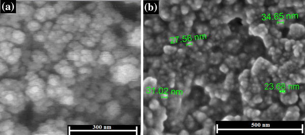
The magnetite nanoparticles were synthesized through the co-precipitation of ferrous and ferric hydroxides by addition of NH 4 OH to a solution of Fe(II) and Fe(III) salts at a 1:2 molar ratio under nitrogen atmosphere to prevent oxidation. Scanning electron microscopy was used to illustrate the morphology of magnetite nanoparticles. It could be seen that the MNPs took a spherical morphology and the average diameter calculated from SEM image was approximately ~20 nm (Fig.  1 a).

Fig. 1

SEM images of MNPs ( a ) and MNPs-mPEG ( b )

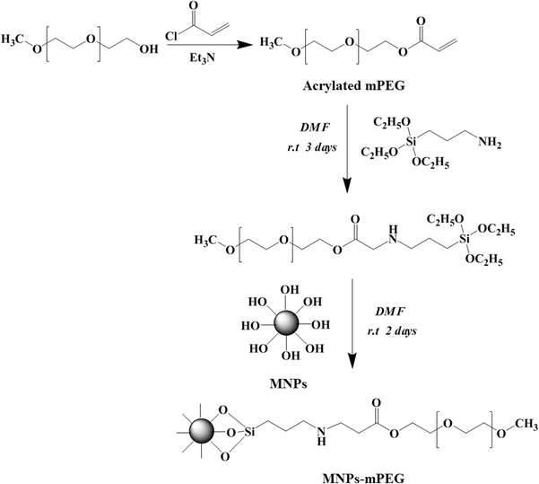
Acrylated mPEG (AmPEG) was synthesized by following the same method reported previously, using acryloyl chloride (Scheme  1 ) [ 25 ].

Scheme 1

Synthesis of acrylated methoxy poly(ethylene glycol) and MNPs-mPEG

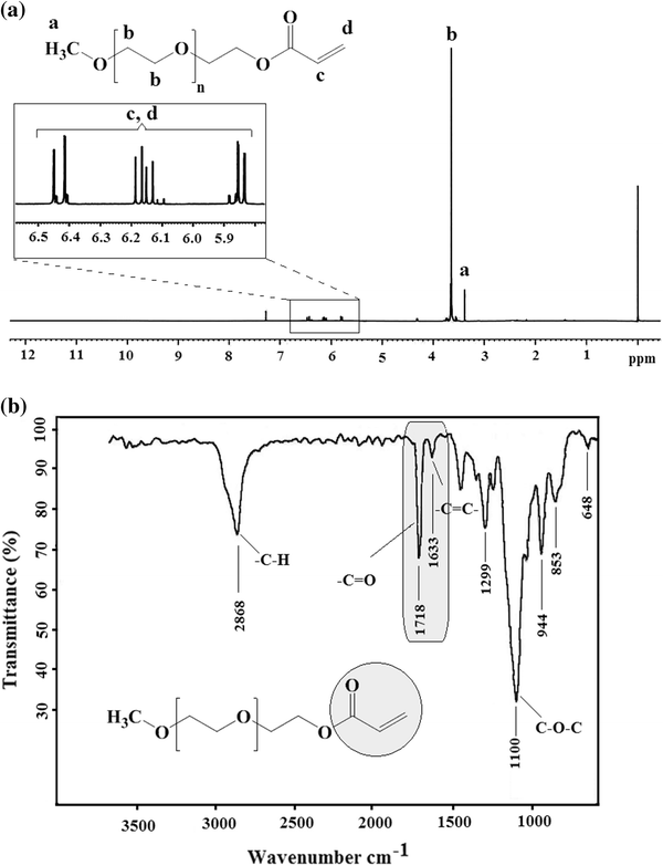
The structure of the synthesized AmPEG was determined by 1 HNMR spectroscopy in CDCl 3 . Vinylic protons of AmPEG were splitted into three different peaks which were located at 6.15 (–COC H C = C–), 5.85 ppm (–COCHC = C H –, cis -proton) and 6.45 ppm (–COCHC = C H –, trans -proton) (Fig.  2 a). FT-IR spectroscopy was also used to confirm the formation of acrylate group. The presence of a strong peak at 1,718 cm −1 corresponding to the C = O stretching bond and a characteristic peak at 1,633 cm −1 related to the C = C of acrylate group indicated the formation of vinylic linkage. This result confirms the reaction between hydroxyl end group of polyethylene glycol and acryloyl chloride (Fig.  2 b).

Fig. 2

1 HNMR ( a ) and FT-IR ( b ) spectra of acrylated methoxy poly(ethylene glycol)

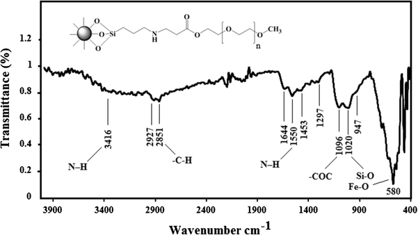
PEG-modified magnetite nanoparticle was synthesized via a two-step method. In the first step, acrylate end groups (–CH = CH 2 ) of AmPEG are reacted with amine groups of APTS through Michael addition reaction at room temperature to produce (EtO) 3 -Si-mPEG (Scheme  1 ). In the next step, the (EtO) 3 -Si-mPEG was condensed at the outer surface of the MNPs via sol–gel reaction. The presence of mPEG layer on MNPs surface was characterized by FT-IR spectroscopy. Fe–O stretching band as the characteristic peak of magnetite nanoparticle (MNPs) was located at 580 cm −1 , important IR bands at 2,927, 2,851, 1,644, 1,550, 1,453, 1,278, 1,096, 1,020 and 947 cm −1 can be assigned to mPEG groups and clearly indicating that mPEG has been grafted onto the surface of MNPs (Fig.  3 ) [ 26 , 27 ]. The peak at 1,096 cm −1 most likely derives from the C–O stretching vibration of ether groups, the peaks at 947 cm −1 can be assigned to C–H rocking in the PEG chain and appearance of two new bands around 2,927 and 2,851 cm −1 belonging to C–H stretching vibration implies the presence of PEG chain in the structure of modified MNPs. The peak at 1,020 cm −1 can be assigned to Si–O stretch vibration on the surface of Fe 3 O 4 nanoparticles [ 23 ].

Fig. 3

FT-IR spectrum of MNPs-mPEG

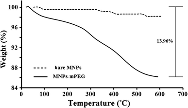
Moreover, the presence of PEG layer on MNPs surface was confirmed by thermogravimetric analysis (TGA). The first step of weight loss approximately at 100 °C is due to the loss of physisorbed water. The second step, starting at about 250 °C, is due to the loss of organic groups that conjugated to the particles surface. A mass loss of about 14 % was found for MNPs-mPEG, attributed to the degradation of mPEG (Fig.  4 ).

Fig. 4

TGA curves of bare MNPs and MNPs-mPEG at a rate of 10 °C/min

Particle size and size distribution of dispersed nanoparticles in water were specified using DLS. The size of the prepared mPEG-modified magnetic nanoparticles is 45 nm and showed narrow size distribution which exhibits zeta potential of +41 mV (data not shown). Figure  1 b, shows the SEM image of MNPs-mPEG. The sizes of MNPs-mPEG were obtained by SEM ~30 nm. MNPs-mPEG nanoparticles show well-dispersed structure than pure magnetic nanoparticles. This may be due to the grafting of mPEG on magnetic nanoparticle surfaces that reduced agglomeration.

Conclusion

Magnetite nanoparticles were synthesized by the co-precipitation of ferrous/ferric mixed salt in aqueous ammonium hydroxide (NH 4 OH) medium. Then, magnetic iron oxide nanoparticles were modified by reaction with biocompatible polyethylene glycol as hydrophilic shell. To prepare the MNPs-mPEG, acrylated mPEG was first reacted with ATPS and then a condensation reaction was carried out between the triethoxysilyl-terminated PEG and hydroxyl groups on the surface of magnetite nanoparticles. Surface modifying of MNP with methoxy polyethylene glycol provides stability and enhanced biocompatibility for MNPs. The presence of the mPEG was confirmed by Fourier transform-infrared spectroscopy and thermogravimetric analysis. These nanoparticles have the potential to be used in different biomedical applications.


References

  1. Weissleder et al. (1995) Long circulating iron oxides for MR imaging (pp. 321-334) 10.1016/0169-409X(95)00033-4
  2. Reimer and Weissleder (1996) Development and experimental application of receptor-specific MR contrast media (pp. 153-163)
  3. Chouly et al. (1996) Development of superparamagnetic nanoparticles for MRI: effect of particle size, charge and surface nature on biodistribution (pp. 245-255) 10.3109/02652049609026013
  4. Gupta and Hung (1989) Magnetically controlled targeted micro-carrier systems (pp. 175-186) 10.1016/0024-3205(89)90593-6
  5. Kumar and Mohammad (2011) Magnetic nanomaterials for hyperthermia-based therapy and controlled drug delivery (pp. 789-808) 10.1016/j.addr.2011.03.008
  6. Xie et al. (2011) Nanoparticle-based theranostic agents (pp. 1064-1079) 10.1016/j.addr.2010.07.009
  7. Berry et al. (2004) Cell response to dextran-derivatised iron oxide nanoparticles post internalisation (pp. 5405-5413) 10.1016/j.biomaterials.2003.12.046
  8. Zhang et al. (2002) Surface modification of superparamagnetic magnetite nanoparticles and their intracellular uptake (pp. 1553-1561) 10.1016/S0142-9612(01)00267-8
  9. D’Souza et al. (2004) Polyvinylpyrrolidone–drug conjugate: synthesis and release mechanism (pp. 91-100) 10.1016/j.jconrel.2003.09.014
  10. Bae et al. (2012) Chitosan oligosaccharide-stabilized ferrimagnetic iron oxide nanocubes for magnetically modulated cancer hyperthermia (pp. 5266-5273) 10.1021/nn301046w
  11. Allen (1994) The use of glycolipids and hydrophilic polymers in avoiding rapid uptake of liposomes by the mononuclear phagocyte system (pp. 285-309) 10.1016/0169-409X(94)90016-7
  12. Hu et al. (2006) Preparation of biocompatible magnetite nanocrystals for in vivo magnetic resonance detection of cancer (pp. 2553-2556) 10.1002/adma.200600385
  13. Masoudi et al. (2012) The effect of poly(ethylene glycol) coating on colloidal stability of superparamagnetic iron oxide nanoparticles as potential MRI contrast agent (pp. 129-141) 10.1016/j.ijpharm.2012.04.080
  14. Yoo et al. (2012) Folate–PEG–superparamagnetic iron oxide nanoparticles for lung cancer imaging (pp. 3005-3013) 10.1016/j.actbio.2012.04.029
  15. Sun et al. (2006) Folic acid-PEG conjugated superparamagnetic nanoparticles for targeted cellular uptake and detection by MRI (pp. 550-557) 10.1002/jbm.a.30781
  16. García-Jimeno and Estelrich (2013) Ferrofluid based on polyethylene glycol-coated iron oxide nanoparticles: characterization and properties (pp. 74-81) 10.1016/j.colsurfa.2012.12.022
  17. Zhou et al. (2011) A general approach for providing nanoparticles water-dispersibility by grinding with poly (ethylene glycol) (pp. 18-26) 10.1016/j.colsurfa.2011.08.055
  18. Lu et al. (2012) Carboxyl–polyethylene glycol–phosphoric acid: a ligand for highly stabilized iron oxide nanoparticles (pp. 19806-19811) 10.1039/c2jm34327d
  19. Bruce et al. (2004) Synthesis, characterisation and application of silica-magnetite nanocomposites (pp. 145-160) 10.1016/j.jmmm.2004.06.032
  20. McBain et al. (2008) Magnetic nanoparticles for gene and drug delivery (pp. 169-180)
  21. Brullot et al. (2012) Versatile ferrofluids based on polyethylene glycol coated iron oxide nanoparticles (pp. 1919-1925) 10.1016/j.jmmm.2012.01.032
  22. Barrera et al. (2009) Colloidal dispersions of monodisperse magnetite nanoparticles modified with poly (ethylene glycol) (pp. 107-113) 10.1016/j.jcis.2008.09.071
  23. Larsen et al. (2009) Size-dependent accumulation of PEGylated silane-coated magnetic iron oxide nanoparticles in murine tumors (pp. 1947-1951) 10.1021/nn900330m
  24. Mahdavian and Mirrahimi (2010) Efficient separation of heavy metal cations by anchoring polyacrylic acid on superparamagnetic magnetite nanoparticles through surface modification (pp. 264-271) 10.1016/j.cej.2010.02.041
  25. Khoee and Rahmatolahzadeh (2012) Synthesis and characterization of pH-responsive and folated nanoparticles based on self-assembled brush-like PLGA/PEG/AEMA copolymer with targeted cancer therapy properties: a comprehensive kinetic study (pp. 416-427) 10.1016/j.ejmech.2012.02.027
  26. Thangaraja et al. (2010) Preparation and characterization of polyethylene glycol coated silica nanoparticles for drug delivery application (pp. 31-38)
  27. Tomasovicova et al. (2006) Infrared study of biocompatible magnetic nanoparticles (pp. 32-35)