Synthesis of SDS micelles-coated Fe3O4/SiO2 magnetic nanoparticles as an excellent adsorbent for facile removal and concentration of crystal violet from natural water samples
Department of Chemistry, Shahr-e-Qods Branch, Islamic Azad University, Tehran, IR
Abstract
In this research, a simple and rapid method for the separation and preconcentration of trace amounts of crystal violet (CV) from aqueous sample solutions by modified magnetic nano-particles (MNPs) has been developed. The modification of magnetite nanoparticles was conducted by tetra ethoxysilane (TEOS) followed by micelles of anionic surfactant (SDS) to enhance the preconcentration of CV. To characterize the shape and structure of the nanoadsorbent, FT-IR and XRD procedures were used. Also, the average size of the synthesized nanoparticles was achieved between 30 and 40 nm by TEM technique. The effects of some important parameters such as: aqueous solution pH, adsorbent dosage, contact time, temperature and desorption conditions on the separation and concentration of CV were investigated. So, under optimal experimental conditions: aqueous solution pH 6, solution temperature = 20 °C, 7 mg of adsorbent, 1 mL of eluent (0.8 mL of acetonitrile + 0.2 mL of acetic acid), the recovery of CV from river water samples was achieved 98.32 ± 0.056% (
n
= 5) in two short periods of time for extraction (5 min) and elution (2 min). The maximum sorption capacity of the nano-composite was determined to be 16.37 mg/g. Also linear dynamic range and limit of detection were calculated to be 10–2500 ppb and 1.82 ppb, respectively. Finally, the proposed method was successfully applied for the separation and concentration of CV from the real water samples and the results were satisfied.
Graphical abstract
Under optimal conditions, a significant amount of nanocomposite was added to the dye solution. After dye removal in a short time, the nanoadsorbent-containing dye was magnetically separated from the solution and then the adsorbed dye was eluted into low volume of a suitable eluting solution.
Introduction
Natural and synthetic dyes are heavily used in many industries including food, cosmetics, textiles, pharmaceuticals, and leather tanning [
1
]. According to their chemical structures, dyes can be classified as acidic and basic dyes. Basic dyes (cationic dyes) are soluble in water with positive charge which are widely applied to acrylic fibers and paper but rarely applied to wool and silk. Cationic dyes are toxic even at trace levels [
1
]. Usually, cationic dyes are present in natural and treated wastewater and even in potable waters at µg or ng per liter [
2
,
3
].
Crystal violet is one of the cationic dyes which cause adverse effects on human health. It may cause eye burn leading to permanent cornea/eye damage. Its inhalation gives rise to a short period of rapid or difficult breathing, nausea, vomiting, profuse sweating, hypermotility, diarrhea, and abdominal pain [
4
]. Therefore, a preliminary preconcentration step is an essential step before analysis by sensitive instruments.
Several methods for preconcentration of dyes include dispersive liquid liquid microextraction (DLLME), cloud point extraction (CPE), solid phase extraction (SPE), and rotating disk sportive extraction (RDSE) [
5
,
6
,
7
–
8
]. Among these techniques, SPE procedures are considered superior to other procedures, because these techniques have many advantages over others, such as: low running cost, high speed, low solvent consumption, and higher enrichment factors [
9
]. The effectiveness of dyes removal in SPE procedures depends strongly on the nature of adsorbent being used, because the analytical sensitivity, affinity, capacity, and precision of the procedure can be usually estimated by the adsorbent action during separation process[
10
,
11
,
12
–
13
].
Recently, many research groups have explored the application of several nanosized SPE adsorbents such as nanoparticles (NPs) and nanotubes. Meanwhile, the magnetic nanoparticles are one of the most popular materials in analytical biochemistry, medicine, and biotechnology. Also, they have been increasingly applied to immobilize proteins, enzymes, and other bioactive agents [
14
].
Nanomaterials can offer several advantages over traditional SPE adsorbents, such as having a short diffusion route and very high surface areas and extraction capacity, which result in their rapid extraction dynamics and high extraction efficiencies [
15
]. Moreover, by using superparamagnetic nanoparticles (NPs), such as Fe
3
O
4
, a shorter analysis time can be achieved. It is mainly due to their easy isolation by an external magnetic field which is applied outside the extraction container [
16
].
However, pure magnetic particles are prone to form aggregates and their magnetic properties can be altered in complex environmental and biological systems. To solve the above problems, a suitable protective coating on a magnetic core is often used. The core–shell magnetic nanoparticles have high surface areas. Between some protective coatings, silica has been considered as one of the most ideal shell materials. This is mainly due to its reliable chemical stability, biocompatibility, and versatility in surface modification [
17
,
18
].
Recent studies on the use of surfactant-coated mineral oxides have demonstrated that these new adsorbent materials, which are shielded with hemimicelles/or admicelles, can be promising tools for the extraction/preconcentration of ionic and non-ionic compounds [
18
,
19
–
20
]. In comparison with the other ordinary adsorbents, one feature of the surfactant-coated adsorbents is that the outer surfaces of hemimicelles and admicelles are hydrophobic and ionic, respectively. Therefore, it makes them as a proper adsorbent, which offers different mechanisms for extraction processes [
18
]. Moreover, since the surfactants are commercially available, the hydrophobicity and the charge of the adsorbent can be easily modified according to the nature of the analytes. So, using mixed hemimicelles/admicelles in SPE has many advantages, such as, high extraction yields, high breakthrough volumes, and rapid elution of analytes [
18
].
In this study, the chemical synthesis of Fe
3
O
4
MNPs and the surface modification of synthesized MNPs with silica and micelles of sodium dodecyl sulfate (SDS) have been described. So, bilayer coatings of silica and SDS micelle in the first and second layers surrounded the Fe
3
O
4
nanoparticle core, respectively. The shape and structure of the synthesized nanomaterials were studied by FTIR, XRD, and TEM techniques. Also, the capability of SDS micelles-coated Fe
3
O
4
/SiO
2
magnetic nanoparticles to separate crystal violet (CV) from water samples was investigated and then the dye was concentrated into the eluting solution.
Experimental
Reagents and materials
Ferric chloride (FeCl
3
·6H
2
O), ferrous chloride (FeCl
2
·4H
2
O), sodium hydroxide, ammonia, ethanol, acetone, sodium dodecyl sulfate (SDS), tetra ethoxysilane (TEOS) (98%), sodium chloride and hydrochloric acid were all analytical grade from Merck (Darmstadt, Germany). Crystal violet, C
25
H
30
N
3
Cl, was purchased from Sigma-Aldrich Company. The chemical structure of the dye is presented in Fig.
1
.
Fig. 1
Chemical structure of crystal violet (CV)
Instrumentation
The dye concentrations were measured by a double-beam UV/Vis spectrophotometer (Shimadzu, Model UV 1601, Japan) at a wavelength of 582 nm. The phase purity was characterized by X-ray powder diffraction (XRD) (bruker AXS GmbH, Karlsruhe, Germany) using Cu-K
α
radiation (
λ
= 1.5406 Å). Transmission electron microscopy was carried out using a Zeiss-EM10C operated at a 100-kV accelerating voltage. The pH values were measured with a Consort C860 multi-parameter analyzer. The FTIR spectra were recorded on a Perkinelmer FTIR 100 spectrometer and using standard KBr pellet technique.
Adsorbent preparation
Synthesis of Fe3O4 nanoparticles
The chemical co-precipitation method was used in the preparation of the Fe
3
O
4
NPs with some modifications [
16
]. At first for preparing a stock solution, 5.4 g of FeCl
3·
6H
2
O and 2.0 g of FeCl
2·
4H
2
O were dissolved in 25 mL of deionized distilled water and 25 mL of 2 M HCl was added to the solution in a beaker. The stock solution was stirred under argon gas protection for 10 min. Then, its temperature was adjusted in 80 °C in a reactor and 40 mL of concentrated ammonia solution was added dropwise under vigorous stirring (1000 rpm) and argon gas protection in 30 min. After adding ammonia to the solution, it was rapidly stirred for 60 min. Then, the solution was cooled to room temperature and the Fe
3
O
4
NPs were produced. The magnetic precipitate was isolated with a permanent magnet and then washed three times with 150 mL of deionized water and absolute alcohol. Finally, the black powder Fe
3
O
4
nanoparticles were dried under vacuum at 50 °C during 24 h.
Modification of Fe3O4 nanoparticles with SiO2 groups
Fe
3
O
4
/SiO
2
NPs were synthesized according to the previously reported method with some modifications [
21
]. So, 1 g of the Fe
3
O
4
nanoparticles was suspended in 200 mL of pure ethanol under sonication and argon gas protection for 1 h. Then, 22.5 mL of concentrated ammonia, 18.75 mL of deionized water, and 0.5 mL of TEOS were added, in sequence, to the suspension and the mixture was sonicated for 1 h. To achieve complete silica-coated magnetic nanoparticles, the suspension was stirred for another 8 h. The silica-modified nanoparticles were isolated by a permanent magnet and rinsed with deionized water three times and then dried during 24 h.
Preparation of SDS micelle-bound SiO2-coated magnetic NPs
To coat the SiO
2
-modified magnetic nanoparticles by micelles of SDS, 10 mL of 5%w/v SDS solution (> CMC of SDS) was added to about 1.0 g of silica-modified MNPs, in a beaker. Then, the solution was vigorously stirred for 1 min with a glass rod and then the beaker was placed on a magnetic field. After completing the grounding of the SDS micelles-bound silica-coated magnetic nanoparticles (SDS micelle-SCMNPs), the solution was decanted and the dark brown modified NPs were washed three times with distilled water, to eliminate the extra amount of surfactant [
21
].
Procedure
Preconcentration of crystal violet by SDS micelle-coated Fe
3
O
4
/SiO
2
MNPs has been studied in batch experiments. A known amount of nanoadsorbent (7 mg) was added to 20 mL of 1 mg L
−1
aqueous crystal violet solution at pH 6 and it was shaken for 5 min at 20 °C. 5 min and 20 °C are contact time and temperature, respectively. To isolate the modified MNPs containing the adsorbed dye, from the remaining solution, a strong magnetic field was used and so the supernatant liquid phase was magnetically decanted. Finally, the adsorbed target analyte (CV) on the SDS micelle-coated Fe
3
O
4
/SiO
2
MNPs was eluted with 1.0 mL of 1:4 v/v solution of acetic acid: acetonitrile under shaking in 2 min, elution time, and exposed to a permanent magnet. The clear solution of the eluent containing the eluted crystal violet was transferred to the glass tube. Then, analyte absorbance in the eluent was determined by a UV spectrophotometer at 589 nm against blank.
Calculation of extraction recovery (response) and relative recovery
To evaluate the efficiency of the method and obtain optimum conditions, the extraction recovery of the analyte was determined. The extraction recovery (ER%) was defined as the percentage of total analyte (
n0
) that was extracted into the sedimented phase (
nsed
) and presented in the following equation:
ER%=nsedn0×100=Vsed×CsedVaq×C0×100,
where
Csed
is the concentration of analyte in the sedimented phase after extraction, and
C0
is the initial concentration of analyte in the aqueous phase.
Vsed
and
Vaq
are the volume of sedimented phase and the volume of aqueous phase, respectively.
N0
and
nsed
are the initial moles of analyte in the aqueous phase and its extracted moles into the sedimented phase, respectively.
To assess the matrix effect of real samples on the proposed method, the relative recovery (RR) was determined using Eq. (
2
):
RR%=nmeasured-nrealnadded×100,
where
nmeasured
is the moles of analyte after addition of a known amount of standard in the real sample,
nreal
is the initial moles of analyte in real sample and
nadded
is the moles of the spiked standard to the real sample.
Results and discussion
Characterization of the synthesized nanoadsorbent
X-ray powder diffraction (XRD)
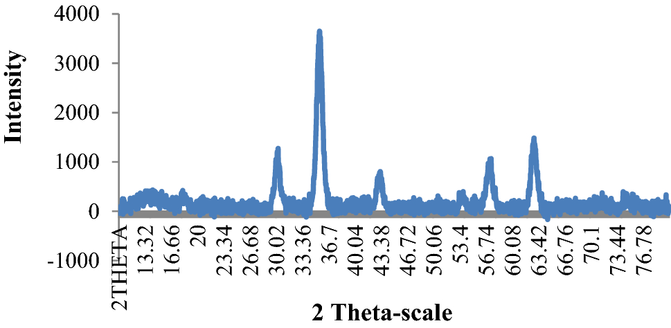
XRD analysis was used to investigate the crystalline structure of synthesized nanoparticles (Fig.
2
). The Joint Committee on Powder Diffraction Standards (JCPDS) reference pattern of magnetite (No. 19-629) was used for comparison. As could be seen in Fig.
2
, the XRD pattern of functionalized magnetite nanoparticles (SDS micelle coated Fe
3
O
4
/SiO
2
MNPs) was matched well with that of the standard Fe
3
O
4
structure. This indicated that the SiO
2
and SDS micelles coating did not cause the phase change in Fe
3
O
4
NPs structure [
22
]. On the other hand, the sharp and narrow peaks also indicate that SDS micelle-coated Fe
3
O
4
/SiO
2
nanoparticles have high crystallinity and purity [
23
,
24
–
25
].
Fig. 2
XRD pattern of synthesized SDS micelle coated Fe
3
O
4
/SiO
2
MNPs
The introduction of SiO
2
and SDS micelle onto the magnetite nanoparticles was approved by TEM results (Fig.
3
). As seen in Fig.
3
, the dark nano-Fe
3
O
4
cores have been surrounded by a gray shell of SDS micelle and SiO
2
coating, and also little agglomeration can be observed [
13
]. Moreover, from TEM report, the particles have an approximate spherical shape and an average size of 30–40 nm.
Fig. 3
TEM micrograph of synthesized SDS micelle coated Fe
3
O
4
/SiO
2
MNPs
FT-IR spectrum
FT-IR is a reliable technique for the monitoring of the variations in the functional groups. So, the structures of the Fe
3
O
4
MNPs, Fe
3
O
4
/SiO
2
and the SDS micelle-coated Fe
3
O
4
/SiO
2
MNPs were characterized by FTIR spectroscopy. The results are shown in Fig.
4
. The main absorption bands of OH (3300–3500 cm
−1
) and Fe–O (589 cm
−1
) groups are observed in all samples. Fe–O stretching band at 589 cm
−1
is the characteristic peak of magnetite. The characteristic band of Si–O is at 1066 cm
−1
, which confirmed that the Fe
3
O
4
NPs were protected by silica coating. On the other hand, the absorption band at 1067 cm
−1
could be attributed to the stretching vibration of S=O band, which showed that the surface of Fe
3
O
4
/SiO
2
MNPs was successfully modified with SDS micelles. Also as reported by T. Liu and co-workers, there are some peaks at 3400–3500 cm
−1
, 550–600 cm
−1
, 1220 cm
−1
, and 1084 cm
−1
for pure SDS and when the SDS adsorbs on the surface of a solid phase (like collophane), these main peaks can shift a little [
26
]. Therefore, the main peak of the streching vibration of S=O at 1220 cm
−1
for pure SDS has shifted a little and is observed at 1067 cm
−1
. Since the peak at 1066–1067 cm
−1
belongs to the stretching vibrations of the both of Si–O and S=O bands and the two peaks overlapped, this peak in the red line is more intense than that of in blue one (Fig.
4
). So, the results in Fig.
4
show that the both of SDS micelles and SiO
2
coatings are adsorbed on Fe
3
O
4
MNPs via purely physical adsorptions [
26
].
Fig. 4
a
IR spectrum of the Fe
3
O
4
MNPs (black line), Fe
3
O
4
/SiO
2
(blue line), and the SDS micelle-coated Fe
3
O
4
/SiO
2
MNPs (red line).
b
Separate IR spectrum of the Fe
3
O
4
MNPs (black line).
c
Separate IR spectrum of the SDS micelle coated Fe
3
O
4
/SiO
2
MNPs (red line)
Optimization of various parameters
Effect of pH
It is well known that the pH of the sample solution is one of the prime factors influencing on the states of species (as ions or neutral forms). Also, adsorption behavior of an adsorbent system can be affected by pH aqueous solution. For instance, at different working pH values, there is different charge density on the Fe
3
O
4
/SiO
2
MNP surface [
27
]. In this study, the sorption behavior of crystal violet on the modified nanoadsorbent from water samples was investigated, in the solution pH range of 3.0–10.0. The pH of the solution was adjusted at the required value by the addition of 1.0 mol L
−1
NaOH or 1.0 mol L
−1
HCl. As shown in Fig.
5
a, b, by increasing the solution pH, from 3 to 6, the adsorption efficiency of dye was improved and reached a maximum at pH about 6 and then decreased at higher pH values. At low pH (about 3), the negative charge density on the SDS micelle coating Fe
3
O
4
/SiO
2
MNPs is decreased [
28
]. This is mainly due to the high free proton ion concentration in the solution, which makes the neutralization of the negative charge on the SDS micelle-coated Fe
3
O
4
/SiO
2
MNPs. So, the tendency of the cationic dye toward the nanoadsorbent is reduced. Then, by increasing the pH value up to 6, H
+
ions concentration is diminished and so the removal percentage of CV on the surface of the nanocomposite is improved. But at higher pH (> 6), a significant decrease in the dye removal and recovery percentage is observed (Fig.
5
). Since the point of zero charge (PZC) of Fe
3
O
4
/SiO
2
was reported about 3.3 [
29
], at higher pH and free hydroxide ion concentration, the SDS micelle-surrounded Fe
3
O
4
/SiO
2
MNPs may be substituted by OH
−
ions. So, under this circumstance, the prolapse of SDS micelle from the Fe
3
O
4
/SiO
2
MNPs may happen [
30
,
31
]. On the other hand, the positive charge of the cationic dye can be neutralized by OH
−
groups in basic conditions. Thus, pH of the solution was adjusted at pH 6.0, as optimum value, for further experiments.
Fig. 5
Effect of pH on the recovery (
a
) and removal (
b
) of crystal violet. Experimental conditions: 20 mL of 1 mg L
−1
dye solution at different pHs (3.0–10.0) in 25 °C, 7 mg of nanoadsorbent, and contact time was 5 min, elution conditions: mixture of 0.8 mL of acetonitrile + 0.2 mL of acetic acid, as eluting solution, and elution time was 2 min
Effect of nanoadsorbent dosage
Compared to ordinary adsorbents (micron-size particle adsorbents), NP adsorbents have higher surface areas. Therefore, satisfactory results can be achieved by fewer amounts of NP adsorbents [
27
,
32
,
33
]. In this study, the effect of the amount of modified nanoparticles on the quantitative extraction of crystal violet was investigated, by varying the amounts of the modified nanoparticles from 0.5 to 17 mg. It can be seen from Fig.
6
a, b that the highest recovery and removal of crystal violet has been obtained with increasing of modified nanoparticles amount up to 5 mg and remained constant at higher amounts of nanocomposite. This is mainly due to the higher collision probability between CV and SDS micelles-coated Fe
3
O
4
/SiO
2
MNPs in higher dosage of nanocomposite. Therefore, 7 mg of the modified nanoparticles was selected as the optimum value for further experiments.
Fig. 6
Effect of nanosorbent dosage on the recovery (
a
) and removal (
b
) of crystal violet. Experimental conditions: 20 mL of 1 mg L
−1
dye solution at pH 6 in 25 °C, different amounts of nanoadsorbent were used and contact time was 5 min, elution conditions: mixture of 0.8 mL of acetonitrile + 0.2 mL of acetic acid, as eluting solution, and elution time was 2 min
Effect of contact time
The effect of contact time on the overall extraction efficiency of the crystal violet was studied, by varying the contact time between adsorbent and dye sample solution in the range of 1–15 min. As shown in Fig.
7
a, b, the adsorption efficiency of crystal violet has been improved by increasing the contact time up to 5 min and remained nearly constant in more than 5 min. Therefore, a short contact time of 5 min was selected for the adsorption of CV on the SDS micelle-coated Fe
3
O
4
/SiO
2
MNPs.
Fig. 7
Effect of contact time on the recovery (
a
) and removal (
b
) of crystal violet. Experimental conditions: 20 mL of 1 mg L
−1
dye solution at pH 6 in 25 °C, 7 mg of nanoadsorbent and contact time was varied between 1 and 15 min, elution condition: mixture of 0.8 mL of acetonitrile + 0.2 mL of acetic acid, as eluting solution, and elution time was 2 min
Effect of temperature
In this study, the effect of temperature on the adsorption of crystal violet on the SDS micelle-coated Fe
3
O
4
/SiO
2
NPs was investigated, in the range of 5–40 °C under optimal conditions (i.e., contact time: 5 min, pH 6 and adsorbent dosage: 7 mg). It can be seen from Fig.
8
a, b, by increasing the temperature from 5 to 20 °C, the adsorption and recovery efficiencies of the crystal violet have been increased nearly 100% and 98.32%, respectively. But at higher temperatures (25–40 °C), the separation and recovery efficiencies of CV have been diminished. This might be due to the fact that the critical micelle concentration (CMC) of SDS and consequently its micellization can be varied as a function of temperature [
34
]. For ionic surfactant, the temperature has strong effect on the hydrophobic and hydrophilic interaction behavior of micelles. According to the CMC versus temperature studies for ionic surfactants, CMC decreased to a minimum value and then increased, by increasing the temperature. Therefore, a U-shape behavior was observed [
34
]. Since the minimum CMC value of SDS was achieved at 21 °C, the maximum micellization could be occurred around this temperature [
34
]. So, maximum recovery and extraction efficiencies of the crystal violet (98% and 100%) can be seen at 20 °C, because of the maximum micellization of SDS at this temperature [
34
].
Fig. 8
Effect of temperature on the recovery (
a
) and removal (
b
) of crystal violet. Experimental conditions: 20 mL of 1 mg L
−1
dye solution at pH 6 in different temperatures, 7 mg of nanoadsorbent and contact time was 5 min, elution condition: mixture of 0.8 mL of acetonitrile + 0.2 mL of acetic acid, as eluting solution, and elution time was 2 min
Desorption condition
Organic solvents can rapidly and completely disrupt the micelles and therefore the analyte is removed from the surface of NPs [
14
,
35
]. By carrying out some experiments we found that desorption ability of acetonitrile was improved, when acetic acid was added into the acetonitrile [
18
]. Thus, different volumes of the mixed solutions of acetonitrile and acetic acid were studied for desorption of analytes from the SDS micelle coated Fe
3
O
4
/SiO
2
MNPs. The results are presented in Table
1
. It was resulted that among the testing solvents, the highest recoveries (91.32%) were achieved using 1 mL of the mixture of acetic acid: acetonitrile (1:4, v/v) at ambient temperature (~ 25 °C). Therefore, it was selected as the elution solvent for desorption of the analyte from the surface of the modified magnetic nanoparticles. Also, the elution time in the range of 1–7 min was investigated and the results showed that the recovery of CV was quantitative (91.32%) in 2 min. So, 2 min was selected as the optimal elution time in other experiments.
Table 1
Effect of different mixture of eluting solution on the recovery efficiency of crystal violet. Experimental conditions: 20 mL of 1 mg L
−1
CV at pH 6 in ambient temperature, 7 mg of nanoadsorbent, and contact time was 5 min. Elution conditions: different mixtures of acetonitrile and acetic acid solutions, elution time was 2 min
Eluent
Volume (mL)
Recovery (%)
Acetonitrile + CH3COOH
0.7 + 0.3
85.27
Acetonitrile + CH3COOH
0.8 + 0.2
91.32
Acetonitrile + CH3COOH
0.3 + 0.7
91.0
Acetonitrile + CH3COOH
0.2 + 0.8
88.2
Acetonitrile + CH3COOH
0.5 + 0.5
79.2
Acetonitrile + CH3COOH
1.0 + 1.0
70.75
Acetonitrile + CH3COOH
0.5 + 1.5
63.5
Acetonitrile + CH3COOH
1.5 + 0.5
60.15
Adsorption capacity and isotherms
To study the adsorption capacity and isotherms of SDS micelle-coated Fe
3
O
4
/SiO
2
MNPs, 7 mg of the adsorbent was added to 20 mL of different concentrations of dye, in the range of 1–20 mg L
−1
, at pH 6.0 and 20 °C. After shaking for 5 min, the supernatant was decanted by applying an external magnetic field and the remaining crystal violet in the supernatant solution was determined by a UV spectrophotometer at 589 nm against blank. The adsorption capacity (
qE
, mg g
−1
) was calculated as:
qE=(C0-CE)Vm,
where
C0
and
CE
are the initial and equilibrium concentrations (mg L
−1
) of crystal violet in the solution, respectively.
V
is the volume of dye solution in mL and
m
is the amount of adsorbent in mg.
Two more common adsorption isotherms, Langmuir and Freundlich, were studied [
36
]. The results are summarized in Table
2
. As seen in Table
2
, the Langmuir model has been fitted better (
R2
= 0.9992) than the Freundlich model (
R2
= 0.9473). Therefore, monolayer adsorption of dye on the homogeneous surface of SDS micelle-coated Fe
3
O
4
/SiO
2
MNP has been considered. Since the constant 1/
n
is 0.174, proper physical adsorption of dye on the nanoadsorbent is concluded [
13
]. On the other hands, the results showed that the maximum capacity of the SDS micelle-coated Fe
3
O
4
/SiO
2
MNPs was 16.37 mg g
−1
for the removal of crystal violet.
Table 2
Adsorption isotherm parameters of crystal violet onto SDS micelle-coated Fe
3
O
4
/SiO
2
MNPs
Freundlich model
Langmuir model
R2
Log KF
1/n
R2
qm (mg g−1)
KL (L mg−1)
0.9473
0.56
0.174
0.9992
16.37
− 0.23
Also, the effect of initial dye concentration, in the range of 1–20 mg L
−1
, on its removal percentage was studied. The results are shown in Fig.
9
. As indicated in Fig.
9
, by increasing the initial concentration of crystal violet in the aqueous solution, the dye removal is diminished. This is mainly due to the finite capacity of nanoadsorbent [
13
].
Fig. 9
Effect of initial concentration of crystal violet (CV) on its removal. Experimental conditions: 20 mL of different concentrations of dye solution (1-20 mg L
−1
) at pH 6 in 20 °C, 7 mg of nanoadsorbent and contact time was 5 min, elution condition: mixture of 0.8 mL of acetonitrile + 0.2 mL of acetic acid, as eluting solution, and elution time was 2 min
Analytical performance
Under optimal conditions: 20 mL of 1 mg L
−1
of CV solution at pH 6 in 20 °C, 7 mg of nanoadsorbent, contact time = 5 min, eluting solution = mixture of 0.8 mL of acetonitrile + 0.2 mL of acetic acid, and elution time = 2 min, the analytical parameters such as: linear dynamic range (LDR), correlation coefficient (
R2
), limit of detection (LOD), and relative standard deviation (RSD) were determined and summarized in Table
3
. The calibration curve of crystal violet was constructed in the range of 10–2500 µg L
−1
and characterized with a high correlation coefficient (
R2
= 0.9991) under optimum conditions. Based on
CLOD
= 3
Sb/m
equation,
CLOD
,
Sb
and
m
are limit of detection, standard deviation of blank, and slope of the calibration curve, respectively. The limit of detection (LOD) of crystal violet was achieved 1.82 µg L
−1
. Standard deviation (SD) of the method was 0.056% (
n
= 7) under optimal conditions. After measuring the amount of crystal violet in the 1 mL of eluent, preconcentration/or enrichment factor was achieved 20.
Table 3
Analytical figures of merit of the proposed method
Crystal violet
LDR (μg L−1)a
10–2500
R2
0.9991
LOD (μg L−1)b
1.82
SD % (n = 7)c
0.056
Enrichment factord
20
Also, the ultraviolet/visible spectrums of crystal violet before and after removal and also after concentration, via proposed method, were investigated, under optimal conditions (Fig.
10
).
Fig. 10
UV/Vis spectra of crystal violet (CV) before and after removal and concentration under optimal conditions: 20 mL of 0.7 mg L
−1
dye (CV) solution at pH 6 in 20 °C, 7 mg of nanosorbent and contact time was 5 min, elution condition: mixture of 0.8 mL of acetonitrile + 0.2 mL of acetic acid, as eluting solution, and elution time was 2 min
Real sample analysis
The proposed method was applied to determine the extraction and concentration of crystal violet from river water samples. Since the concentration of CV in river water samples was at very low levels and could not be determined, two different amounts of standard dye solution were spiked to two river water samples, 0.5 and 1 mg L
−1
, and the recovery percentage of dye was investigated. The results are shown in Table
4
. As it is seen in Table
4
, recovery percentage of dye from matrices was obtained about 97.1% and 97.7% when 0.5 and 1 mg L
−1
crystal violet was spiked into two river water samples, respectively. As a result, the complexity of the medium did not have significant effect on the efficiency of dye recovery with the proposed method under optimum conditions.
Table 4
The recovery of crystal violet from river water samples. Experimental conditions: 0.5 and 1 mg L
−1
crystal violet were spiked into two river samples at pH 6 in 20 °C, 7 mg of nanoadsorbent, and the contact time was 5 min, and elution conditions: mixture of 0.8 mL of acetonitrile + 0.2 mL of acetic acid, as eluting solution, and elution time was 2 min
Sample
Added (mg L−1)
Founda (mg L−1)
Recovery (%)
River waterb
0
n.d.c
–
0.50
0.49 ± 0.05 (n = 5)
97.1
1.00
1.01 ± 0.05 (n = 5)
97.7
Comparison with other preconcentration methods
A comparison of the proposed method with other reported preconcentration methods for extraction of crystal violet is given in Table
5
. As seen in Table
5
, the LOD (1.8 µgL
−1
), standard deviation (SD %) and contact time of the proposed method are comparable to or better than those of some previously reported preconcentration methods.
Table 5
Comparison of the proposed method with other reported preconcentration methods for extraction and concentration of crystal violet
Sorbent
Timea (min)
PFb
LOD (µg L−1)
SD %
Detection
References
SDS micelle coated Fe3O4/SiO2
5
20
1.82
0.056
UV–Vis
This work
Nanoporous silica SBA-3
2
200
1.3
< 1.3
FO-LADSc
[37]
Diatomite
–
89–96
28
2.3
UV–Vis
[7]
GOd
15
19.6
9
1.99
UV–Vis
[38]
RDSEe
100
–
1.8
< 8.4
UV–Vis
[8]
Conclusion
This study proved that SDS micelle-coated Fe
3
O
4
/SiO
2
nanoadsorbent, with average size of 30–40 nm, can act as an effective solid phase for the adsorption of crystal violet (CV) from water samples. This may be due to the strong electrostatic interactions between negatively charged SDS micelle-coated Fe
3
O
4
/SiO
2
nanoparticles and the positively charged dye under optimal conditions. One of the most important characteristics of the synthesized SDS micelle-coated Fe
3
O
4
/SiO
2
nanoadsorbent is its magnetically assisted separation of analytes from the aqueous sample solutions, while shorter analysis time can be achieved. On the other hand, the larger surface area of nanomaterials results in the higher adsorption capacity. So, lower amounts of SDS micelle-coated Fe
3
O
4
/SiO
2
nanoadsorbent are needed than those of other ordinary adsorbents and the extraction process was done in less time. Also, crystal violet was concentrated into low volume of eluting solution in a very short time. Therefore, fast removal and concentration of dye was performed via proposed method, in less than 10 min.
The SDS micelle-coated Fe
3
O
4
/SiO
2
NPs, as an efficient adsorbent, were used to remove the crystal violet dye from river water samples and the results proved the reliability of the proposed method.
Acknowledgements
The authors gratefully acknowledge use of the services, facilities and financial support of Islamic Azad University, Shahr-e-Qods branch, Tehran, Iran.
Publisher's Note
Springer Nature remains neutral with regard to jurisdictional claims in published maps and institutional affiliations.
References
Crini (2006) Non-conventional low-cost adsorbents for dye removal: a review (pp. 1061-1085) 10.1016/j.biortech.2005.05.001
Šafařı́k and Šafařı́ková (2002) Detection of low concentrations of malachite green and crystal violet in water (pp. 196-200) 10.1016/S0043-1354(01)00243-3
Sharma et al. (2019) Efficient capture of eosin yellow and crystal violet with high performance xanthan-acacia hybrid super-adsorbent optimized using response surface methodology (pp. 314-323) 10.1016/j.colsurfb.2018.12.017
Otero et al. (2003) Elimination of organic water pollutants using adsorbents obtained from sewage sludge (pp. 55-65) 10.1016/S0143-7208(03)00005-6
Sadeghi and Nasehi (2018) Simultaneous determination of brilliant green and crystal violet dyes in fish and water samples with dispersive liquid–liquid micro-extraction using ionic liquid followed by zero crossing first derivative spectrophotometric analysis method (pp. 134-142) 10.1016/j.saa.2018.04.061
An et al. (2010) Simultaneous spectrophotometric determination of trace amount of malachite green and crystal violet in water after cloud point extraction using partial least squares regression (pp. 883-888) 10.1016/j.jhazmat.2009.10.092
Al-Degs and Sweileh (2012) Simultaneous determination of five commercial cationic dyes in stream waters using diatomite solid-phase extractant and multivariate calibration (pp. 219-224) 10.1016/j.arabjc.2010.08.016
Manzo et al. (2013) Determination of crystal violet in water by direct solid phase spectrophotometry after rotating disk sorptive extraction (pp. 305-308) 10.1016/j.talanta.2012.11.004
Korn et al. (2006) Separation and preconcentration procedures for the determination of lead using spectrometric techniques: a review (pp. 16-24) 10.1016/j.talanta.2005.10.043
Tian et al. (2009) Solid-phase extraction of tanshinones from Salvia Miltiorrhiza Bunge using ionic liquid modified silica sorbents (pp. 738-742) 10.1016/j.jchromb.2009.02.012
Falaki and Fakhri (2013) Study of the adsorption of methyl orange from aqueous solution using nickel oxide nanoparticles: equilibrium and kinetics studies 10(2) (pp. 117-124)
Falaki and Fakhri (2014) Adsorption properties of nickel oxide nanoparticles for removal of congo red from aqueous solution 10(4) (pp. 255-262)
Helmi et al. (2017) Highly facile removal of acid red 88 from aqueous samples by using synthesized iron oxide magnetic nanoparticles and 1-butyl-3-methylimidazolium tetrachloroferrate magnetic ionic liquid (pp. 331-340) 10.5004/dwt.2017.21321
Zhao et al. (2008) Cetyltrimethylammonium bromide-coated magnetic nanoparticles for the preconcentration of phenolic compounds from environmental water samples (pp. 1201-1206) 10.1021/es071817w
Faraji et al. (2010) Magnetic nanoparticles: synthesis, stabilization, functionalization, characterization, and applications10.1007/BF03245856
Falaki and Berijani (2016) Aspartic acid-modified magnetic nanoparticles as an ideal sorbent for solid phase extraction of Pb in water samples prior to ICP-OES determination (pp. 25765-25772) 10.1080/19443994.2016.1157706
Zhang et al. (2006) Synthesis of novel porous magnetic silica microspheres as adsorbents for isolation of genomic DNA (pp. 514-518) 10.1021/bp050400w
Zhu et al. (2010) Mixed hemimicelles SPE based on CTAB-coated Fe3O4/SiO2 NPs for the determination of herbal bioactive constituents from biological samples (pp. 1873-1880) 10.1016/j.talanta.2009.10.037
Ezoddin et al. (2010) Application of modified nano-alumina as a solid phase extraction sorbent for the preconcentration of Cd and Pb in water and herbal samples prior to flame atomic absorption spectrometry determination (pp. 900-905) 10.1016/j.jhazmat.2010.02.023
Keyhanian et al. (2016) Magnetite nanoparticles with surface modification for removal of methyl violet from aqueous solutions (pp. S348-S354) 10.1016/j.arabjc.2011.04.012
Davudabadi Farahani and Shemirani (2014) Mixed hemi-micelle solid-phase extraction based on modified magnetic nanoparticles for extraction of cadmium and lead from food and water samples 97(6) (pp. 1682-1688) 10.5740/jaoacint.12-407
Unknown ()
Ramimoghadam et al. (2012) The effect of sodium dodecyl sulfate (SDS) and cetyltrimethylammonium bromide (CTAB) on the properties of ZnO synthesized by hydrothermal method (pp. 13275-13293) 10.3390/ijms131013275
Sharma et al. (2019) Response surface methodology directed synthesis of luminescent nanocomposite hydrogel for trapping anionic dyes (pp. 380-390) 10.1016/j.jenvman.2018.10.038
Bhalla et al. (2019) One-pot green synthesis of polymeric nanocomposite: biodegradation studies and application in sorption-degradation of organic pollutants (pp. 345-356) 10.1016/j.jenvman.2018.12.117
Sun et al. (2017) Application and mechanism of anionic collector sodium dodecyl sulfate (SDS) in phosphate beneficiation 7(29) (pp. 1-13)
Bavili Tabrizi and Dehghani Teymurlouie (2016) Application of sodium dodecyl sulfate coated iron oxide magnetic nanoparticles for the extraction and spectrofluorimetric determination of propranolol in different biological samples (pp. 108-116)
Rahman (1983) Effect of pH on the critical micelle concentration of sodium dodecyl sulphate (pp. 1331-1334) 10.1002/app.1983.070280407
Esmaeili-Shahri and Es’haghi (2015) Superparamagnetic Fe3O4@SiO2 core–shell composite nanoparticles for the mixed hemimicelle solid-phase extraction of benzodiazepines from hair and wastewater samples before high-performance liquid chromatography analysis (pp. 4095-4104) 10.1002/jssc.201500743
Shariati et al. (2011) Fe3O4 magnetic nanoparticles modified with sodium dodecyl sulfate for removal of safranin O dye from aqueous solutions (pp. 160-165) 10.1016/j.desal.2010.11.040
Ghanbari Adivi et al. (2019) Agarose-coated Fe3O4@SiO2 magnetic nanoparticles modified with sodium dodecyl sulfate, a new promising sorbent for fast adsorption/desorption of cationic drugs 75(3) (pp. 1239-1256) 10.1007/s00289-018-2418-7
Bagheri et al. (2012) Extraction of fluoxetine from aquatic and urine samples using sodium dodecyl sulfate-coated iron oxide magnetic nanoparticles followed by spectrofluorimetric determination (pp. 61-65) 10.1016/j.aca.2011.10.033
Tavakoli et al. (2014) Magnetic mixed hemimicelles solid-phase extraction of three food colorants from real samples (pp. 100-108) 10.1007/s12161-013-9603-y
Dehghan Noudeh et al. (2007) The effect of temperature on thermodynamic parameters of micellization of some surfactants (pp. 47-52) 10.3923/jas.2007.47.52
Zhao et al. (2008) Preparation of silica-magnetite nanoparticle mixed hemimicelle sorbents for extraction of several typical phenolic compounds from environmental water samples (pp. 140-147) 10.1016/j.chroma.2008.02.069
Sharma et al. (2019) Selective removal of cationic dyes using response surface methodology optimized gum acacia sodium alginate blended superadsorbent (pp. 331-345) 10.1016/j.ijbiomac.2018.11.213
Unknown ()
Razi-Asrami et al. (2017) Simultaneous spectrophotometric determination of crystal violet and malachite green in water samples using partial least squares regression and central composite design after preconcentration by dispersive solid-phase extraction10.1007/s10661-017-5898-2