P. G. Department of Physics, Smt. KRP Kanya Mahavidyalaya, Islampur, Maharashtra, 415409, IN
Department of Physics, PDVP Mahavidyalaya, Tasgaon, Maharashtra, 416 312, IN
Department of Physics, Arts, Science and Commerce College, Ramanandnagar, Maharashtra, 416308, IN
Abstract
The ferrite material with compositions Ni
0.7
Cu
0.1
Zn
0.2
La
x
Fe
2−
x
O
4
(where
x
= 0, 0.015, 0.025, and 0.035) was synthesized by oxalate co-precipitation method. The ferrite samples were characterized by thermo-gravimetric and differential temperature analysis (TG–DTA), energy-dispersive X-ray analysis (EDAX), X-ray diffraction (XRD), Fourier transform infrared spectroscopy (FTIR), field-emission scanning electron microscopy (FE-SEM), and vibrating sample magnetometer (VSM) techniques. The EDAX analysis confirmed the formation of required stoichiometric ferrite samples. The formation of cubic spinel structure with the presence of weak ortho-ferrite phases was confirmed from X-ray diffraction analysis. The lattice constant of all the ferrites was found to be increase with increase in La
3+
content. The presence of main two recognized strong absorption bands in the frequency range 400–600 cm
−1
in the FTIR spectra shows the formation of well spinel ferrite. Morphological study shows that grain size of the ferrites lies in the range 16.23–24.21 nm. It is observed that the saturation magnetization and magnetic moment of Ni–Cu–Zn ferrites decrease with La
3+
content.
Introduction
Soft-ferrite materials are mostly useful material because of its technological and industrial applications. These applications are depending on their properties such as high resistivity, moderate permeability, low dielectric loss, low permittivity, etc. These properties play an important role in the fabrication of components such as a transformer core, antenna rods, multi-layer chip inductor, micro-inductors, electromagnetic filters, etc. [
1
,
2
,
3
–
4
]
Recently, researchers synthesized ferrites in the form of nanoscale range because of its growing applications such as production of bio-diesel [
5
], nano-catalyst [
6
], humidity sensor [
7
], gas sensor [
8
], super-capacitor [
9
], electrode material for Li-ion battery [
10
], etc. Various methods such as sol–gel auto-combustion, co-precipitation, citrate precursor, wet chemical route, hydrothermal [
11
,
12
,
13
,
14
–
15
], etc. were used for the preparation of nano-ferrite materials.
In the last decade, researchers investigated various properties of Ni–Zn ferrites due to their interesting properties such as high resistivity, high permeability, and low eddy current losses. Recently, Ni–Zn ferrite material was used in high-frequency applications such as multi-layer chip inductors and electromagnetic interference filters. Das and Singh [
16
] investigated the structural, magnetic, and dielectric properties of Cu-substituted Ni–Zn ferrites. They reported that the coercivity and saturation magnetization of Ni–Zn ferrites improved by substituting Cu content. Avati et al. [
17
] illustrated that the poor densification and slow grain growth rate of Ni–Zn ferrites can be greatly improved by the substitution of Cu
2+
ions. In last few years, physicists synthesized Ni–Cu–Zn ferrites for the fabrication of electronic devices instead of Ni–Zn and Mg–Zn ferrites. Recently, it was found that properties of Ni–Cu–Zn nano-ferrites are extremely improved due to incorporation of rare earth metals. Shirsath et al. [
18
] investigated magnetic properties of Dy
3+-
substituted Ni–Cu–Zn ferrite nanoparticles. They reported that the saturation magnetization, initial permeability, and Curie temperature of the ferrites enhance by Dy
3+
substitution. Chaudhari et al. [
19
] investigated the crystallographic, magnetic, and electrical properties of Ni
0.5
Cu
0.25
Zn
0.25
La
x
Fe
2−
x
O
4
nano-ferrites. They found that the lattice constant, porosity, and specific surface area were increased, whereas particle size, bulk density, saturation magnetization, and coercively of Ni–Cu–Zn ferrites were decreases with increasing La
3+
content. On La
3+
substitution, the structural parameters such as relative density, grain size, crystallite size, and density increase, and on the other hand, porosity, anisotropy, and compressive macro-stress of Ni–Cu–Zn ferrites decrease [
20
]. Roy [
21
] et al. studied electromagnetic properties of La-substituted Ni–Cu–Zn ferrites. They reported that the initial permeability and saturation magnetization of Ni–Cu–Zn ferrites increase on substitution of small fraction of La
3+
ions. Gabal et al. [
22
] synthesized La-substituted Ni–Cu–Zn ferrites by sol–gel technique. They were studied its structural and magnetic properties and reported that the saturation magnetization and Curie temperature of ferrites decrease with increase in La
3+
content. They attribute it to the decreasing of Fe
3+
–Fe
3+
interactions on the octahedral (B) sites. Structure and magnetic properties of Ni–Cu–Zn ferrite materials with La doping were investigated by Yuan-Xun et al. [
23
]. They showed that with La
3+
substitution, saturation magnetization and complex permeability of Ni–Cu–Zn ferrites increase. Most of the researches [
19
,
23
] have been prepared La
3+
-substituted Ni–Cu–Zn ferrites by sol–gel method and ceramic method.
On literature survey, it was found that there is no any work carried out on La
3+
-substituted Ni–Cu–Zn ferrites prepared by oxalate co-precipitation method at lower sintering temperature. The oxalate co-precipitation technique is important and attractive technique used for the preparation of nano-sized ferrite materials because of its advantages such as good stoichiometric control, the production of ultrafine particles with low sintering temperature, and smaller duration as compared to ceramic method.
The purpose of this work is to synthesize La
3+
-substituted Ni–Cu–Zn nano-ferrites at lower sintering temperature followed by oxalate co-precipitation method and study its structural and magnetic properties.
Experimental
Synthesis of La3+-substituted Ni–Cu–Zn ferrites
Lanthanum-substituted Ni–Cu–Zn nano-ferrites with chemical formula Ni
0.7
Cu
0.1
Zn
0.2
La
x
Fe
2−
x
O
4
(where
x
= 0, 0.015, 0.025, and 0.035) were prepared by oxalate co-precipitation method. The AR grade nickel sulphate (NiSO
4
6H
2
O), copper sulphate (CuSO
4
5H
2
O), zinc sulphate (ZnSO
4
7H
2
O), iron sulphate (FeSO
4
7H
2
O), and lanthanum sulphate octa-hydrate [La
2
(SO
4
)
3
8H
2
O] were used as a starting materials supplied by Thomas Becker.
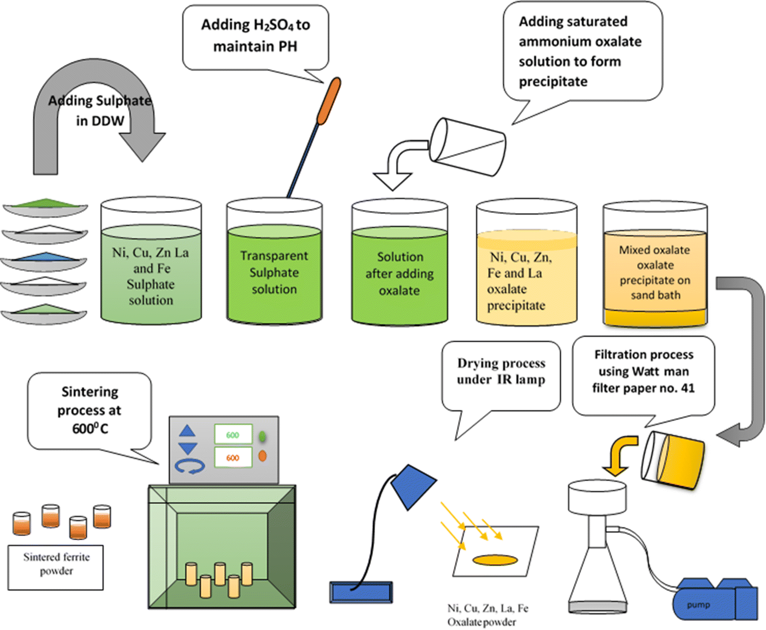
The required stoichiometric sulphates were dissolved in the double-distilled water. The pH of the solution was maintained at 4 by adding dropwise conc. H
2
SO
4
and heated for 2 h for proper mixing of all the sulphate. After cooling, super-saturated solution of ammonium oxalate was added in the solution until to complete the process of precipitation. A solution with precipitation placed on the sand bath for shrinkage. Due to this, the precipitates settle down at the bottom of beaker. The precipitate was filtered by Whatman filter paper no 41 using Buckner funnel and vacuum pump. The precipitate was washed several times to remove sulphate ions. The removal of sulphate ions in the precipitate was confirmed by barium chloride test and dried under radiation of IR light to remove water content. Pre-sintering and sintering temperatures in the investigation were decided by analyzing the precipitated powder by thermo-gravimetric and differential temperature analysis (TG–DTA). The resulting oxalate powder was pre-sintered at 300 °C for 2 h. After cooling, the pre-sintered powder was milled in agate mortal with acetone base. The powder was then sintered at 600 °C for 4 h. The sintered powder was again milled to have a fine powder. Pre-sintering and sintering process were carried out in the muffle furnace. Heating and cooling rate of the furnace were maintained at 80 °C/h.
The process of synthesis of La
3+
-substituted Ni
0.7
Cu
0.1
Zn
0.2
La
x
Fe
2−
x
O
4
(where
x
= 0, 0.015, 0.025, and 0.035) by oxalate co-precipitation method is shown diagrammatically in Fig.
1
.
Fig. 1
Diagrammatic synthesis procedure of La
3+
substituted Ni–Cu–Zn nano-ferrites by oxalate co-precipitation method
Characterization
The thermo-gravimetric (TG) and differential thermal analysis (DTA) of prepared oxalate nano-powder was carried out by SDT Q600 V20.9 build 20 instrument in air atmosphere with heating rate of 10 °C/min in the range 25–1000 °C by ramp method. The crystal structure, phase purity, crystallite size, and other parameters were studied by X-ray powder diffractometer (Phillips, Model 3710) with Cu-Kα radiation (
λ
= 1.5425 Å). The elemental composition of prepared ferrite was tested by Energy-Dispersive X-ray spectroscopy (EDAX) model ASTM E1508-98 Oxford. The Fourier transform infrared spectroscopy (FTIR) analysis was carried out in the range 350–800 cm
−1
by compositing the ferrite powder into KBr discs by Bruker ALPHA 100508 at room temperature. The morphology and grain size of nano-ferrite powder were studied with Zeiss Ultra 55 field effect-scanning electron microscopy (FE-SEM). The magnetic parameters such as saturation magnetization, remanence magnetization, coercivity, magnetic moment, and anisotropy constant of all the ferrites were obtained from vibrating sample magnetometer (PFM-Magneto make model) at room temperature.
Results and discussion
Thermo-gravimetric and differential temperature analysis
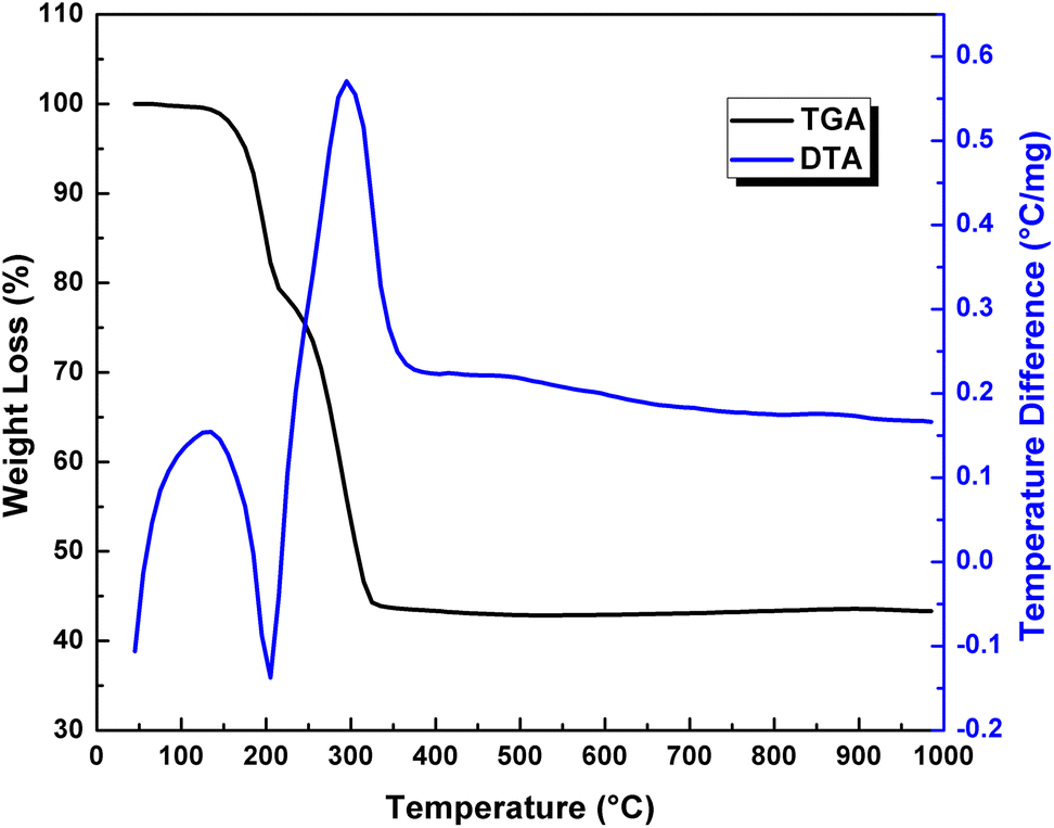
A typical TG–DTA curve for the oxalate powder prepared for Ni
0.7
Cu
0.1
Zn
0.2
La
0.035
Fe
1.965
O
4
is shown in Fig.
2
. The TGA curve exhibits two distinct weight loss steps corresponding to endothermic peak and exothermic peaks of DTA curve.
Fig. 2
TG-DTA plot for Ni
0.7
Cu
0.1
Zn
0.2
La
0.035
Fe
1.965
O
4
system
The TGA curve initially shows 19.95% weight loss in the range 116.13–212.98 °C. During this loss, endothermic peak exhibits at 204.91 °C in DTA curve and represents the evaporation of water and other elements is completed. Further TGA curve shows 36.15% weight loss in the range 212.98–366.33 °C with existence of an exothermic peak at 298.73 °C of DTA curve. This shows that decomposition of oxalate was completed at 366.33 °C and confirms the formation of phase in the material. Above 366.33 °C, TGA plot shows no further weight loss.
Murbe and Topfer [
24
] synthesized Ni–Cu–Zn ferrites by oxalate precursor method using Ni, Cu, and Zn acetate hydrate as starting materials. They observed quite similar behavior for TG–DTA plot. They found that thermal decomposition of the oxalate hydrate was completed at 300 °C. DTA and TG analysis of Ni–Cu–Zn ferrites prepared by sol–gel auto-combustion method was reported by Yue et al. [
25
]. They observed exothermic peak at about 220 °C and attributed to the reaction of nitrates with citric acid, whereas the exothermic peak at about 400 °C corresponds to the decomposition of citric acid. Wei-chi-Hsu et al. [
26
], Ramakrishna et al. [
27
], Shifeng-yan et al. [
28
], and Humbe et al. [
29
] also studied the thermo-gravimetric and differential temperature analysis for Ni–Cu–Zn ferrites.
The EDAX of Ni
0.7
Cu
0.1
Zn
0.1
La
x
Fe
2−
x
O
4
(where
x
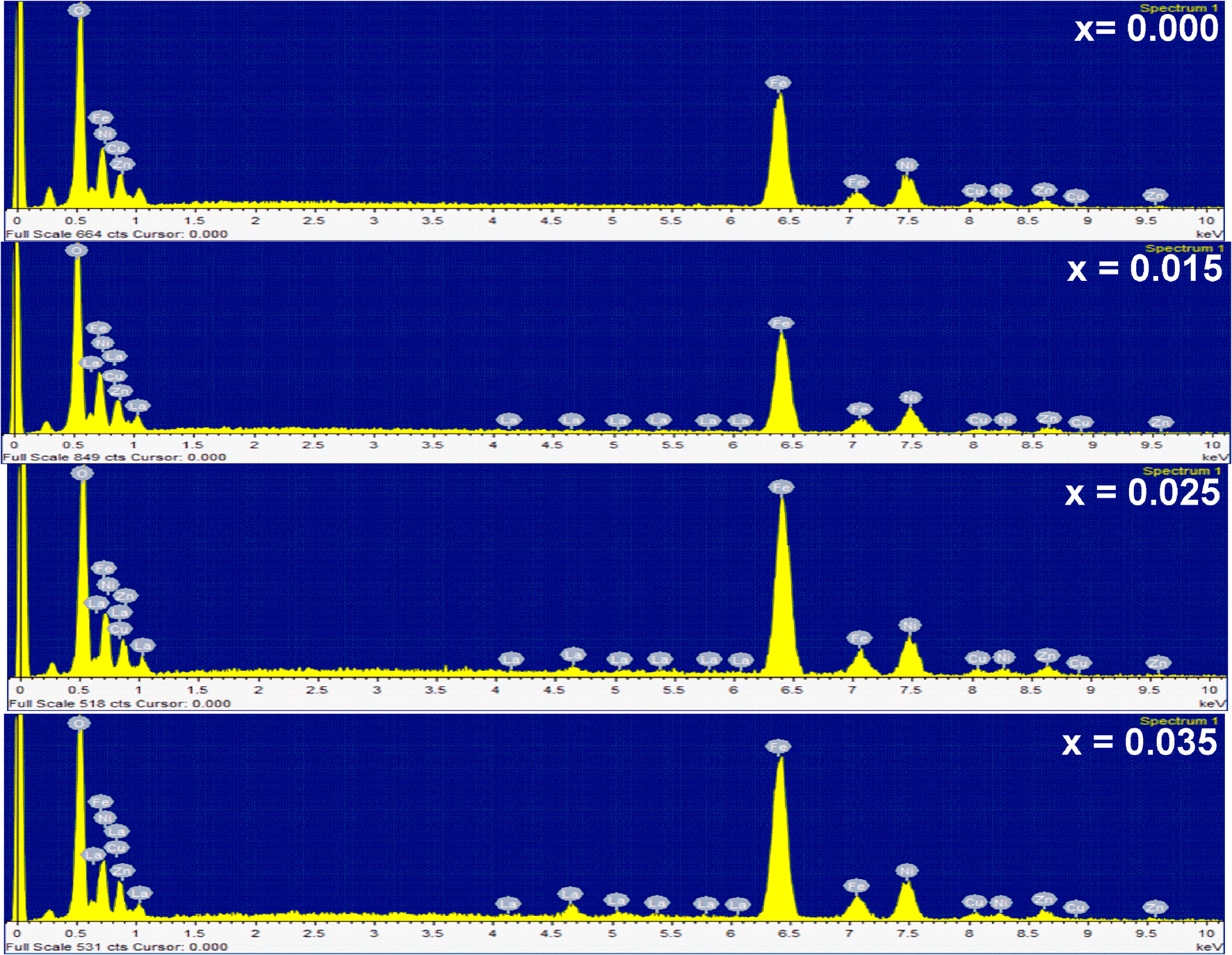
= 0.000, 0.015, 0.025, and 0.035) system is shown in Fig.
3
a–d. The presence of peaks corresponding to the elements Ni, Cu, Zn, La, Fe, and O in the spectra confirms the formation of appropriate respective ions in the material as per the initial stoichiometric considered without any other impurity ions. The compositional atomic percentage of Ni
2+
, Cu
2+
, Zn
2+
, La
3+
, Fe
3+
, and O
2−
ions in the all ferrites are tabulated in Table
1
. From this table, it is found that the molar ratio of tetrahedral-to-octahedral site metal ions is approximately agree with theoretical ratio of respective ions and confirms the formation of required homogeneous stoichiometric ferrite under investigation.
Fig. 3
EDAX spectra for Ni
0.7
Cu
0.1
Zn
0.2
La
x
Fe
2−
x
O
4
(where
x
= 0.0, 0.015, 0.025, and 0.035) system
Table 1
Atomic % of metal ions of Ni
0.7
Cu
0.1
Zn
0.2
La
x
Fe
2−
x
O
4
(where
x
= 0.000, 0.015, 0.025, and 0.035) system
Content x
Atomic % of metal ions
Ni
Cu
Zn
La
Fe
O
0.00
9.16
1.90
2.59
–
22.19
64.16
0.015
7.57
1.15
2.19
0.03
21.50
67.56
0.025
9.18
1.55
2.91
0.50
27.86
58.01
0.035
9.50
1.40
2.99
0.94
26.71
58.46
XRD analysis of La3+-substituted Ni–Cu–Zn ferrites
XRD patterns of Ni
0.7
Cu
0.1
Zn
0.2
La
x
Fe
2−
x
O
4
(where
x
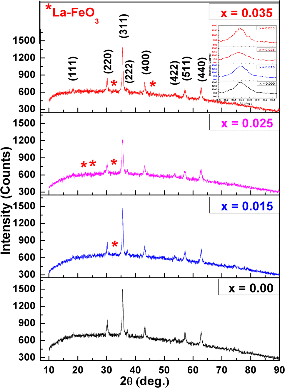
= 0.000, 0.015, 0.025, and 0.035) are shown in Fig.
4
. The presence of (111), (220), (311), (222), (400), (422), (511), and (440) planes in the diffraction pattern confirmed the formation of cubic spinel structure [
19
,
20
,
22
] having Fd-3 m space group in addition with weak appearance of ortho-ferrite phase shown by the Star (*). The formation of ortho-ferrite phase is due to La–FeO
3
. Chaudhary et al. [
19
] also observed ortho-ferrite phase for La
3+-
substituted Ni–Cu–Zn ferrites synthesized by sol–gel method. From diffraction pattern, it is seen that the intensity of peaks decreases with increasing La
3+
content up to
x
= 0.025 and thereafter increases for
x
= 0.035 (Table
2
). It means that the lighter percentage of La
3+
content diluted completely in ferrites having compositions
x
≤ 0.025, but thereafter, there may be possibility of inappropriate dilution for the composition
x
= 0.035 content. The undiluted La
3+
ions form the LaFeO
3
phase on the grain boundaries. Due to this, there may be insufficient diffraction centers in the nano-sized particles, which results increase in peak intensity for the composition
x
= 0.035 [
30
]. Chaudhary et al. [
19
] reported that the maximum limit for replacement of Fe
3+
by La
3+
in Ni–Cu–Zn ferrites is up to
x
= 0.025.
Fig. 4
X-ray diffraction patterns for Ni
0.7
Cu
0.1
Zn
0.2
La
x
Fe
2−
x
O
4
(where
x
= 0.0, 0.015, 0.025, and 0.035) system
Table 2
Crystallite size (
D
), lattice constant (
a
), X-ray density (
ρx
) grain size (
G
), and intensity (
I
) of Ni
0.7
Cu
0.1
Zn
0.2
La
x
Fe
2−
x
O
4
(where
x
= 0.000, 0.015, 0.025, and 0.035) system
Composition
Crystallite size D (nm)
Lattice constant a (Å)
X-ray density ρX (gm/Cm3)
Gain size G (nm)
Intensity I (A.U.)
Ni0.7Cu0.1Zn0.2Fe2O4
25.98
8.3619
5.3659
24.21
1504.68
Ni0.7Cu0.1Zn0.2 La0.015Fe1.985O4
20.14
8.3725
5.3729
23.40
1469.37
Ni0.7Cu0.1Zn0.2 La0.025Fe1.975O4
15.93
8.3843
5.3690
17.69
1218.74
Ni0.7Cu0.1Zn0.2La0.035Fe1.965O4
23.77
8.3936
5.3698
16.23
1387.34
The lattice parameter ‘a’ of all the ferrites was calculated for most intense (311) peak using the formula:
a=dobsh2+k2+l2,
where d
obs
is the observed inter-planner spacing.
Lattice constant of all the ferrites is tabulated in Table
2
. From this table, it is seen the lattice constant of Ni–Cu–Zn ferrites increases with increase in La
3+
content. This is attributed to replacement of smaller ionic radii Fe
3+
ions (0.67 Å) by larger ionic radii La
3+
ions (1.05 Å). This indicates that La
3+
ions occupy octahedral (B) site. Similar type of result was reported by Angari [
31
] and Gabal et al. [
22
] for La
3+
-substituted Ni ferrites and Ni–Cu–Zn ferrites synthesized by egg-white precursor method. Roy et al. [
20
] synthesized La
3+
substituted Ni–Cu–Zn ferrites by citrate nitrate auto-combustion method. They found that there is no any remarkable trend for lattice constant of La
3+
-substituted Ni–Cu–Zn ferrites.
The X-ray density (
ρx
) of all the ferrites was calculated using the formula [
32
]:
ρx=8MNa3,
where
M
is the molecular weight,
N
is an Avogadro’s number.
It is seen that the X-ray density of La
3+
-substituted ferrites are higher than pure Ni–Cu–Zn ferrite, but shows fluctuation trend with La
3+
content. In our investigation, it is observed that M as well as lattice constant (a) increases with increasing La
3+
content. Thereby, the ratio (M/a
3
) shows fluctuation trend with La
3+
contents. That is why, X-ray density shows fluctuation trend with La
3+
contents (Table
2
).
The average crystallite size (D) of the ferrite powder was estimated for the most intense (311) peak of XRD using the Debye Scherer formula [
32
]:
D=0.9λβcosθ,
where
λ
is wavelength of used X-ray beam,
β
is the full-width at half-maxima,
θ
is diffracting angle.
Obtained crystallite size of the ferrites is presented in Table
2
. From this table, it is seen that the crystallite size of the ferrites decreases with increasing lanthanum content up to
x
= 0.025 and thereafter increases. It is known that the induced crystalline anisotropy increases with replacement of smaller ionic radii Fe
3+
ions by larger ionic radii La
3+
ions, which creates the strain inside the volume of the crystal [
38
]. This may result decrease in crystallites size with increase in La
3+
contents except
x
= 0.035 composition. However, a crystallite size for the composition
x
= 0.035 is higher as compared to compositions
x
= 0.015 and 0.025. This may be due to inappropriate dilution of La
3+
ions (
x
= 0.035) in Ni–Cu–Zn ferrite. Chaudhari et al. [
19
] also reported that the crystallite size of Ni–Cu–Zn ferrites decreases with increase in La
3+
contents. The crystallite size for pure Ni–Cu–Zn ferrite is higher than the La
3+
-substituted Ni–Cu–Zn ferrite. Augustin et al. [
33
] reported the similar behavior for La
3+
substituted strontium ferrite prepared by citrate combustion method.
Hopping lengths (
LA
and
LB
) are the distance between the magnetic ions in the respective sites. These lengths are calculated using the relations [
19
]:
LA=a34LB=a24.
Bond lengths (A–O and B–O) and ionic radii (
rA
and
rB
) on A-site and B-sites were calculated using the relations suggested by Standley’s:
A-O=u-14a3B-O=58-uarA=u-14a3-r0rB=58-ua-r0.
The oxygen ionic radii
ro
= 1.32 Å [
34
] and oxygen ion parameter
u
= 0.381 are used for calculations. The hopping lengths, bond lengths, and ionic radii on A-site and B- site of all the ferrites are presented in Table
3
. From this table, it is seen that the bond lengths, ionic radii, and hopping lengths on A-site as well as on B-sites are increased with incorporation of small amount of La
3+
ions in pure Ni–Cu–Zn ferrites. This may be attributed to higher lattice constant for La
3+
-substituted ferrites.
Table 3
Ionic radii, bond lengths, hopping lengths, wave numbers, and force constants of Ni
0.7
Cu
0.1
Zn
0.2
La
x
Fe
2−
x
O
4
(where
x
= 0.000, 0.015, 0.025, and 0.035) system
La3+ content x
Ionic radii (Å)
Bond length (Å)
Hopping length (Å)
Wave numbers (cm−1)
Force constant (dyne/cm)
rA A-site
rB B-site
A–O A-site
B–O B-site
LA A-site
LB B-site
ϑ1 A-site
ϑ2 B-site
Kt A-site
Ko B-site
0.000
0.5787
0.7195
1.8987
2.0395
7.2416
5.9128
587.36
403.81
268663
100487
0.015
0.5811
0.7221
1.9011
2.0421
7.2508
5.9202
599.60
401.77
280652
99476
0.025
0.5838
0.7249
1.9038
2.0445
7.2610
5.9286
595.52
401.77
277142
99476
0.035
0.5859
0.7272
1.9059
2.0472
7.2691
5.9352
595.52
401.77
277455
99476
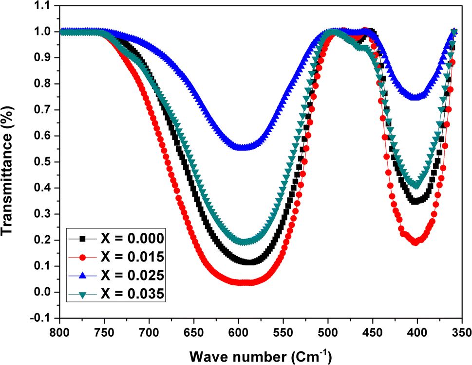
Fourier transform infrared spectroscopy
The position of absorption bands corresponding to tetrahedral (A), octahedral (B) sites, and the information about chemical changes in structure of ferrites is obtained from FTIR analysis. FTIR spectra of Ni
0.7
Cu
0.1
Zn
0.2
La
x
Fe
2−
x
O
4
(0.00 ≤
x
≤ 0.035) system recorded in the range 350–800 cm
−1
are shown in Fig.
5
. The higher frequency absorption band (
υ1
) corresponding to tetrahedral (A) site lies in the range 587–599 cm
−1
, whereas lower frequency absorption band (υ
2
) corresponding to octahedral (B) site lies in the range 401–403 cm
−1
. The change in the band position of
υ1
arises due to the change in the bond length of Fe
3+
-O
2−
at octahedral site, whereas the change in band position of
υ2
may be due to increasing concentration of Fe
2+
ions during synthesis of ferrite material. The observed wave numbers
υ1
and
υ2
corresponding to tetrahedral and octahedral sites are presented in Table
3
. From this table, it is seen that
υ1
becomes higher and
υ2
becomes lower than pure Ni–Cu–Zn ferrite. This trend observed in the wave numbers may be due to formation of Fe
2+
ions during the synthesis process [
19
].
Fig. 5
FTIR spectra for Ni
0.7
Cu
0.1
Zn
0.1
La
x
Fe
2−
x
O
4
(where
x
= 0.0, 0.015, 0.025, and 0.035) system
It is also observed that the absorption as well as broadening of bands for both tetrahedral and octahedral sites are greater for
x
= 0.015 composition as compared to other compositions. Absorption of
υ1
and
υ2
bands is very lower for
x
= 0.025 composition as compared to other compositions. In XRD analysis, we have reported similar type of interpretation for intensity.
The force constant on tetrahedral site (
Kt
) and on octahedral site (
K0
) was calculated using the relations suggested by Waldron’s [
35
]. The values of K
t
and
K0
of all the ferrites are presented in Table
3
. From this table, it is observed that
Kt
is lies in the range 2.686 × 10
5
to 2.806 × 10
5
dyne/cm and
Ko
lies in the range 0.9994 × 10
5
to 1.0048 × 10
5
dyne/cm. It is clear that
Kt
is greater than
K0
. Kabbur et al. [
34
] evaluated values of
Kt
and
Ko
for Dy
3+
-substituted Ni–Cu–Zn ferrites prepared by sol–gel auto-combustion method. They also observed that
Kt
is greater than
Ko
for all the compositions. It is also seen that the trends of K
t
and
K0
of ferrites are similar to that observed for
υ1
and
υ2
.
Field-emission scanning electron microscopy
FE-SEM micro-photographs of Ni
0.7
Cu
0.1
Zn
0.2
La
x
Fe
2−
x
O
4
(where
x
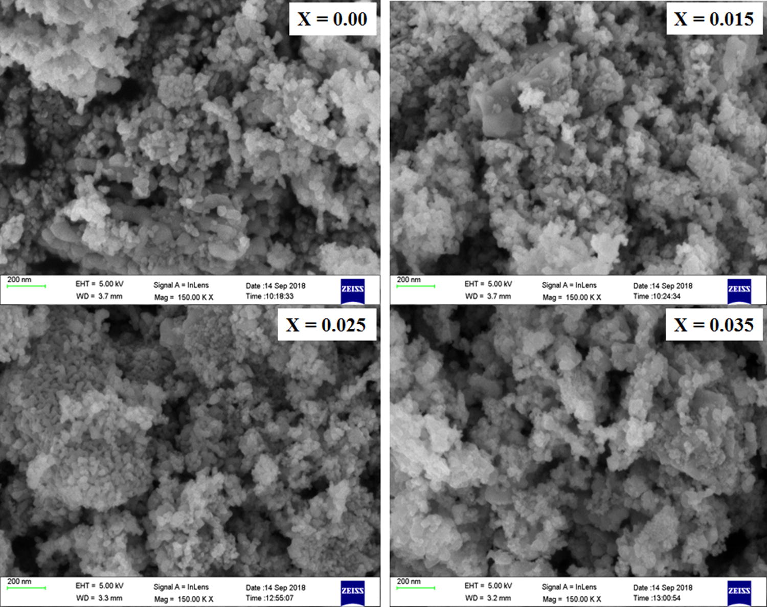
= 0.0, 0.015, 0.025, and 0.035) system are presented in Fig.
6
. From these micrographs, it is clear that the formation of ferrite particles is approximately spherical. The grain size of the ferrite was calculated using linear intercept method [
32
] and its values are presented in Table
2
. It is seen that the grain size of the Ni–Cu–Zn ferrites decreases with increase in La
3+
content. Similar type of result was reported by Chaudhary et al. [
19
] and Ikram et al. [
36
] for La
3+
-substituted Ni–Cu–Zn ferrite and La
3+
-substituted Ni–Cd–Zn ferrite nanoparticles prepared by sol–gel method.
Fig. 6
FE-SEM micro-photographs for Ni
0.7
Cu
0.1
Zn
0.1
La
x
Fe
2−
x
O
4
(where
x
= 0.0, 0.015, 0.025, and 0.035) system
VSM analysis
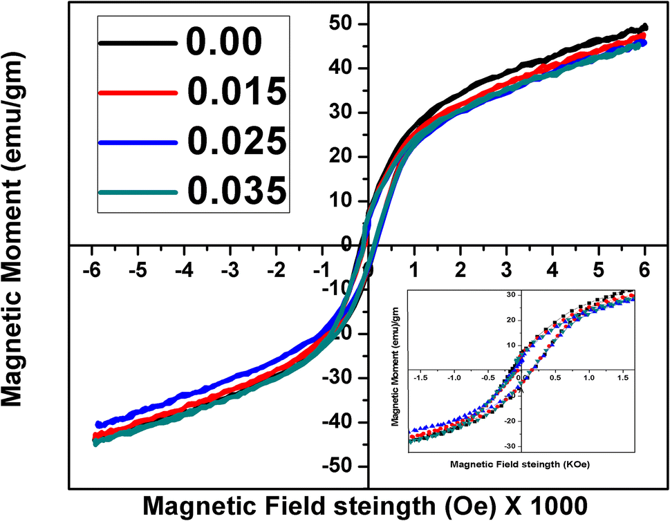
Figure
7
shows the hysteresis loops of Ni
0.7
Cu
0.1
Zn
0.2
La
x
Fe
2−
x
O
4
(where
x
= 0.000, 0.015, 0.025, and 0.035) system. The magnetic parameters such as saturation magnetization (
MS
), retentively (
Mr
), and coercivity (
HC
) obtained from the loops are listed in Table
4
. These parameters are depending on the chemical composition, grain size, porosity, density, method of preparation, sintering process, and heat treatment conditions of ferrites [
34
]. From Table
4
, it is seen that on La
3+
substitution, the saturation magnetization of Ni–Cu–Zn ferrites decreases. It may be attributed to the replacement of magnetic element (Fe
3+
) by non-magnetic element (La
3+
) on B-site, thereby decreasing B–B interaction and hence net magnetic moment reduces. Furthermore, non-magnetic La
3+
ions substituted in the ferrite would weaken the interactions between magnetic particles and reduce resistance of the domain-wall motion, which caused the decrease in coercivity. Ren and Xu [
37
] reported similar type of result for La
3+
-doped Ni–Co–Zn ferrites prepared by sol–gel method. They found that the addition of La
3+
ions results in the decrease of saturation magnetization and coercivity. The effect of La
3+
substitution on the structural and magneto-crystalline anisotropy of nano-crystalline cobalt ferrite synthesized by citrate precursor method was reported by Kumar and Kar [
38
]. They found that the saturation magnetization, magnetic coercivity, and magneto-crystalline anisotropy constants of ferrites were decreased with increase in La
3+
concentration. From Table
4
, it is observed that the Mr decreases with increase in La
3+
content except
x
= 0.035 composition. This is attributed to lowering the concentration of magnetic Fe
3+
ions by non-magnetic La
3+
ions. For the composition
x
= 0.035, there may be possibility of increasing the concentration of Fe
2+
ions during the synthesis process, and therefore, Mr is higher as compared to compositions
x
= 0.015 and 0.025. It is also seen that Mr and H
c
of La
3+
-substituted Ni–Cu–Zn ferrites are lower than pure Ni–Cu–Zn ferrite [
39
,
40
].
Fig. 7
Hysteresis loop for Ni
0.7
Cu
0.1
Zn
0.2
La
x
Fe
2−
x
O
4
(where
x
= 0.0, 0.015, 0.025, and 0.035) System
Table 4
Magnetic parameters of Ni
0.7
Cu
0.1
Zn
0.2
La
x
Fe
2−
x
O
4
(where
x
= 0.000, 0.015, 0.025, and 0.035) system
Composition
Saturation magnetization Ms (emu/gm)
Remanence magnetization Mr (emu/gm)
Coercivity Hc (Oe)
Squareness ratio R
Magnetic moment nB(obs) μB
Anisotropy constant k1 erg/cc
Ni0.7Cu0.1Zn0.2Fe2O4
49.7877
6.88
157
0.1381
2.105614
8142.36
Ni0.7Cu0.1Zn0.2La0.015Fe1.985O4
47.6917
6.71
146
0.1407
2.027302
7253.11
Ni0.7Cu0.1Zn0.2La0.025Fe1.975O4
46.2013
6.05
155
0.1308
1.970818
7459.58
Ni0.7Cu0.1Zn0.2La0.035Fe1.965O4
45.4952
6.63
129
0.1456
1.947464
6113.41
The squareness ratio (
R
), magnetic moment (
ηB
), and anisotropy constant (
k1
) for all compositions were calculated using the relations [
34
]:
R=MrMSηB(Obs)=MXMS5585K1=HCXMS0.96.
The values of
R
,
ηB (obs
) and
K1
are listed in Table
4
. From this table, it is cleared that η
B
goes on decreasing with increase in La
3+
content. This is may be due to the decreasing saturation magnetization of ferrites with increasing La
3+
content. Crystallographic, magnetic, and electrical properties of La
3+
-substituted Ni–Cu–Zn nano-ferrites have been studied by Chaudhari et al. [
19
]. They reported that both observed and theoretical magnetic moments of Ni–Cu–Zn ferrites decrease with increasing La
3+
content. Lenin et al. [
41
] synthesized La
3+
-substituted nickel ferrite by sono-chemical reaction method. They reported that
ηB
of nickel ferrite found to be decreasing with increasing La
3+
content.
Conclusion
La
3+
-substituted Ni–Cu–Zn nano-ferrites were successfully synthesized by oxalate co-precipitation method at a low sintering temperature. EDAX shows the formation of appropriate stoichiometric ferrite under investigation. The formation of cubic spinel ferrite with weak ortho-ferrite phases due to LaFeO
3
was confirmed from XRD. The lattice constant of Ni–Cu–Zn ferrites increases with increase in La
3+
content. The average crystallite size as well as grain size of all the ferrites are lies in nano-particle range and it verifies the formation of nano-ferrites. On La
3+
substitution, grain size of the ferrites was decreased. The presence of absorption bands in the desired region of FTIR spectra confirms the formation of ferrites. Saturation magnetization and magnetic moment of ferrites decrease with increase in La
3+
content. It is observed that the ferrite having composition Ni
0.7
Cu
0.1
Zn
0.2
La
0.025
Fe
1.975
O
4
has smaller crystallite size, lower intensity, lower retentivity, and comparatively higher coercivity as compared to other La
3+
-substituted compositions.
Publisher's Note
Springer Nature remains neutral with regard to jurisdictional claims in published maps and institutional affiliations.
References
Hossain and Roy (2017) Structural and electro-magnetic properties of low temperature co-fired BaSrTiO3 and NiCuZn ferrite composites for EMI filter applications (pp. 18136-18144) 10.1007/s10854-017-7759-8
Unknown ()
Unknown ()
Su et al. (2006) Sintering characteristics and magnetic properties of NiCuZn ferrites for MLCI applications (pp. 172-175) 10.1016/j.mseb.2006.01.008
Dantas et al. (2017) Magnetic nanocatalysts of Ni0.5Zn0.5Fe2O4 doped with Cu and performance evaluation in transesterification reaction for biodiesel production (pp. 463-471) 10.1016/j.fuel.2016.11.107
Lin et al. (2011) Synthesis and characterization of nickel ferrite nano-catalysts for CO2 decomposition (pp. 88-96) 10.1016/j.cattod.2011.02.013
Jeseentharani et al. (2012) Nanocrystalline spinel NixCu08−xZn0.2Fe2O4: a novel material for humidity sensing (pp. 3529-3534) 10.1007/s10853-011-6198-9
Kumbhar et al. (2012) Chemical synthesis of spinel cobalt ferrite (CoFe2O4) nano-flakes for supercapacitor application (pp. 39-43) 10.1016/j.apsusc.2012.06.034
Fu et al. (2012) Nickel ferrite–graphene hetero architectures: Toward high-performance anode materials for lithium-ion batteries (pp. 338-342) 10.1016/j.jpowsour.2012.04.039
Shinde et al. (2017) Structural and magnetic properties of Cr3+ substituted nickel zinc copper nano ferrites (pp. 433-442)
Arulmurugan et al. (2005) Effect of zinc substitution on Co–Zn and Mn–Zn ferrite nanoparticles prepared by co-precipitation (pp. 470-477) 10.1016/j.jmmm.2004.09.138
Assar et al. (2017) Effect of γ-ray’s irradiation on the structural, magnetic, and electrical properties of Mg–Cu–Zn and Ni–Cu–Zn ferrites (pp. 355-367) 10.1016/j.jmmm.2016.08.028
Sakar et al. (2013) Annealing temperature mediated physical properties of bismuth ferrite (BiFeO3) nanostructures synthesized by a novel wet chemical method (pp. 2878-2885) 10.1016/j.materresbull.2013.04.008
Nejati and Zabihi (2012) Preparation and magnetic properties of nano size nickel ferrite particles using hydrothermal method10.1186/1752-153X-6-23
Das and Singh (2016) Structural, magnetic and dielectric study of Cu substituted NiZn ferrite nanorod (pp. 918-924) 10.1016/j.jmmm.2015.10.132
Awati et al. (2013) Fabrication of Cu2+ substituted nanocrystalline Ni–Zn ferrite by solution combustion route: Investigations on structure, cation occupancy and magnetic behavior (pp. 157-162) 10.1016/j.jallcom.2012.11.045
Shirsath et al. (2012) Enhanced magnetic properties of Dy3+ substituted Ni–Cu–Zn ferrite nanoparticles10.1063/1.3679688
Chaudhari et al. (2013) Crystallographic, magnetic and electrical properties of Ni0.5Cu0.25Zn0.25LaxFe2−xO4 nanoparticles fabricated by sol–gel method (pp. 213-220) 10.1016/j.jallcom.2012.09.060
Roy and Bera (2007) Enhancement of the magnetic properties of Ni–Cu–Zn ferrites with the substitution of a small fraction of lanthanum for iron (pp. 77-83) 10.1016/j.materresbull.2006.05.009
Roy et al. (2008) Study on electro-magnetic properties of La Substituted Ni–Cu–Zn ferrite synthesized by auto-combustion method (pp. 1128-1132) 10.1016/j.jmmm.2007.10.025
Gabal et al. (2011) On the structural and magnetic properties of La-substituted NiCuZn ferrites prepared using egg-white (pp. 2625-2630) 10.1016/j.ceramint.2011.04.007
Li et al. (2017) Structure and magnetic properties of NiCuZn ferrite materials with La doping (pp. 202-204) 10.1007/s12598-014-0376-2
Mürbe and Töpfer (2012) High permeability Ni–Cu–Zn ferrites through additive-free low-temperature sintering of nanocrystalline powders (pp. 1091-1098) 10.1016/j.jeurceramsoc.2011.11.021
Yue et al. (1999) Preparation and characterization of NiCuZn ferrite nanocrystalline powders by auto-combustion of nitrate–citrate gels (pp. 68-72) 10.1016/S0921-5107(99)00152-X
Hsu et al. (2004) Preparation of NiCuZn ferrite nanoparticles from chemical co-precipitation method and the magnetic properties after sintering (pp. 142-149) 10.1016/j.mseb.2004.04.009
Ramakrishna et al. (2017) Investigation of cation distribution and magnetocrystalline anisotropy of NixCu0.1Zn0.9−xFe2O4 nanoferrites: Role of constant mole percent of Cu2+ dopant in place of Zn2+ (pp. 7984-7991) 10.1016/j.ceramint.2017.03.078
Yan et al. (2004) Preparation of nanocrystalline NiZnCu ferrite particles by sol–gel method and their magnetic properties (pp. 84-89) 10.1016/j.jmmm.2003.10.014
Humbe et al. (2018) Nanocrystalline Ni0.70−xCuxZn030Fe2O4 with 0 ≤ x ≤ 025 prepared by nitrate-citrate route: structure, morphology and electrical investigations (pp. 3467-3481) 10.1007/s10854-017-8281-8
Zhou et al. (2007) Preparation and magnetic properties of La-substituted Zn–Cu–Cr ferrites via a rheological phase reaction method (pp. 7-10) 10.1016/j.jmmm.2007.02.030
Al-Angari (2011) “Magnetic properties of La-substituted NiFe2O4 via egg-white precursor route (pp. 1835-1839) 10.1016/j.jmmm.2011.02.003
Shinde et al. (2010) Saturation magnetization and structural analysis of Ni0.6Zn0.4NdyFe2−yO4 by XRD, IR and SEM techniques (pp. 120-124) 10.1007/s10854-009-9878-3
Augustin et al. (2005) Effect of La3+ substitution on the structural, electrical and electrochemical properties of strontium ferrite by citrate combustion method (pp. 406-411) 10.1016/j.matchemphys.2004.09.028
Kabbur et al. (2018) Effect of Dy3+ substitution on structural and magnetic properties of nano-crystalline Ni–Cu–Zn ferrites (pp. 665-675) 10.1016/j.jmmm.2017.12.006
Ikram et al. (2019) Tailoring the structural, magnetic and dielectric properties of Ni–Zn–CdFe2O4 spinel ferrites by the substitution of lanthanum ions (pp. 3563-3569) 10.1016/j.ceramint.2018.11.015
Ren and Guangliang (2014) Electromagnetic and microwave absorbing properties of NiCoZn-ferrites doped with La3+ (pp. 44-48) 10.1016/j.jmmm.2013.10.056
Kumar and Kar (2012) Effect of La3+ substitution on the structural and Magneto-crystalline anisotropy of nanocrystalline cobalt ferrite (CoFe2−xLaxO4).” (pp. 4771-4782) 10.1016/j.ceramint.2012.02.065
Gholizadeh and Jafari (2017) Effects of sintering atmosphere and temperature on structural and magnetic properties of Ni–Cu–Zn ferrite nano-particles: magnetic enhancement by a reducing atmosphere (pp. 328-336) 10.1016/j.jmmm.2016.09.029
Ribeiro et al. (2018) Li quid phase sintering of ferrite of NiCuZn with low magnetic permeability for miniaturization (pp. 723-727) 10.1016/j.ceramint.2017.09.236
Lenin et al. (2018) Structural, electrical and magnetic properties of NiLaxFe2-xO4 nanoferrites (pp. 385-393) 10.1016/j.matchemphys.2018.03.062