Preparation of inorganic–organic nanohybrid compound based on NiII, MoO3 and p-phenylenediamine by hydrothermal method and its photoluminescence property
Department of Inorganic Chemistry, Faculty of Chemistry, Tehran North Branch, Islamic Azad University, Tehran, 1651153311, IR
Abstract
The new nanocrystalline composition was synthesized using NiSO
4
and MoO
3
and PPDA (
p
-phenylenediamine) via the hydrothermal method. The structure, morphology and photoluminescence property of nanocrystal were studied using X-ray diffraction (XRD), Fourier transform infra red (FT-IR) spectroscopy, scanning electron microscopy (SEM), photoluminescence (PL) spectra, thermogravimetric analysis (TGA–DTA) and energy-dispersive X-ray spectroscopy (EDX). The effect of factors such as type and concentration of initial materials, pH values, temperature and reaction duration on the structure and morphology of nanocrystals was investigated in the preparation of composition. The results show that the pH of the initial solution affects the size of the prepared nanocrystals; the size of the crystals is increased and the morphology of the nanostructures is changed with the increment in pH value. According to the obtained results, neutral or low alkaline conditions of pH are more favorable for the formation of the nanocrystals. The obtained nanocrystal shows an intense PL emission at room temperature with a maximum peak at 461 nm and excitation at the wavelength of 300 nm. TGA and DTA analysis display a total weight loss of 13.42%.
Introduction
Due to the unique properties and characteristics of inorganic–organic nanohybrid materials in various industries, there is a great desire to prepare and study their applications. Surface and volumetric properties of inorganic–organic nanohybrids and the surface-to-volume ratio of these materials in the scale of nanometers have produced significant changes in them [
1
,
2
,
3
,
4
–
5
].
The engineering of nanocrystals of inorganic–organic hybrid materials is focused on the two aspects in terms of their structural flexibility [
6
] and, secondly, their application as a catalyst, as gas storage, as ion sieve, in photochemistry and in electromagnetism [
7
,
8
,
9
,
10
,
11
,
12
,
13
–
14
]. Hydrothermal crystallization in the presence of multifunctional organic amines is one of the methods widely used for the synthesis of these materials. Multifunctional organic nitrogen-containing ligands such as bipyridine (4,4׳-byp) [
15
,
16
–
17
], ethylenediamine (en) [
18
,
19
] and 1, 10-phenanthroline (phen) [
20
,
21
] have large possibilities for the formation of 3D frameworks containing metal atoms. On the other hand, these frameworks can be coupled with oxomolybdates and can form semiconductor nanostructures [
22
] with novel applications in photoluminescence [
23
,
24
], light harvesting [
25
,
26
] and so on. PPDA as multifunctional organic ligand can be placed between structural blocks of oxomolybdates which are widely used in the structure of inorganic–organic nanohybrids as agents for structural directing to facilitate the formation of different networks [
27
]. These ligands can not only be used as coordination factors to transition metals, but also cause the formation of one-dimensional or multi-dimensional networks by forming intermolecular interactions, such as hydrogen bonding [
28
,
29
,
30
–
31
]. These compounds are commonly synthesized by the hydrothermal method in solid state at temperatures above 150 °C. The advantage of this method is that chemical homogeneity, morphology purity, shape and composition phase are controlled under moderate conditions and thus a nanocrystalline structure with different capabilities such as electrical properties is obtained.
Inorganic–organic hybrid materials composed of Mo and PPDA have been studied mostly in terms of crystal structure [
32
,
33
–
34
], but preparation of their nanoparticles has not been reported. Therefore, we were interested in synthesizing these nanoparticles and studying their structure and properties. In this article, the hydrothermal method has been used for the synthesis of NiSO
4
and MoO
3
nanostructure crystals using PPDA spacers. Herein, PPDA is chosen as the spacer group because it can be easily polymerized under neutral conditions, and the NH
2
donor sites within PPDA makes it suitable for the construction of an inorganic–organic nanohybrid. The effect of factors influencing the hydrothermal process and pH values on the morphology of nanocrystals as well as their luminescence properties has also been discussed.
Materials and methods
Materials and spectral measurements
All initial materials were purchased from Aldrich and Merck and used without further purification. Powder X-ray diffraction (PXRD) spectra were measured on a STOE-STADIP. Fourier transform infrared (FT-IR) was recorded in KBr discs on a Perkin-Elmer 78 spectrophotometer. Scanning electron microscopy (SEM) and energy-dispersive X-ray spectroscopy (EDX) were observed with EM 3200-KYKY. Photoluminescence (PL) spectra were obtained with a Perkin Elmer LS55 spectrometer using a 150 W xenon lamp as the excitation source. Thermogravimetric analysis (TGA–DTA) was performed on a BAHR STA 503 Thermal analyzer in air with a heating rate of 5 °C/min.
General method to prepare nanocrystals
A mixture of NiSO
4
.6H
2
O (0.05 g, 0.190 mmol), MoO
3
(0.015 g, 0.104 mmol), PPDA (
p
-phenylenediamine) (0.011 g, 0.102 mmol) and distilled water (10 g, 555.5 mmol) with molar ratio of 1.82:1:1:5555 (and repeat with 0.96:1:1:5555) was stirred for 30 min at room temperature. The pH value of the mixture was set exactly at 2, 4, 6, 8 and 10 using aqueous solutions of NaOH and HCl (1 M). The mixture was transferred to a Teflon-lined stainless steel autoclave (25 mL) and the temperature was increased at the speed of 5 °C/10 min so that it became fixed at 180 °C. After 4 days, the temperature was lowered at the rate of 5 °C/10 min until it reached room temperature. At the end of the reaction, the pH of the solution did not change. The black precipitate was washed with distilled water and dried at room temperature in a desiccator. The product has a melting point of 1800 °C and is insoluble in water and other common solvents.
Results and discussions
Many factors affect the formation of nanocrystalline products. These factors generally are: the type of initial materials, molar ratio, pH values, reaction duration and temperature during the hydrothermal synthesis process. Changes of these factors have led to many reactions in many different situations. Temperature was evaluated in the range of 150–200 °C and the reaction time was evaluated in the range of 2 to 6 days and we finally concluded that suitable nanocrystals are formed at the temperature of 180 °C in 4 days. Changing the initial material from molybdenum trioxide to ammonium heptamolybdate tetrahydrate did not lead to formation of any nanocrystals.
PXRD was used for te evaluation of phase changes of the prepared nanocrystals. On the basis of X-ray diffraction pattern data, it was observed that powders synthesized by the Ni/Mo = 0.96 molar ratio of raw materials forms amorphous crystals, whereas the sample is completely crystallized using Ni/Mo = 1.82 molar ratio. This results show that synthesized nanocrystals are formed with an additional amount of NiSO
4
.
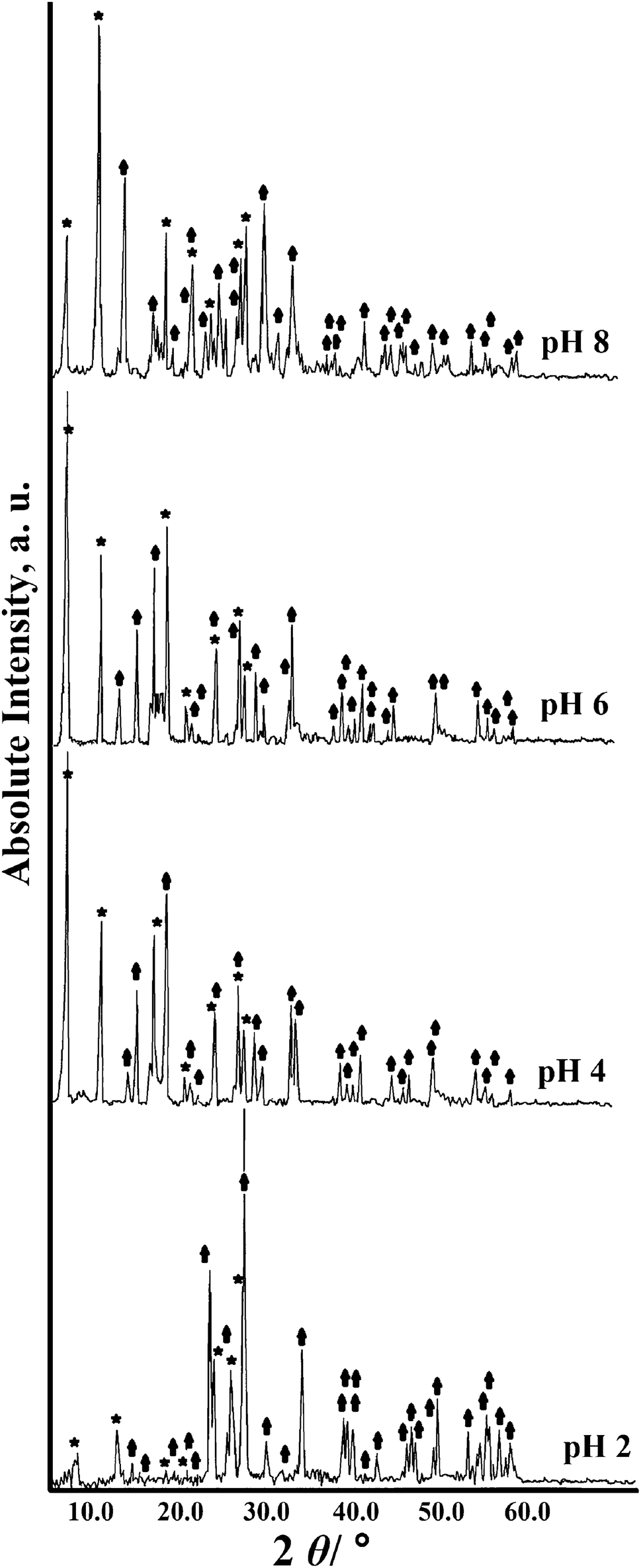
Figure
1
shows the X-ray diffraction patterns of nanocrystals which have been synthesized with molar ratio of Ni/Mo = 1.82 at different pH values. Nanocrystals obtained at pH values less than 10 consist of two phases. The first phase is the main phase of nickel bis(
p
-phenylenediamino) [formula: Ni(C
6
H
4
(NH)
2
)
2
] related to JCPDS No. 00-025-1554 and the second phase is the smaller phase of NiMoO
4
related to JCPDS No. 00-031-0902 (the main and minor phase peaks are indicated with the * and
↑
sign, respectively, in Fig.
1
). The main phase at 4, 6 and 8 pH values is monoclinic, with its first high peak at 2
θ
≈ 7.010, its second high peak at 2
θ
≈ 20.498 and its third high peak at 2
θ
≈ 23.772. At pH value of 2, the smaller phase is the dominant phase with its first high peak at 2
θ
≈ 28.852, its second high peak at 2
θ
≈ 14.320 and its third high peak at 2
θ
≈ 33.762. Small peaks which are observed to be more than 28 at 2
θ
are attributed to diffraction peaks of smaller phase. Existence of this phase leads to a small shift to greater angle of peaks in the main phase and, as a result, there is a little overlap in the peaks of these two phases. The intensity of the diffraction peaks increases by increasing pH values from 2 to 6 and, in contrast, the intensity of the diffraction peaks in smaller phases decreases. The intensity of peaks in the main phase is slightly reduced again at pH value of 8 and ultimately broadening of peaks at pH value of 10 indicates that the powders have become amorphous. The results suggest that neutral or low alkaline conditions of pH are more favorable for formation of the main phase, while the smaller phase forms in acidic conditions.
Fig. 1
XRD patterns of synthesized nanocrystals with molar ratio of Ni/Mo=1.82 at different pH values (The Ni(C
6
H
4
(NH)
2
)
2
and NiMoO
4
phase peaks are indicated with the * and
↑
sign, respectively)
The average size of crystals has been estimated using Scherrer equation:
D=0.9λ/βCosθ,
where
D
is the average diameter of the particle,
λ
is the CuKα radiation (
λ
= 1.54060 Å),
β
is the FWHM and
θ
is the Bragg’s diffraction angle. The results of the highest five diffraction peaks at different pH values have been calculated using Scherrer equation and have been listed in Table
1
. According to this equation, the average size of nanocrystals at different pH values was obtained in the range of 34–54 nm. The XRD results indicate that the size of crystals is increased with the increment in pH value and the pH of the initial solution affects the size of the prepared nanocrystals.
Table 1
The average crystallite sizes of the five highest diffraction peaks at different pH values
Peak no.
D(pH=2) (nm)
D(pH=4) (nm)
D(pH=6) (nm)
D(pH=8) (nm)
1
42.25
45.22
52.21
47.92
2
42.48
40.03
44.11
40.28
3
54.03
39.08
43.20
41.88
4
51.67
49.32
48.19
53.98
5
34.33
37.28
40.97
43.85
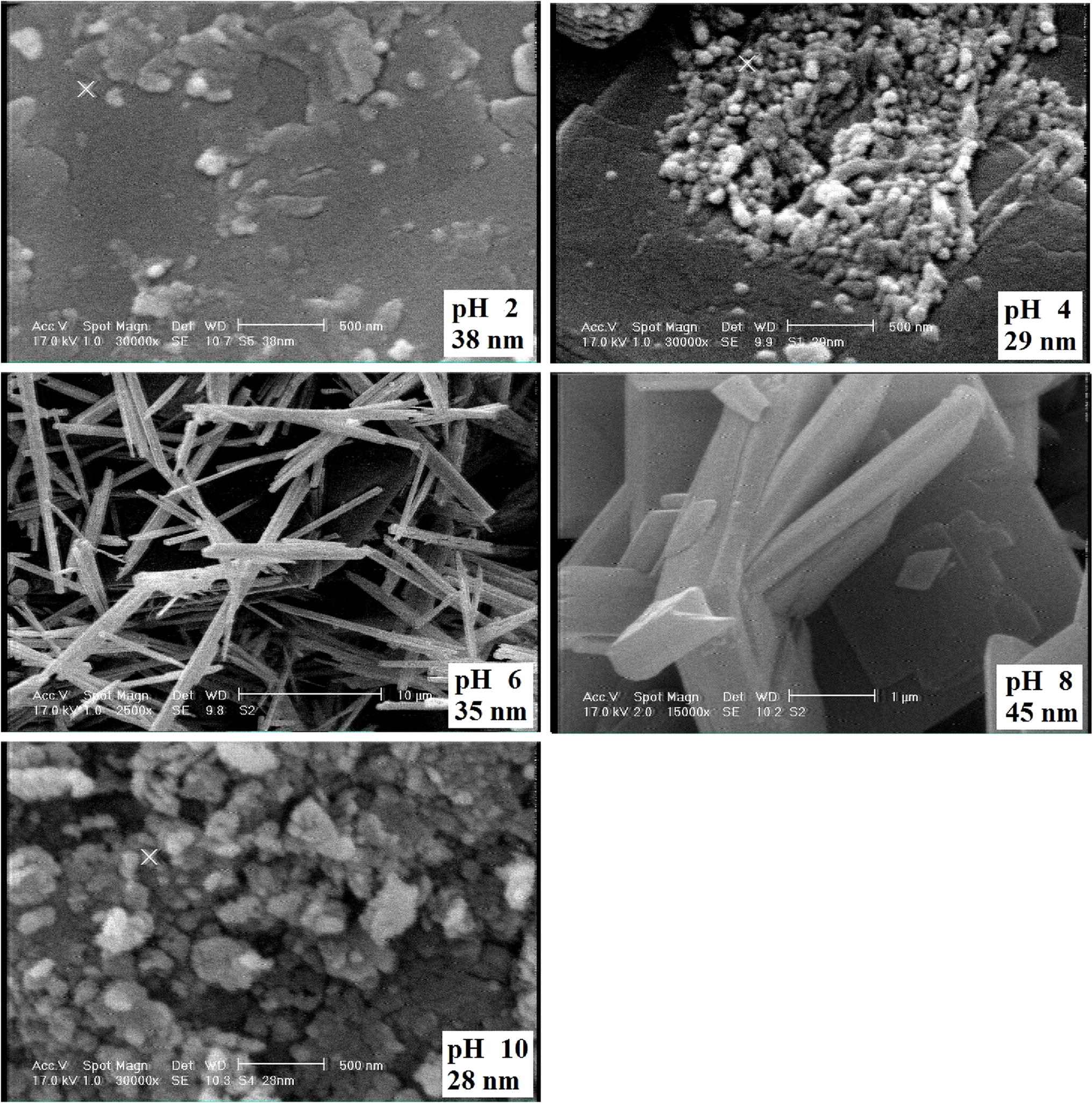
Figure
2
shows the micrographs of SEM crystals prepared with molar ratio of Ni/Mo = 1.82 at different pH values. The results shows that the morphology of the crystals changes at different pH values. The morphology of the nanocrystals is spherical with the size of 28–38 nm at pH values of 2, 4 and 10 and they are wired with the size of 35 nm at the pH value of 6. Thus, the smallest size of the crystals can be observed at pH 10 (28 nm). The SEM results suggest that the morphology of the nanostructures is changed with the increment in the pH value.
Fig. 2
SEM images of nanocrystals prepared with molar ratio of Ni/Mo = 1.82 at different pH values
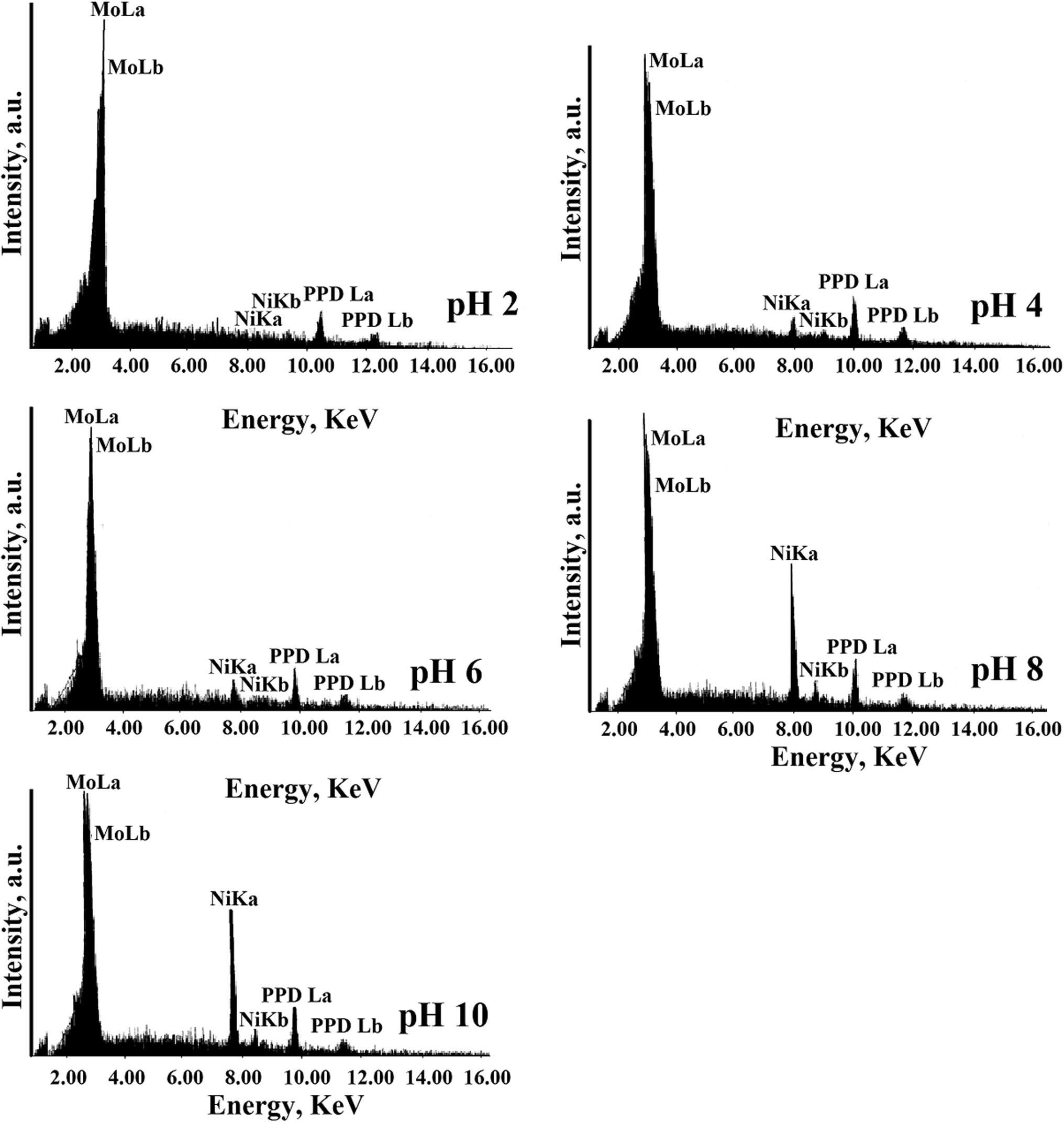
The EDX elemental analysis of products at the pH values 2–10 has been shown in Fig.
3
, in which both the major and minor phases confirm the presence of Ni, Mo and PPDA. It can be concluded that the structure of the prepared compound includes PPDA spacer groups, MoO
4
units act as bridges among PPDA spacer groups and, as a result, a 3D network is formed.
Fig. 3
EDX elemental analysis of prepared nanocrystals at the pH values 2–10
The percentages and weight ratios derived from EDX elemental analysis have been listed in Table
2
. The results indicate that the ratio of Ni/Mo weight percentages is increased by raising the pH value. The ratio of Ni to Mo at pH 4 and 6 are approximately 1–4.
Table 2
The percentages and weight ratios obtained from EDX elemental analysis of products at pH 2–10
pH
2
4
6
8
10
Mo (Wt%)
27.12
17.65
18.04
14.99
5.99
Ni (Wt%)
1.38
3.98
5.01
18.04
45.23
PPDA (Wt%)
71.36
83.72
76.99
66.98
46.59
Ni/Mo
0.05
0.22
0.28
1.20
7.55
Ni/PPDA
0.02
0.05
0.06
0.27
0.97
The IR spectra obtained from pH values 2–10 (Table
3
) show bands related to NH and water stretching vibration in the range of 3322–3731 cm
−1
, C–N in the range of 1238–1288 cm
−1
and C=C of PPDA ring in the range of 1509–1518 cm
−1
. Stretching vibration bands of Ni–N and Ni–O can be observed in the ranges of 518–590 and 429–498 cm
−1
. Symmetric and asymmetric stretching vibrations of MoO
2
groups were identified in the ranges of 819–880 and 922–969 cm
−1
. It shows that the crystallization of nanocrystals at neutral pH environment is optimized.
Table 3
IR characteristic band frequencies (cm
−1
) of the obtained compounds at different pH values
Characteristic bands
pH = 2
pH = 4
pH = 6
pH = 8
pH = 10
ν(C=C)
1510.23
1511.07
1510.01
1518.91
1509.25
ν(C–N)
1292.88
1259.99
1290.33
1240.01
1270.18
ν(NH)
3434.02
3376.08
3368.54
3322.93
3435.04
ν(OH)
3731.44
3429.75
3429.03
3397.07
3639.09
ν(MoO2)
819.22, 965.53
862.65, 928.98
869.17, 922.13
880.03, 969.02
869.82, 935.62
ν(Ni–N)
590.35
560.16
563.81
588.07
518.87
ν(Ni–O)
498.12
494.04
494.52
490.98
429.99
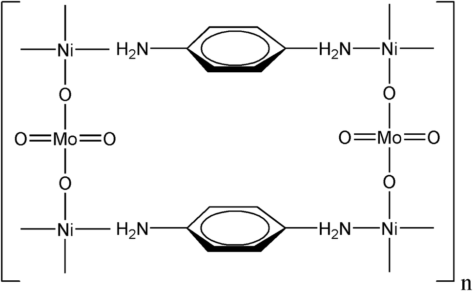
Based on the obtained results, the structure of synthesized nanocrystals is shown in Fig.
4
, which indicates a three-dimensional structure. In this structure, MoO
4
acts as building blocks, linked not only through direct coordination into oxo-bridged arrays but also through secondary metal sites acting as bridge in coordination complex subunits. The structure-directing role of the metal–ligand subunit shows the coordination priority of the metal and the geometric constraints ligand.
Fig. 4
The proposed structure of prepared nanocrystals at pH 6
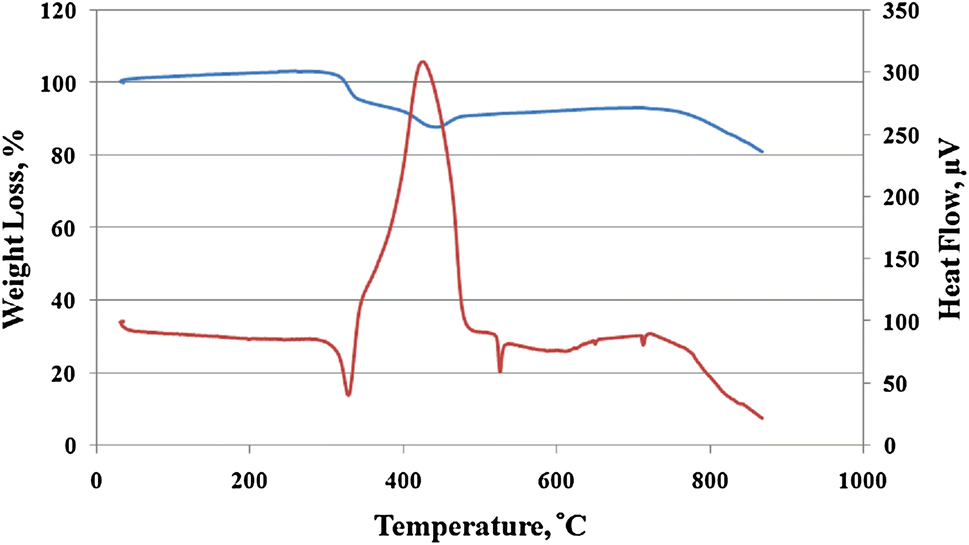
Figure
5
shows the TGA and DTA of the prepared nanocrystals at pH 6. TGA and DTA diagrams of the product are obtained at a heating rate 5 °C/min in the atmosphere and contain a two-step weight loss. The first weight loss equals to 5.42% at the temperature range of 259–330 °C related to the fracture of coordinated water molecules which match the endothermic peak of 325.01 °C DTA. The second weight loss equals to 8.00% at the temperature range of 337–448 °C along with decomposition of the PPDA ligand and complex structure, which is confirmed by the exothermic peak of 420.90 °C DTA. The total weight loss of 13.42% is in agreement with the calculated data and so it has thermal resistance.
Fig. 5
The TGA-DTA thermal analysis of prepared nanocrystal at pH 6
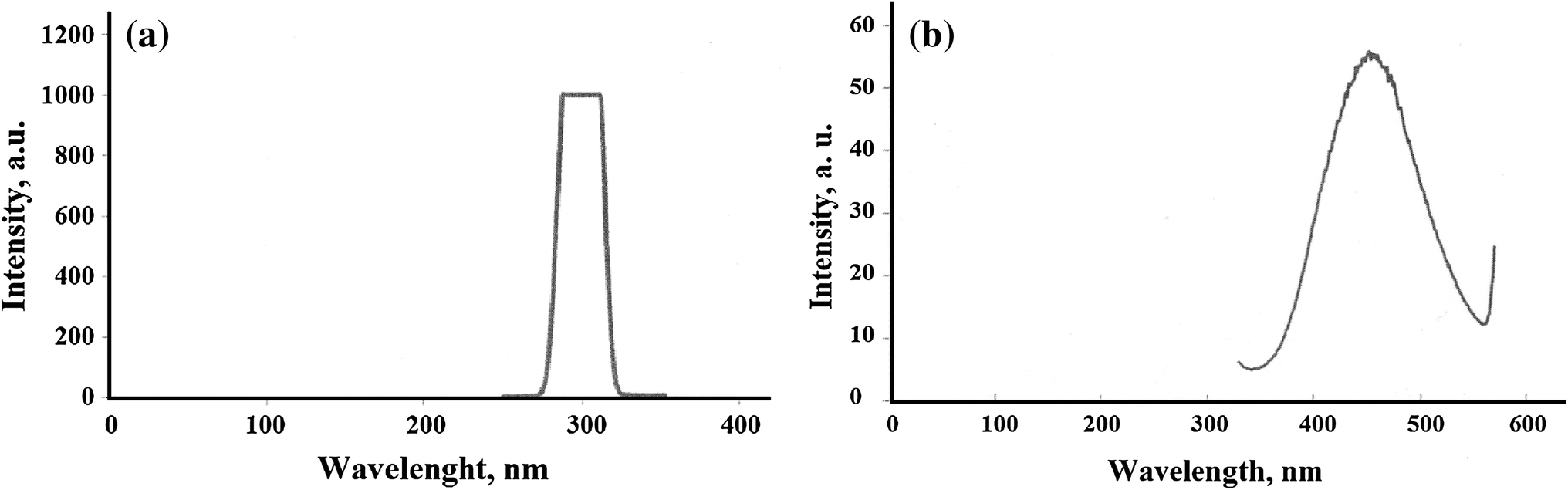
The absorbance and emission spectra of nanocrystals optimized at pH 6 in solid state at room temperature have been shown in Fig.
6
. The obtained nanocrystals show an intense PL by emission peak at the wavelength of 461 nm based on excitation at the wavelength of 300 nm. Dispersing the PL light to form the spectrum indicates the structural purity of the prepared nanocrystals as well as confirms its optical properties which depend on the energy difference between the valence and conduction bands. Placement of PPDA groups between inorganic materials increases the hardness of the structure and leads to observation of PL emission as a result. In general, PL is a characteristic of the sample and is sensitive to size; therefore, the intensity of PL peak decreases by increasing particle sizes related to nanoparticles [
35
].
Fig. 6
The
a
absorbance and
b
emission spectra of prepared nanocrystals at pH 6 in solid state at room temperature
Conclusion
A new inorganic–organic hybrid compound was synthesized via the hydrothermal method using NiSO
4
, MoO
3
and PPDA (
p
-phenylenediamine). The results indicate that MoO
4
inorganic group can be covalently connected to complexes of transition metals such as Ni-PPDA to obtain multi-dimensional structures. It was observed that the molar ratios of Ni/Mo, pH values, temperature and reaction duration effectively influence the morphology and nanostructure of final crystals. The crystal sizes increase when the pH value is increased, and particles with sizes smaller than 38 nm are formed while other parameters are fixed. On the basis of the obtained results, the crystallization of nanocrystals is controlled by a random nucleation process and is optimized at the pH value of 6. The results indicate that the ratio of Ni/Mo weight percentages is increased by raising the pH value.
Using PL spectroscopy, the optical properties of the synthesized samples, originating from the energy difference between conduction and valence bands, are confirmed. Room temperature PL, which was excited at the wavelength of 300 nm, indicates a photoluminescence emission peak at the wavelength of 461 nm. The fabricated structure displays an increase in hardness as well as a photoluminescence emission due to the fact that PPDA groups locate between inorganic species. This novel inorganic–organic hybrid compound can find applications as excellent thermal stability and in nanoscience as fluorescent nanofillers with controlled functionality, shape and size.
Publisher's Note
Springer Nature remains neutral with regard to jurisdictional claims in published maps and institutional affiliations.
References
Samiey et al. (2014) Organic-inorganic hybrid polymers as adsorbents for removal of heavy metal ions from solutions: a review 7(2) (pp. 673-726) 10.3390/ma7020673
Zhang et al. (2013) Fabrication and characterization of macroporous epichlorohydrin cross-linked alginate beads as protein adsorbent 43(5) (pp. 431-444) 10.1080/10826068.2012.745872
Sanchez et al. (2005) Applications of hybrid organic–inorganic nanocomposites 15(35–36) (pp. 3559-3592) 10.1039/b509097k
Hatton et al. (2005) Past, present, and future of periodic mesoporous organosilicas the PMOs 38(4) (pp. 305-312) 10.1021/ar040164a
Michinobu et al. (2009) Two-dimensionally extended aromatic polyamines for optimization of charge-transporting properties by partial oxidation 47(18) (pp. 4577-4586) 10.1002/pola.23510
Xiang and Cao (2012) Synthesis of Luminescent covalent-organic polymers for detecting nitroaromatic explosives and small organic molecules 33(14) (pp. 1184-1190) 10.1002/marc.201100865
Chang et al. (2004) Nanoporous metal-containing nickel phosphates: a class of shape-selective catalyst 43(21) (pp. 2819-2822) 10.1002/anie.200353502
Perles et al. (2005) Metal − organic scandium framework: useful material for hydrogen storage and catalysis 17(23) (pp. 5837-5842) 10.1021/cm051362e
Sun et al. (2015) Highly selective capture of the greenhouse gas CO2 in polymers 3(12) (pp. 3077-3085) 10.1021/acssuschemeng.5b00544
Chang et al. (2013) Microporous organic polymers for gas storage and separation applications 15(15) (pp. 5430-5442) 10.1039/c3cp50517k
Coronado et al. (2004) New conducting radical salts based upon Keggin-type polyoxometalates and perylene 14(12) (pp. 1867-1872) 10.1039/B402630F
Coronado et al. (2004) Metallic conductivity down to 2 K in a polyoxometalate-containing radical salt of BEDO-TTF 43(23) (pp. 3022-3025) 10.1002/anie.200453985
Shi et al. (2006) Influence of metal ions on the structures of Keggin polyoxometalate-based solids: hydrothermal syntheses, crystal structures and magnetic properties 179(1) (pp. 253-265) 10.1016/j.jssc.2005.09.051
Lu et al. (2002) Three polymeric frameworks constructed from discrete molybdenum oxide anions and 4,4′-bpy-bridged linear polymeric copper cations 14(6) (pp. 2649-2655) 10.1021/cm011725k
Han et al. (2005) Hydrothermal synthesis and characterization of a new hybrid organic–inorganic compound [Cd(en)3]MoO4 741(1) (pp. 31-35) 10.1016/j.molstruc.2005.01.064
Duan et al. (2005) Synthesis and characterization of a new compound based on mixed Mo/V polyoxometalates connected and modified by [Ni(en)2]+2 groups 15(2) (pp. 79-80) 10.1070/MC2005v015n02ABEH001937
Wu et al. (2009) Synthesis, structure and property of a new inorganic–organic hybrid compound [Cu(phen)2][Cu(phen)H2O]2[Mo5P2O23]·3.5H2O 11(1) (pp. 43-48) 10.1016/j.solidstatesciences.2008.05.016
Ma et al. (2010) Hydrothermal synthesis of two Anderson POM-supported transition metal organic–inorganic compounds 967(1–3) (pp. 15-19) 10.1016/j.molstruc.2009.12.008
Niu et al. (2004) 1D Polyoxometalate-based composite compounds derived from the Wells–Dawson subunit: synthesis and crystal structure of [{Ce(DMF)4(H2O)3}{Ce(DMF)4(H2O)4}(P2W18O62)]·H2O 4(2) (pp. 241-247) 10.1021/cg034131y
Yuan et al. (2008) Superwetting nanowire membranes for selective absorption 3(6) (pp. 332-336) 10.1038/nnano.2008.136
Benedetto et al. (2008) Patterning of light-emitting conjugated polymer nanofibres 3(10) (pp. 614-619) 10.1038/nnano.2008.232
Zhao et al. (2008) Novel polyfluorinated polyimides derived from α, α-bis(4-amino-3,5-difluorophenyl)phenylmethane and aromatic dianhydrides: synthesis and characterization 44(3) (pp. 808-820) 10.1016/j.eurpolymj.2007.12.016
Zhu et al. (2007) Panchromatic conjugated polymers containing alternating donor/acceptor units for photovoltaic applications 40(6) (pp. 1981-1986) 10.1021/ma062376o
Nandia et al. (2011) Fe(III)-containing mesoporous poly-(p-phenylenediamine): synthesis, characterization and magnetic properties 142(2–3) (pp. 557-563) 10.1016/j.micromeso.2010.12.048
Dolbecq et al. (2003) Hybrid organic–inorganic 1D and 2D frameworks with ε-Keggin polyoxomolybdates as building blocks 9(12) (pp. 2914-2920) 10.1002/chem.200204670
Burkholder and Zubieta (2004) A two-dimensional bimetallic oxide constructed from ζ-octamolybdate clusters and Ag(I)-tpyprz cationic polymer components (tpyprz = tetra-2-pyridylpyrazine) 6(12) (pp. 1421-1428) 10.1016/j.solidstatesciences.2004.07.016
Bu et al. (2001) One- and two-dimensional framework materials constructed from the mixed Mo/V tetra-capped Keggin structure clusters and M(en)2 (M=Ni, Cu) complexes groups 4(1) (pp. 1-4) 10.1016/S1387-7003(00)00179-9
Hagrman et al. (2000) Polyoxomolybdate clusters and copper–organonitrogen complexes as building blocks for the construction of composite solids (pp. 212-224) 10.1016/S0020-1693(99)00566-6
Nezamzadeh-Ejhieh and Salimi (2010) Heterogeneous photodegradation catalysis of o-phenylenediamine using CuO/X zeolite 390(1–2) (pp. 110-118) 10.1016/j.apcata.2010.09.038
Wang et al. (2008) The preparation and characterization of poly(o-phenylenediamine)/gold nanoparticles interface for immunoassay by surface plasmon resonance and electrochemistry 63(2) (pp. 254-261) 10.1016/j.colsurfb.2007.12.007
Palys et al. (2007) Poly-o-phenylenediamine as redox mediator for laccase 52(24) (pp. 7075-7082) 10.1016/j.electacta.2007.05.029
Liou et al. (2005) Synthesis, luminescence and electrochromism of aromatic poly(amine–amide)s with pendent triphenylamine moieties 15(18) (pp. 1812-1820) 10.1039/b419183h