Graphical abstract

Cobalt oxide/zinc oxide core/shell nanoparticles (CZ NPs) were synthesized at low temperature by the sol–gel method. X-ray diffraction (XRD) was used for the detection of crystalline phases. Energy-dispersive analysis X-ray (EDAX) results for the sample as prepared showed that where only Co, Zn, C, and O was found, in forming CZ core/shell NPs. Transmission electron microscopy images indicated that the particles size of core/shell sample CZ NPs and the particles size were around 12 nm. The optical properties study by UV–Vis spectroscopy to estimate the bandgap of core/shell NPs. Frequency dependence dielectric was observed in core/shell NPs were prepared using the sol–gel method. Dielectric constant ε ′ and dielectric loss ε ″ for CZ NPs were found to decrease with increasing frequency.
To purvey novel functionalities and properties massive efforts have been made to fabricate metal-based composites as metal/metal or metal oxide composites [ 1 ]. Core/shell nanoparticles (NPs) are a particular type of nanostructured material and its properties depend on the core–shell volume ratio [ 2 , 3 ]. Core/shell nanoparticles are one of the solutions to obstacles in which an amendment in properties cannot be accomplished using one type of nanoparticles [ 4 ]. Employing a magnetic core can guide particles using an outer magnetic field. If a core/shell composite contains a magnetic core and semiconductor shell, the transmission and purging of the particles becomes potential [ 5 ]. Cobalt oxide Co 3 O 4 magnetic nanoparticles are interesting due to its numerous oxidation states [ 6 ]. Zinc oxide (ZnO) semiconductor nanoparticles are on the forefront of research due to their special properties and massive usage [ 7 ]. ZnO-based materials are used in different applications, due to the photocatalytic nature, environmental sustainability, and low cost [ 8 ].
In the present work, we synthesized Co 3 O 4 /ZnO core/shell NPs using the sol–gel method. Structure, morphology, optical properties, dielectric, and magnetization were studied using EDAX, XRD, UV–Vis, TEM, and dielectric properties, respectively.
Ammonium hydroxide Solution, Cobalt (ɪɪ) Chloride purum p.a., anhydrous, ≥ 98%, Zinc chloride 97.6% obtained from Holyland (Saudi Arabia), Ethanol absolute was purchased from Sigma Aldrich Co. Ltd (USA). Acetic acid, glacial biochemical grade 99.86% purchased from ACROS.
For ZnO (NPs) prepared by a sol–gel method, we dissolved (1.35 g) of ZnCl 2 in a mixture of 20 mL of water and 5 mL NH 4 OH and then added 80 mL of ethanol using a dropper for 60 min at 60 ºC while stirring for 2 h. The precipitate was filtered and washed several times with alcohol and deionized water then dried at 60 °C.
To prepare Co 3 O 4 /ZnO core/shell NPs, 1.29 g of CoCl2 and ZnO NPs (as-synthesized) were dissolved in a mixture of 20 mL of water and 5 mL NH 4 OH and then added 80 mL of ethanol drop by drop for 60 min at 60 °C with stirring for 3 hours until the sol was converted to gel. Finally, the dried gel was calcined at 300 °C for 4 h to obtain Co 3 O 4 /ZnO core/shell NPs.
Co 3 O 4 /ZnO core/shell NPs nanoparticles were characterized by X-ray diffraction using Shimadzu 7000 Diffractometer operating with CuKα ( λ = 0.15406 nm) with a scan rate of 2 min −1 for 2 θ values between 20° and 80°.
In EDAX attaching the particles to 12.5 mm diameter Al when accelerating voltage was 20.0 kV, working distance = 10 mm, Spot size = 4.5 (EDX). In secondary electron imaging mode at different magnifications, the images were digitally recorded at a resolution setting of 1024 × 884 pixels. EDX analysis was performed using EDAX Genesis XM4 system. Standardless ZAF options were used to determine elemental contents.
The Transmission Electron Microscopy (TEM) studies were performed [High Resolution Transmission Electron Microscope (HRTEM) JEOL–JEM-1011, Japan]. The samples for TEM were prepared by making suspension from the powder in deionized water. A drop of the suspension was put into the carbon gride and left to dry. The optical properties of Co 3 O 4 /ZnO core/shell NPs structures were characterized by UV–Vis absorption (UV–Vis spectrophotometer Model JASCO V-670, Japan instrument). The electrical properties were measured at room temperature by Hioki (LCR Hitester 3532–50). The frequency dependence of electrical properties for prepared samples was measured from 50 Hz to 5 MHz.
The elemental contents of CZ core/shell NPs were determined using standard less ZAF option, as shown in Fig.
1
. The spectra obtained during EDAX studies were used to carry out the quantitative analysis. Quantitative analysis in Fig.
1
proved high Cobalt contents (36.16%) in the examined samples. The presence of zinc, oxygen, and carbon, the contents of which amounted to 32.63, 23.59, and 7.62%, respectively, was also shown.
EDAX images for CZ core/shell NPsFig. 1
XRD patterns of Co
3
O
4
/ZnO core/shell NPs are detailed in Fig.
3
. The XRD for Co
3
O
4
/ZnO core/shell NPs showed that the major peaks refer to ZnO hexagonal wurtzite structure in agreement with JCPDS card: 36-1451. The XRD patterns of the CZ core/shell NPs in Fig.
2
are located at (2
θ
) match with the reflection of (100), (002), (101), (102), (110), (103), (200), (112), and (201) crystal planes, respectively [
9
,
10
]. Co
3
O
4
peaks appear as secondary phase attributed to cubic spinel Co
3
O
4
JCPDS card: 42-1467. These peaks located at (2
θ
) match with the reflection of (220), (311), and (400) crystal planes [
11
]. In XRD, the core NPs are fading due to a considerable amount of ZnO NPs in the shell [
12
].
XRD pattern in CZ core/shell NPFig. 2
TEM images are used to study the morphological aspects of the CZ NPs. Figure
3
a, b presents results which confirm the formation of CZ core/shell NPs and the images show nearly spherical nanoparticles. The TEM micrograph showed CZ core/shell NPs. Figure
3
a, b shows that the Co
3
O
4
cores appear darker than the ZnO shell. The result of TEM shows that the particles size of our sample is around 12 nm.
a
,
b
TEM images for CZ core/shell NPsFig. 3
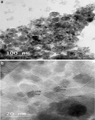
In TEM images, a clear interface between the core and the shell can be observed. The edge of the shell shows lower intensity than the core (Fig. 3 ). It can occur as a result of [ 13 ]:
The molar mass of the material in the shell being smaller than in the core
The electron beam finding the smaller amount of material at the edge of NPs.
The optical absorption spectra of CZ core/shell NPs were obtained in the wavelength range from 200 to 800 nm using UV–Vis spectrophotometer (JASCO V-670, Japan instrument), as shown in Fig.
4
a. The bulk zinc oxide (ZnO) has wide bandgap (WBG) of 3.37 eV, the semiconductors (WBG) are semiconductor materials which have a relatively large bandgap compared to conventional semiconductors [
14
], and the bulk Co
3
O
4
has a bandgap between 2.85 and 1.70 eV [
15
]. The optical absorption spectrum of CZ core/shell NPs has been carried out using UV–Vis spectrophotometer. Figure
4
a also shows the absorption peaks at 410 and 262 nm for CZ core/shell NPs. The first band at 410 nm indicates change transfer or the second band appears at 262 nm; this absorption peak around 262 nm was observed. These absorption peaks in the UV region can be attributed to the local Plasmon resonance [
16
]. To determine the optical bandgap of the CZ core/shell NPs, we plotted (
αhѵ
)
2
versus
hѵ
, where α is the absorption coefficient, and
hѵ
is the photon energy [
17
]. A typical plot is shown in Fig.
4
b. The bandgap of the CZ core/shell NPs was evaluated from the plot to be 4.97 eV, which showed blue shift compared to bulk ZnO and bulk Co
3
O
4
. The blue shift is due to a reduction in the particles size, as TEM result showed that the ZnO shells are fragile and Co
3
O
4
p-type magnetic semiconductor at core next to n type of ZnO in shell influences on the properties of NPs and makes a blue shift in CZ core/shell NPs [
18
]. The p–n-type core–shell heterojunction can reinforce the charge separation effect under UV [
19
]. In addition, we observed another beak with gap energy 2.75 eV.
Optical absorption spectra of the CZ NPs core/shell in the wavelength range from 200 to 800 nm (
a
) and plot of (
αhѵ
)
2
versus
hѵ
for the CZ NPs core/shell (
b
)Fig. 4
Dielectric constant (
ε
′) and dielectric loss (
ε
″) frequency of CZ core/shell NPs as a function of frequency range (50 Hz–5 MHz) at room temperature are shown in Fig.
5
. The spectrum for CZ core/shell NPs sample show that dielectric constant (
ε
′) decreases exponentially with the increase of frequency. The dielectric constant (
ε
′) proximate a constant value at high frequencies due to the shortage of dipoles to twirl quickly leading to a delay between applied field and frequency of wobbling dipole [
20
]. The high values of
ε
′ at low frequencies may be attributed to the interfacial polarization which is appearing in materials composed of different phases [
21
]. In addition, in Fig.
5
, it is clearly shown that dielectric loss (
ε
″) at room temperatures decreases with the frequency of CZ core/shell NPs.
Dielectric constant
ε′
and dielectric loss
ε″
as a function of frequency for CZ NPs core/shell at room temperaturesFig. 5
The frequency dependence of ε′′ in Fig.
5
can be interpreted, by the following equation approbating to the free electron theory:
ε
″ =
δ
/2
π ε
0
f
, where
f
is frequency,
δ
is the electrical conductivity, and
ε
0 is the dielectric constant in a vacuum. Figure
6
shows the relationship between conductivity and frequency. The conductivity of CZ core/shell NPs generates from its free electrons. In addition,
ε
″ is critical to the absorbing frequency which becomes a defiance for technological applications [
22
].
σac
presents a flat frequency plateau at lower frequencies as is shown in Fig.
6
. However, the conductivity, as a frequency-dependent behavior, shows itself at higher frequencies. It may be due to hopping-type conduction at a high-frequency range [
23
].
log σ
ac
as a function of frequency for CZ core/shell NPs at room temperaturesFig. 6
The decrease in particles size to nanometric scale has been found to produce massive reduction in the tan
δ
(
D
) value, as shown in Fig.
7
. The value of tan δ depends on many factors such as synthesis methods and sample composition.
Dielectric loss tangent
D
as a function of frequency
log f
for CZ core/shell NPs at room temperaturesFig. 7
ε ′, ε ″, and tan of CZ core/shell NPs exhibit the electrical energy storage capacity and average loss in the materials [ 24 ]. The dielectric constant ( ε ′) and the tan δ values of investigated CZ core/shell NPs at 1 kHz are 7.8 and 1.77, respectively. It was also observed that the dielectric constant ( ε ′) of CZ core/shell NPs at 1 kHz under room temperature is less than the values of ZnO (40 nm) [ 25 ] and higher than ε ′ value of Co 3 O 4 (36 nm) [ 26 ] that measured in the previous studies where the ε ′ value were about 40 and 7 for ZnO and Co 3 O 4 at 1 kHz under room temperature, respectively.
CZ core/shell NPs with nearly spherical nanoparticles have been synthesized successfully by the sol–gel method. The particles size CZ core/shell was around 12 nm. The optical properties study by UV–Vis spectroscopy to estimate the bandgap of core/shell NPs, and band-gap value of CZ core/shell NPs is higher than bandgap of bulk ZnO and Co 3 O 4 . The dielectric measurements indicate that the dielectric constant of CZ core/shell NPs decreases, in the high-frequency range.
The author would like to show her gratitude towards Professor Magdah Dawy of the National Research Center in Cairo, Egypt, whose expertise greatly assisted the research.
Springer Nature remains neutral with regard to jurisdictional claims in published maps and institutional affiliations.