Graphical abstract

We have proposed an optimized homogeneous precipitation method for the synthesis of spherical and well-dispersed pure Ln 2 O 2 SO 4 (Ln = Gd, Ho, Dy and Lu) nanoparticles (NPs) using synthesized lanthanide sulfates and urea as starting materials. Ln 2 O 2 SO 4 NPs can be easily transformed to Ln 2 O 2 S by reduction under H 2 flow at 650 °C for Gd, Dy and Ho whereas the reduction with Lu gave only Lu 2 O 3 . Particle size was between 500 nm and 3.5 µm. This reduction method allows to obtain Ln 2 O 2 S without using H 2 S which is highly toxic and uncomfortable gas. Ln 2 O 2 SO 4 /Ln 2 O 2 S synthetized can be used for release and oxygen storage or doped with luminescent centers for multimodal medical imaging applications.
During this last decade, lanthanide oxysulfate and oxysulfide compounds (Ln = Gd, Ho, Dy and Lu) have been widely used due to their applications in phosphorescent material design, X-ray-computerized tomography, oxygen storage, and radiation detection [ 1 , 2 – 3 ]. The other remarkable features of lanthanide oxysulfide/oxysulfate are their large accurate sensing capability over a wide range of temperatures, as well as their ability to withstand a harsh environment [ 4 ]. Moreover, the crystal structure of Ln 2 O 2 SO 4 is usually the alternative stacking of a [Ln 2 O 2 ] 2+ layer and a layer of sulfate [SO 4 ] 2− and that of Ln 2 O 2 S is the alternative stacking of a [Ln 2 O 2 ] 2+ layer and a layer of sulfide (S 2− ) [ 5 ]. Thus, Ln 2 O 2 SO 4 is an intermediate phase for the synthesis of lanthanide oxysulfide Ln 2 O 2 S and is usually used as a precursor for the synthesis of Ln 2 O 2 S in reduction atmosphere.
The Ln 2 O 2 SO 4 materials for oxygen storage and release reported so far have been prepared by several methods, such as calcination of Ln(SO 4 ) 3 ·nH 2 O [ 6 , 7 – 8 ], solid-state reaction method using lanthanide oxide and diammonium hydrogen sulfate as the starting materials [ 9 , 10 ], utilization of precursors of layered Ln-dodecyl sulfate mesophases’ [ 11 ], Ln-precipitation [ 12 , 13 ], thermal decomposition of nanodroplets formed by Ln-acetylacetonates (Ln(acac) 3 ) [ 14 ] and hydrothermal decomposition of hydroxyl sulfates of Ln 2 (OH) 4 SO 4 (Ln = Eu-Lu and Y) [ 15 ] .
All of these methods result to bulk materials with an irregular morphology. The synthesis of NPs intended for applications in imaging and medical therapy imposes the control of many physicochemical characteristics such as the crystal structure and the morphology of the particles. The soft chemistry processes for preserving texture, morphology and structure are, therefore, well adapted for the development of these NPs [ 16 ]. The homogeneity of the particles obtained, the control of the various steps of germination and growth of the crystallites originating from the liquid are the main advantages of these synthesis processes. Homogeneous precipitation synthesis is one of the most promising techniques because of its advantages such as the relatively simple synthetic route, low cost, ease of mass production.
Lian et al. [ 17 ]. have synthesized (Gd 1 − x ,Eu x ) 2 O 2 SO 4 sub-microphosphors by homogeneous precipitation method from commercially available Gd 2 O 3 , Eu 2 O 3 ,H 2 SO 4 and (NH 2 ) 2 CO (urea) starting materials. After precipitation reaction, the precursor which is mostly composed of gadolinium hydroxyl, carbonate and sulfate groups with some crystal water; can be transformed into pure Gd 2 O 2 SO 4 phase after heating at 900 °C for 2 h in air. They reported that it is only when the molar ratio of urea to Gd 2 (SO 4 ) 3 is equal to 5, there is formation of pure Gd 2 O 2 SO 4 phase. The particles were quasi-spherical with a mean size of about 500 nm. The same authors have also reported [ 18 ] the photoluminescence of (Gd 1 − x ,Dy x ) 2 O 2 SO 4 using the same method. In that work, they use molar ratio of urea to Gd 2 (SO 4 ) 3 equal to 400, to form pure Gd 2 O 2 SO 4 phase. Thus, there is no clearly in literature the optimal conditions about molar ratio of urea to Ln 2 (SO 4 ) 3 that can be used to synthesize Ln 2 O 2 SO 4 NPs by homogeneous precipitation method.
This paper suggested an optimized homogeneous precipitation method to prepare spherical and well-dispersed pure Ln 2 O 2 SO 4 and Ln 2 O 2 S (Ln = Gd, Ho, Dy and Lu) NPs using synthesized lanthanide (Ln = Gd, Dy, Ho and Lu) sulfates and urea as starting materials. Then, we studied the reduction reaction of Ln 2 O 2 SO 4 under H 2 to form Ln 2 O 2 S to avoid the use of dangerous sulfur-based gases.
Gadolinium, dysprosium, holmium and lutetium sulfate were synthesized at our laboratory. Urea (99%) was purchased from Reaktiv OAO (Saint-Petersburg, Russia).
Precursors were prepared by coprecipitation method as previously described [ 1 , 2 , 19 ] with some modifications using lanthanide (Ln = Gd, Dy, Ho and Lu) sulfates in deionized water and ethanol, using a protocol based on the decomposition of urea at temperatures around 85 °C. The optimum concentration employed in this study was [Ln 3+ ] = 5.6 × 10 −3 M. The concentration of urea was varied (0.125, 0.25 and 0.5 M). So molar ratio of urea to Ln 3+ was equal to r = 22.3, 44.6 and 89.3. Solvent employed was deionized water–ethanol with 20 vol% of ethanol. Gd, Dy, Ho and Lu sulfates and urea were dissolved in the solvent, and the solution was placed in a round-bottom flask at reflux for aging at 85 °C in an oil bath, during 120 min for a total volume of 2L under vigorous stirring. The suspension was then centrifuged for 10 min at 6000 rpm. The supernatant of the solution is removed and the solid phase is suspended in deionized water for washing. The solid is dried in an oven at 80 °C overnight.
The precursors were heated at 800 °C during 2 h (5 °C/min) in argon to form Ln 2 O 2 SO 4 (Ln = Gd, Ho, Dy and Lu) NPs.
In our previous work [ 1 , 20 , 21 ], lanthanide oxysulfides were prepared by sulfuration using Ar-H 2 S gas or H 2 -S mixture. Unfortunately, H 2 S is highly toxic and uncomfortable to work with. Therefore, for preparation of oxysulfides we adopted reduction of lanthanide oxysulfates in H 2 flow. Ln 2 O 2 S (Ln = Gd, Ho, Dy and Lu) NPs were synthesized by heating Ln 2 O 2 SO 4 (Ln = Gd, Ho, Dy and Lu) NPs at 650 °C during 5 h in H 2 to form Ln 2 O 2 S.
X-ray diffractometry (XRD) measurements were performed on a DRON3 X-ray diffractometer using Co-Kα radiation (1.79021 Ȧ). The data were collected with 2 θ value from 5° to 80°.
The determination of the different chemical bonds was made by infrared spectroscopy, using a PerkinElmer 100 Series spectrometer. The pellets were prepared by mixing the powders with potassium bromide (1/100 by weight).
Thermal analysis, i.e., thermogravimetry (TG) and differential thermal analysis (DTA), was performed using a Netzsch STA 449 F 3 Jupiter system. DTA and TG with 10 °C/min heating rate in the argon were done on the sample of 50 mg at the same time.
The particle morphology of the synthesized products was observed by a JEOL JSM-6510LV scanning electron microscope (SEM).
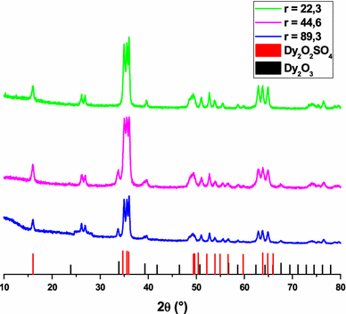
The XRD pattern of precursor (not shown here) did not reveal diffraction peaks. This observation is specific to amorphous or disordered compounds. Figure
1
shows the diffractograms of the precursors with various molar ratio of urea to Ln
3+
after annealing in argon at 800 °C. When the molar ratio
r
is equal to 89.3 [(urea) = 0.5 M], the final products obtained are mainly composed of Ln
2
O
2
SO
4
and a small amount of Ln
2
O
3
. With decreasing of
r
values to 22.3 [(urea) = 0.125 M], the disappearance of Ln
2
O
3
diffraction peaks can be observed, indicating that more OH
-
or CO
3
2−
anions are substituted by SO
4
2−
anions as a result of excess of Ln
2
(SO
4
)
3
dosage during homogeneous precipitation. Thus, the concentration of urea (
r
= 22.3) is chosen as the best condition in the rest of our work.
XRD patterns of product obtained from Dy precursor after annealing at 800 °C during 2 h (5 °C/min) in argonFig. 1
The XRD patterns (Fig.
2
) of the final products of all lanthanides (Ln = Gd, Ho, Dy and Lu) exhibit well-developed peaks, characteristic of lanthanide oxysulfate phases without precipitation of impurities.
XRD patterns of Ln
2
O
2
SO
4
NPs obtained after annealing of precursor in Ar at 800 °C (2 h)Fig. 2

The lattice parameters were calculated and could be indexed to the orthorhombic phase of Ln
2
O
2
SO
4
matched with the standard of the literature values [
22
] and presented in Table
1
.
Lattice parameters of Ln
2
O
2
SO
4
(Ln = Gd, Ho, Dy and Lu) Samples Standardized parameters Calculated parameters a (Ȧ) b (Ȧ) c (Ȧ) a (Ȧ) b (Ȧ) c (Ȧ) Gd2O2SO4 4.062 4.188 12.96 4.0638 4.1791 12.9840 Dy2O2SO4 4.152 4.026 12.76 4.1497 3.9741 12.7508 Ho2O2SO4 4.142 4.012 12.73 4.1451 3.9580 12.7213 Lu2O2SO4 4.108 3.961 12.42 4.1065 3.9571 12.4207Table 1
Figure
3
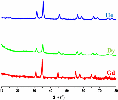
shows XRD patterns of reduction of Ln
2
O
2
SO
4
(Ln = Gd, Dy and Ho) particles in H
2
at 650 °C during 5 h. The patterns show well-developed peaks, characteristic of hexagonal lanthanide oxysulfide phases for Gd, Dy and Ho. All peaks are indexed according the ICDD data in the hexagonal phase, of Gd
2
O
2
S(File card No 020-1422), Dy
2
O
2
S(File card No 026-0592) and of Ho
2
O
2
S(File card No 025-1143), respectively, for Gd, Dy and Ho. No additional peak from impurities can be detected.
XRD patterns of: Ln
2
O
2
S particles obtained after reduction in H
2
at 650 °C during 5 h (5 °C/min)Fig. 3
For Lu, reduction in H 2 gave only lutetium oxide: Lu 2 O 3 (XRD patterns not shown here) even though the reduction temperature was changed to 650, 690, and 800 °C.
Transformation of Ln
2
O
2
SO
4
to Ln
2
O
2
S is commonly classified as a redox reaction and can be described by the equation:
For Lu, the presence of only lutetium oxide: Lu
2
O
3
can be described by the equation:
These results are consistent with those reported by Andreev et al. [ 20 , 21 ] on the transformation of Ln 2 O 2 SO 4 bulk materials obtained by solid-state reaction to Ln 2 O 2 S under H 2 flow.
SEM images corresponding to each Ln
2
O
2
SO
4
before and after reduction are showed in Fig.
4
. Before reduction, NPs are spherical and monodispersed in size with a mean diameter of 500 nm, 3.5 µm, 1.7 µm and 1.2 µm respectively for Gd, Dy, Ho and Lu. After reduction, the morphology of particles is conserved with a reduction in size with a mean diameter of 310 nm, 2.8 µm, 830 nm and 1 µm respectively for Gd, Dy, Ho and Lu. The difference in morphology observed could be explained by a difference in the reactivity of these lanthanides during the precipitation reaction and the effect of the famous “contraction of the lanthanides”. In fact, the particle size is significantly affected by a decrease in the effective ionic radius of the lanthanide ions. Reducing the radius with a simultaneous increase in the nuclear charge leads to an increase in the tendency of the particles to agglomerate. In our SEM pictures observation, two types of particles were identified. The particles sizes differ from nanoscale and monodisperse (Gd) to microscale (Ho, Dy, Lu). This division is not accidental. The synthesized compounds of gadolinium enter into the region of crystal-chemical instability and in their properties are similar to compounds of light rare-earth elements. While the elements from terbium to lutetium are heavy. Within the series of heavy rare earth elements, two families are distinguished: similar terbium (Tb-Tm, Y) and similar ytterbium (Yb-Lu). Thus, the differences in particle morphology are caused by the fact that compounds are formed by rare earth elements being representatives of different families.
SEM images of Ln
2
O
2
SO
4
: before (left) and after (right) reduction in H
2
at 650 °C (5 h)Fig. 4

Figure
5
showed FT-IR analysis spectra of precursor and its annealed product for each lanthanide.
FT-IR spectra of:
a
precusor and
b
Ln
2
O
2
SO
4
particlesFig. 5
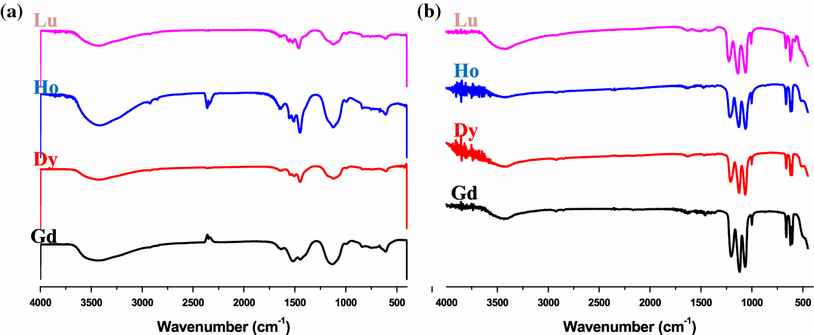
The FT-IR spectrum of the precursor for each lanthanide (Fig. 5 a) shows bands attributed to the absorption peaks of physically absorbed water, crystal water, hydroxyl groups [ 23 ] (near 3500 and 1610 cm −1 ), the CO 3 2− (near 1450 cm −1 ) and the SO 4 2− anions (near 1125 and 680 cm −1 ), which shows that the precursor is consisted of lanthanide, hydroxyl, carbonate and sulfate groups with some crystal water. After calcination under argon at 800 °C (Fig. 5 b), spectrum showed that the peak centered at 3500 cm −1 becomes weak, indicating that the removal of crystal water from the precursor. The SO 4 2− absorption bands (near 1125 and 625 cm −1 ) split into some narrow sharp peaks, the absorption bands become weaker, and a new small absorption peak centered about 500 cm −1 appears, which corresponds to the characteristic vibration peaks of Ln–O bond in the prepared product. These observations are very similar to the ones already discussed in the previous work [ 1 , 17 , 18 ]. We, thus, conclude that the formulation of the NPs elaborated in a water–EtOH (80–20 vol%) mixture is Ln 2 (OH) 2 CO 3 SO 4 ·xH 2 O (Ln = Gd, Dy, Ho and Lu), the lanthanide hydroxycarbonate sulfate. This precursor has been well transformed into Ln 2 O 2 SO 4 (Ln = Gd, Ho, Dy and Lu) after annealing in argon at 800 °C.
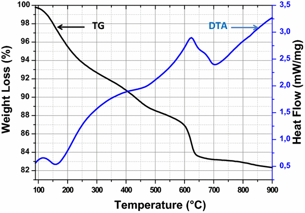
The TG/DTA curves (Fig.
6
) show the thermal decomposition under argon atmosphere of precursor Dy precursor and offer interesting result to complete the determination of its formula. The TG curve presents a continuous weight loss between 80 and 900 °C with an overall weight loss of approximately 17 wt%. This total weight loss mainly consists of the following steps in the whole temperature range. The weight loss in the temperature range from room temperature to ≈ 300 °C is about 7.5% by mass and represents the progressive removal of physically absorbed water from the precursor. This mass loss corresponds to a weak endothermic peak at around 160 °C in DTA curve. The weight loss between 300 and 500 °C is about 4.1% by mass, which is associated with the complete dehydroxylation of the precursor with the progressive decomposition of CO
3
2−
anions and emission CO
2
. This weight loss corresponds to an endothermic peak at 450 °C in the DTA curve. The weight loss in the temperature range from 500 to 693 °C is about 5.4% by mass, which is attributed with the full decomposition of CO
3
2−
anions and release CO
2
of the precursor. Moreover, as shown in Fig.
6
, small variations in mass are observed at temperatures higher than 750 °C on the TG curve. This is why we used 800 °C as the optimal heat treatment temperature for the synthesis of Ln
2
O
2
SO
4
particles. The same thermal decomposition (Curves not shown here) was observed for Gd, Ho and Lu precursor. Thus, the formula of precursor is Ln
2
(OH)
2
CO
3
SO
4
·xH
2
O (Ln = Gd, Dy, Ho and Lu). These results are in a good agreement with those reported previously [
17
,
18
] and with those obtained by FTIR analyses and XRD patterns.
TG-DTA curves of Dy precursor in argonFig. 6
We have proposed an optimized homogeneous precipitation method to synthesize spherical and well-dispersed pure Ln 2 O 2 SO 4 (Ln = Gd, Ho, Dy and Lu) NPs with size between 500 nm and 3.5 µm. If Ln 2 O 2 SO 4 NPs can be easily transformed to Ln 2 O 2 S by reduction under H 2 flow at 650 °C for Gd, Dy and Ho, Lu gave only Lu 2 O 3 under H 2 flow even though the reduction temperature was changed to 650, 690, and 800 °C. This reduction method allows to obtain Ln 2 O 2 S without using H 2 S which is highly toxic and uncomfortable gas. Ln 2 O 2 SO 4 /Ln 2 O 2 S synthesized can be used for release and oxygen storage or doped with luminescent centers for multimodal medical imaging applications.
This research was supported by Tyumen State University (Russia) by offering PostDoc Grant-2017.
Springer Nature remains neutral with regard to jurisdictional claims in published maps and institutional affiliations.