Department of Chemistry, NIT, Agartala, 799046, IN
Abstract
Graphene quantum dots (GQDs) as a new series of nanomaterials have drawn great attention in recent years owning to their unique properties. Here we report the single-step synthesis of GQDs using pyrolysis of citric acid which produced GQDs at different pH. The effect of different pH was studied in detail to optimize the conditions of the formation of GQDs. UV–Visible absorption and normalized fluorescence spectra were applied to analyze the optical properties of GQDs. The particle size distribution of the GQDs in case of varying pH was determined to optimize the synthesis conditions. The surface morphology and microstructures were studied by atomic force microscope (AFM).
Introduction
Emerging graphene quantum dots (GQDs) have received enormous attention because of nanometer-scaled graphene particles with sp
2
–sp
2
carbon bonds. Graphene being one-atom-thick layer of extended sp
2
carbons represents the limit of the thinnest possible 2D conductive surface [
1
,
2
], for which they exhibit very fast electron mobility and high charge carrier density [
3
]. Due to having remarkable properties derived from 2D confinement at the nanoscale caused by the effect of the edges, graphene quantum dots (GQDs) exhibit new properties such as emission and their behavior as spin qubit with collective spin states [
4
,
5
]. Like zero-dimensional carbon-based material, GQDs were planar nanomaterials with lateral dimension ranging from 2 to 20 nm, showing intrinsic luminescence as a result of quantum confinement, surface defects and edge structure. Recent reviews have summarized the unique properties of GQDs [
6
–
13
]. Again, most applications of GQDs have been focused on the photoluminescence (PL)-related fields. Recently, additional excellent properties of GQDs such as high transparency and high surface area have been proposed for energy and display applications [
14
,
15
].
Electrodes of GQDs are applied for capacitors [
16
], batteries [
17
,
18
], where the conductivity of GQDs is higher than that of graphene oxide (GO) [
19
], because of the large surface area of GQDs. GQDs form persistent dispersions in different solvents and may have large biomedical application due to the possibility to cross cellular membranes [
20
]. In addition, GQDs have attracted considerable attention as emerging fluorescent dots for bioimaging, sensing [
21
], pollutant removal [
22
] and even in photovoltaic devices [
15
]. GQDs also hold promise in catalysis due to their large surface area and accessibility of the active sites [
23
].
There are many synthetic methods to prepare GQDs [
24
–
26
], but most of these methods need several steps and post-treatment with surface passivating agents to improve their water solubility and luminescence property. Recently, blue luminescent graphene quantum dots and graphene oxide were reported by tuning the carbonization degree of citric acid. However, during the enlarged syntheses, reproducibility is critically important for the potential technological applications of GQDs. In continuation of our research work [
27
–
29
], in this work we have reported that the pyrolysis of citric acid was carried out which on further addition of sodium hydroxide for maintaining the pH produced GQDs. The effect of different pH on the formation of GQDs was studied in detail.
Experimental
Materials and methods
Citric acid and sodium hydroxide were purchased from E Merck. Double distilled water was used throughout the experiment. The absorption spectra of GQDs obtained in each case was characterized by UV–Vis spectrophotometer (Shimadzu 1800). The steady-state photoluminescence spectra were measured using florescence spectrophotometer (Perkin Elmer LS 55). The particle size distribution analysis was carried out using dynamic light scattering (DLS: Nanotrac wave W3222). FT-IR spectra were recorded with KBr pellets with a Perkin Elmer Spectrum 100 FT-IR spectrometer. The nano-morphology of GQDs was studied by atomic force microscopy (AFM, multimode V8).
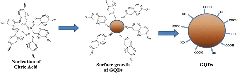
Pyrolysis of citric acid to prepare graphene quantum dots (GQDs)
Five grams of citric acid was heated and melted, which then converted into dark orange color within 25–30 min. 1.5 M solution of NaOH was added dropwise in the melted dense solution of citric acid at room temperature, to prepare the solutions of different pH ranging from 8 to 12. The effect of different pH on the yield and size of the graphene quantum dots (GQDs) was studied in detail. The mechanism of formation of GQDs from the citric acid has been shown in the Scheme
1
.
Scheme 1
Preparation of GQDs from citric acid
Results and discussion
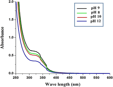
Citric acid when heated at its melting temperature decomposes and the hydronium ion formed from the acid acts as a catalyst in subsequent decomposition reaction stages. The polymerisation and condensation of the products certainly gave rise to soluble polymers. Aromatization and formation of aromatic clusters take place via aldol condensation and cycloaddition. When concentration of aromatic clusters reaches a critical supersaturation point, a burst nucleation takes place and GQDs are formed. GQDs typically showed optical absorption in the UV region with a tail extending to the visible range (Fig.
1
). It may be attributed to some absorption shoulders for the p–p* transition of the C=C bonds, n–p* transition of C=O bonds and/or others. The UV–Vis spectra clearly indicated that on increasing the pH, the absorption peaks becomes broader which suggested the increase of distribution of particle size along with the pH. At pH 9, the absorption peak was found to be more intense and after this pH, the intensity of the peaks decreases up to 12.
Fig. 1
UV–Visible spectra of GQDs at different pH
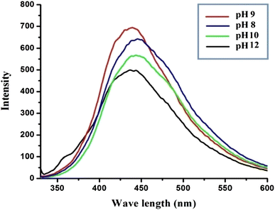
One of the most fascinating features of GQDs, both from fundamental and application-oriented perspectives, is their fluorescence. The requirement for surface passivation is only partially understood, but appears to be linked to the synthetic method. However, more and more cases have emerged with
λex
independent emission position, which may be attributed to their uniform size and surface chemistry. Our experimental results showed that the intensity of the fluorescence decreases with the increase of pH (Fig.
2
).
Fig. 2
Fluorescence curve of GQDs at different pH
Interestingly, in case of fluorescence also, the intensity of the peak was found to be more at pH 9 as compared to other pH.
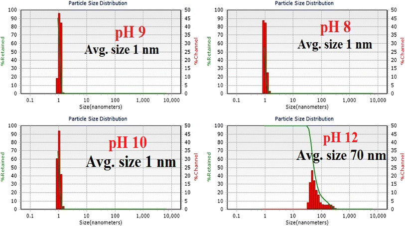
The particle size distribution of the GQDs was carried out at 25 °C using dynamic light scattering (DLS). Figure
3
shows the particle size distribution of the solutions formed by varying the pH of the solution at room temperature. It was found that at pH 8, the average particles size was found to be around 1 nm, and the yield was found to be 85%. In case of pH 9, 95% of the GQDs were found to be around 1.5 nm in size. On further increasing the pH up to 10 of the solution, the yield of the nanoparticles decreased up to 90%, retaining the average particle size of 1 nm only. At pH 12, the yields of GQDs decreased dramatically to 65%. The average particle size was found to be around 70 nm. It may be attributed to the fact that in case of lower pH, the presence of hydroxyl groups hindered the aggregation of the carbon nanoparticles. But, on increasing the pH, the increasing number of hydroxyl groups get adhered to the nanoparticles and hence resulted in the increase of the size of the nanoparticles. Thus, the pH of the solution of citric acid played an important role in the synthesis of GQDs with size below 5 nm.
Fig. 3
Particle size distribution curve of GQDs at different pH
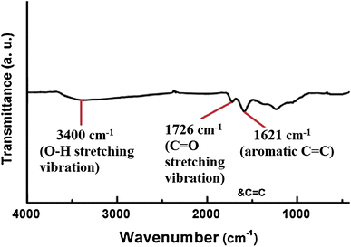
The FT-IR spectra of GQDs showed the broadening of several peaks related to oxygen functional groups, viz. at 3400 cm
−1
(O–H stretching vibration), 1726 cm
−1
(C=O stretching vibration), and 1621 cm
−1
(aromatic C=C, skeletal ring vibrations from the graphitic domain). The intensity of these peaks which were very low in the spectra of GQDs confirmed that the ketone, hydroxyl and epoxide groups have been reduced to form GQDs (Fig.
4
).
Fig. 4
FTIR spectra of GQDs prepared at pH 9
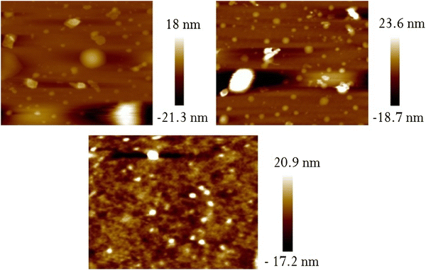
AFM showed the topographic image of well-dispersed GQDs prepared at pH 9 (Fig.
5
). The AFM image confirmed the formation of spherical-shaped GQDs, where the particle sizes were below 2 nm at this pH.
Fig. 5
AFM image of GQDs prepared at pH 9
Conclusions
In this paper, pyrolysis of citric acid was carried out which decomposes and the hydronium ion formed from the acid acts as a catalyst in subsequent decomposition reaction stages. Aromatization and formation of aromatic clusters take place via aldol condensation and cycloaddition which after further addition of sodium hydroxide produced GQDs at different pH. At pH 9, the UV–Vis absorption peak was found to be more intense and after this pH, the intensity of the peaks decreases up to 12. The intensity of the fluorescence was found to decrease with the increase of pH. The particle size distribution of the GQDs, carried out at 25 °C showed that in case of pH 9, 95% of the GQDs was found to be around 1.5 nm in size. On further increasing the pH of the solution up to 10, the yield of the nanoparticles decreased up to 90%, retaining the average particle size of 1 nm only. At pH 12, the yields of GQDs decreased dramatically to 65%. The AFM image confirmed the formation of spherical-shaped GQDs, where the particle sizes were below 2 nm at this pH. Thus, the pH plays an important role for the formation of GQDs from citric acid.
Acknowledgements
We acknowledge the CRF, NIT, Agartala for providing AFM images.
References
Geim and Novoselov (2007) The rise of graphene (pp. 183-191) 10.1038/nmat1849
Novoselov et al. (2004) Electric field effect in atomically thin carbon films (pp. 666-669) 10.1126/science.1102896
Bolotin et al. (2008) Ultrahigh electron mobility in suspended graphene (pp. 351-355) 10.1016/j.ssc.2008.02.024
Ponomarenko et al. (2008) Chaotic Dirac billiard in graphene quantum dots (pp. 356-358) 10.1126/science.1154663
Trauzettel et al. (2007) Spin qubits in graphene quantum dots (pp. 192-196) 10.1038/nphys544
Ahmed et al. (2016) Graphene Quantum Sheets with Multiband Emission: Unravelling the Molecular Origin of Graphene Quantum Dots 120(52) (pp. 21678-21684)
Zhixing et al. (2016) Photon Reabsorption and Nonradiative Energy-Transfer-Induced Quenching of Blue Photoluminescence from Aggregated Graphene Quantum Dots 120(51) (pp. 29432-29438) 10.1021/acs.jpcc.6b10704
Cao et al. (2013) Photoluminescence properties of graphene versus other carbon nanomaterials 46(1) (pp. 171-180) 10.1021/ar300128j
Jiang (2011) Chemical preparation of graphene-based nanomaterials and their applications in chemical and biological sensors 7(17) (pp. 2413-2427) 10.1002/smll.201190064
Rozhkov et al. (2011) Electronic properties of mesoscopic graphene structures: charge confinement and control of spin and charge transport (pp. 77-114) 10.1016/j.physrep.2011.02.002
Sheng et al. (2012) Electronic and optical properties of semiconductor and graphene quantum dots 7(3) (pp. 328-352) 10.1007/s11467-011-0200-5
Zhang et al. (2012) Graphene quantum dots: an emerging material for energy-related applications and beyond (pp. 8869-8890) 10.1039/c2ee22982j
Shen et al. (2012) Graphene quantum dots: emergent nanolights for bioimaging, sensors, catalysis and photovoltaic devices (pp. 3686-3699) 10.1039/c2cc00110a
Liu et al. (2013) Superior micro-supercapacitors based on graphene quantum dots (pp. 4111-4122) 10.1002/adfm.201203771
Chao et al. (2014) Graphene quantum dots coated VO2 arrays for highly durable electrodes for Li and Na ion batteries (pp. 565-573) 10.1021/nl504038s
Pan et al. (2013) Electrophoretic fabrication of highly robust, efficient, and benign heterojunction photoelectrocatalysts based on graphene-quantum-dot sensitized TiO2 nanotube arrays (pp. 3551-3555) 10.1039/c3ta00059a
Ding et al. (2015) Few-layered graphene quantum dots as efficient hole-extraction layer for high-performance polymer solar cells (pp. 186-192) 10.1016/j.nanoen.2015.04.019
Harhaji-Trajkovic et al. (2012) Graphene quantum dots as autophagy-inducing photodynamic agents (pp. 7084-7092) 10.1016/j.biomaterials.2012.06.060
Lu et al. (2012) Economical, green synthesis of fluorescent carbon nanoparticles and their use as probes for sensitive and selective detection of mercury (II) Ions (pp. 5351-5357) 10.1021/ac3007939
Liu et al. (2012) A novel acid-driven, microwave-assisted, one-pot strategy toward rapid production of graphitic N-doped carbon nanoparticles-decorated carbon flakes from N,N-dimethylformamide and their application in removal of dye from water (pp. 4632-4635) 10.1039/c2ra20381b
Liu et al. (2012) A general strategy for the production of photoluminescent carbon nitride dots from organic amines and their application as novel peroxidase-like catalysts for colorimetric detection of H2O2 and glucose (pp. 411-413) 10.1039/C1RA00709B
Fan et al. (2015) Controllable size-selective method to prepare graphene quantum dots from graphene oxide (pp. 55-63) 10.1186/s11671-015-0783-9
Yin et al. (2014) An approach to controlling the fluorescence of graphene quantum dots: from surface oxidation to fluorescent mechanism (pp. 128103-128107) 10.1088/1674-1056/23/12/128103
Dong et al. (2015) Improved solvothermal method for cutting graphene oxide into graphene quantum dots (pp. 855-864)
Sutradhar and Saha (2016) Silver Nanoparticles: Synthesis and Its Nanocomposites for Heterojunction Polymer Solar Cells (pp. 8941-8949) 10.1021/acs.jpcc.6b00075
Sutradhar and Saha (2016) Size-controlled synthesis of silver nanoparticles using Zizyphus mauritiana fruit extract (pp. 47-55) 10.3233/MGC-150182
Sutradhar and Saha (2015) Green synthesis of zinc oxide nanoparticles using tomato (Lycopersicon esculentum) extract and its photovoltaic application (pp. 314-327)