Results and discussion
Preparation of magnetite chlorochromate nanomaterials
The sequence of steps in the immobilization of chlorochromate in the magnetite nanoparticles is illustrated in Fig.
1
. In the first step, hydrothermal method was applied to the synthesis of Fe
3
O
4
magnetic nanoparticles. Second, the external surface of MNPs was coated with silica shell to create SCMNPs. This silica coating protects the magnetic nanoparticles from possible decomposition induced by the surrounding environment and prevents further aggregation. In addition, the silica surface can be functionalized using a variety of known surface modifiers. The action of silanol groups of SCMNPs with triphenylphosphonium (3-chloropropyl) trimethoxysilane generated PCPTSCMNPs. The next step involves reaction of PCPTSCMNPs groups with chlorochromate to yield the magnetite chlorochromate nanomaterials CCr-PCPTSCMNPs. In this step, phosphonium groups give positively charged cations which bounds electrostatically to the chlorochromate. The excess of chlorochromate was removed by washing the resulted materials with water. The attached chlorochromate anions are stable on the surface of modified magnetite and cannot be leached to an aqueous solution [
9
].
Schematic illustration for the preparation of Fe
3
O
4
@SiO
2
@PPh
3
@[CrO
3
Cl]Fig. 1
Characterization of the prepared magnetite–chlorochromate nanomaterials
To demonstrate the surface modification of the magnetite nanoparticles and preparation of magnetite–chlorochromate nanomaterials, the FT-IR spectra of the ready MNPS, SCMNPs, PCPTSCMNPs, and CCr-PCPTSCMNPs are illustrated in Fig.
2
.
FT-IR spectra of
a
Fe
3
O
4
,
b
Fe
3
O
4
@SiO
2
,
c
Fe
3
O
4
@SiO
2
@PPh
3
and
d
Fe
3
O
4
@SiO
2
@PPh
3
@[CrO
3
Cl]Fig. 2
As shown in Fig. 2 a, Fe 3 O 4 typical bands centered at 586 and 640 cm −1 , which related to the Fe–O stretches. The surfaces of Fe 3 O 4 powder contain surface hydroxyl functional groups (Fe–OH) whose characteristic IR peak was at 3,500 cm −1 in FT-IR plot shown in Fig. 2 a [ 12 – 14 , 20 , 21 ]. After the coating of Fe 3 O 4 @SiO 2 core–shells, the silane framework on the surface of the MNPs was confirmed by the FT-IR bands at 1,105, 1,039 and 994 cm −1 assigned to the Si–O–H and Si–O–Si groups [ 18 ] (Fig. 2 b). These results indicate that Fe 3 O 4 @SiO 2 is formed [ 12 , 15 , 16 ]. The presence of anchored propyl and triphenylphosphine groups was confirmed by stretching vibrations appeared at about 2,935, 2,870 cm −1 (C–H stretching vibrations) in the FT-IR spectrum of PCPTSCMNPs (Fig. 2 c) [ 9 , 20 ]. The presence of absorption bands 430, 770, 805, 895, 905 (ν asymmetric Cr=O (A 1 )), 940 (ν symmetric Cr=O (E)) approbated the immobilization of chlorochromate (Fig. 2 d) [ 1 ].
The elemental analysis (XRF) of the Fe
3
O
4
@SiO
2
@PPh
3
@[CrO
3
Cl] is shown in Table
1
. As can be seen in this Table, the quantity of Chromium is approximately 1.04 %, which confirms the immobilization amount of [CrO
3
Cl]
−
which is equal to 0.02 mol/100 g. Loss on ignition (LOI) shows the quantity of organic matter in Fe
3
O
4
@SiO
2
@PPh
3
@[CrO
3
Cl].
Results of chemical analysis of XRF of the Fe
3
O
4
@SiO
2
@PPh
3
@[CrO
3
Cl] Element Fe Si O Cr P LOI (%) 24.46 27.93 33.84 1.04 1.17 11.56Table 1
Figure
3
shows the XRD spectrum of the magnetite chlorochromate nanoparticles. The XRD data show the diffraction peaks at 2
θ
= 29.9765° (220), 35.1295° (311), 42.8283° (400), 56.9694° (511) and 62.56522° (440) for sample. The reflection peak positions and relative intensities of MNPs are well in agreement with the XRD patterns of MNPs in the literature (JCPDS Card No. 19-0629). The results indicate that the shape of the MNPs is inverse spinel Fe
3
O
4
with a face-centered cubic (FCC) structure, suggesting that the sample has a cubic crystal system. Moreover, no characteristic peaks of impurities were observed in the XRD spectrum [
20
,
22
–
24
]. This indicates that chlorochromate species well dispersed on the surface of PCPTSCMNPs and there is no crystalline phase of chlorochromate in the resulted nanomaterials. The D values calculated from the XRD spectrum were indexed to the inverse cubic spinel phase of Fe
3
O
4
. From the width of the peak at 2
θ
= 35.1295° (311), the crystallite size of the Fe
3
O
4
@SiO
2
@PPh
3
@[CrO
3
Cl] nanoparticle is calculated to be 16.3 nm using Scherrer’s equation:
D
= 0.89
λ
/(
β
cos
θ
) where
D
is the particle size,
λ
is the X-ray wavelength (nm),
θ
is Bragg’s angle;
β
is the excess line broadening (radiant) [
4
,
25
].
XRD pattern of Fe
3
O
4
@SiO
2
@PPh
3
@[CrO
3
Cl]Fig. 3
Figure
4
shows the absorption spectra of samples in the solid state. It is clear that the pure Fe
3
O
4
absorbs light with wavelengths mainly below 762 nm (Fig.
4
a). (In comparison with the optical absorption of Fe
3
O
4
, the absorption edge toward shorter wavelengths (blue shift) and increasing intensity in <502 nm was observed in the absorption spectra of Fe
3
O
4
@SiO
2
(Fig.
4
b). Fe
3
O
4
@SiO
2
and Fe
3
O
4
@SiO
2
with triphenylphosphine showed a red shift and decreasing in intensity (Fig.
4
c). The Fe
3
O
4
@SiO
2
@PPh
3
@[CrO
3
Cl] showed a little blue shift with growth intensity (Fig.
4
d).
Solid state UV–Vis spectra of
a
Fe
3
O
4
,
b
Fe
3
O
4
@SiO
2
,
c
Fe
3
O
4
@SiO
2
@PPh
3
and
d
Fe
3
O
4
@SiO
2
@PPh
3
@[CrO
3
Cl]Fig. 4
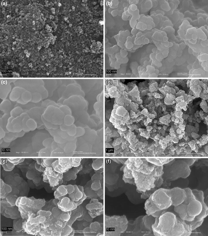
Figure
5
a shows the SEM images of the primary Fe
3
O
4
. The SEM images illustrate that the created Fe
3
O
4
are spherical nanoparticles with an average grain size 16 nm. Aggregation gives rise to enhancing the size of observed nanoparticles as observed in the SEM image. Figure
4
b, c shows that iron oxide nanoparticles have core–shell mode and a spherical morphology. Figure
5
d–f indicates images of obtaining Fe
3
O
4
@SiO
2
@PPh
3
@ [CrO
3
Cl].
Images of SEMs
a
Fe
3
O
4
,
b
,
c
Fe
3
O
4
@SiO
2
,
d
,
e
,
f
Fe
3
O
4
@SiO
2
@PPh
3
@[CrO
3
Cl]Fig. 5
The TEM images shown in Fig.
6
demonstrated that the silica–magnetite compound made as supports of the catalyst had good spherical morphologies.
Magnetization curves of
a
Fe
3
O
4
,
b
Fe
3
O
4
@SiO
2
@PPh
3
@[CrO
3
Cl]Fig. 6
The magnetization measurements of Fe
3
O
4
, Fe
3
O
4
@SiO
2,
and Fe
3
O
4
@SiO
2
@PPh
3
@[CrO
3
Cl] nanoparticles were performed by a vibrating sample magnetometer at room temperature (Fig.
7
). Figure
7
indicates the schemes of the magnetization ‘M’ versus applied field ‘H’ (between −10,000 and +10,000 Oe) of these prepared Fe
3
O
4
, Fe
3
O
4
@SiO
2
, and Fe
3
O
4
@SiO
2
@PPh
3
@[CrO
3
Cl] samples. As can be seen in Fig.
7
, the values of the saturation magnetization were 53 emu/g for Fe
3
O
4
nanoparticles, 37 emu/g for Fe
3
O
4
@SiO
2
, and 25.4 emu/g for Fe
3
O
4
@SiO
2
@PPh
3
@[CrO
3
Cl], respectively. The reduction, nearly 25 emu/g of the saturation magnetization, proposes the presence of some SiO
2
@PPh
3
@[CrO
3
Cl] on the surface of the magnetic supports.
Applied magnetic field (Oe)Fig. 7