Results and discussion
The concept of this research is the combination of two different families of nanomaterials, 2D polymeric nanomembranes and 1D metallic NWs, aiming to incorporate the best properties of each nanomaterial into a new flexible, conformable and electrically conductive nanostructured ultrathin material as a basis for the development of a biointegrated electrochemical H
2
O
2
sensor. The flexible and free-standing 2D polymeric nanomembranes consisted on alternated layers of PLA and PEDOT:PSS. While the PLA layers conferred mechanical stability and contained the nanoperforations to host the nanostructures, PEDOT layers were used for their electrical properties. The PLA/PEDOT multilayered nanomembranes were alternatively deposited using spin coating for PLA layer and electropolymerization for PEDOT one. Figure
1
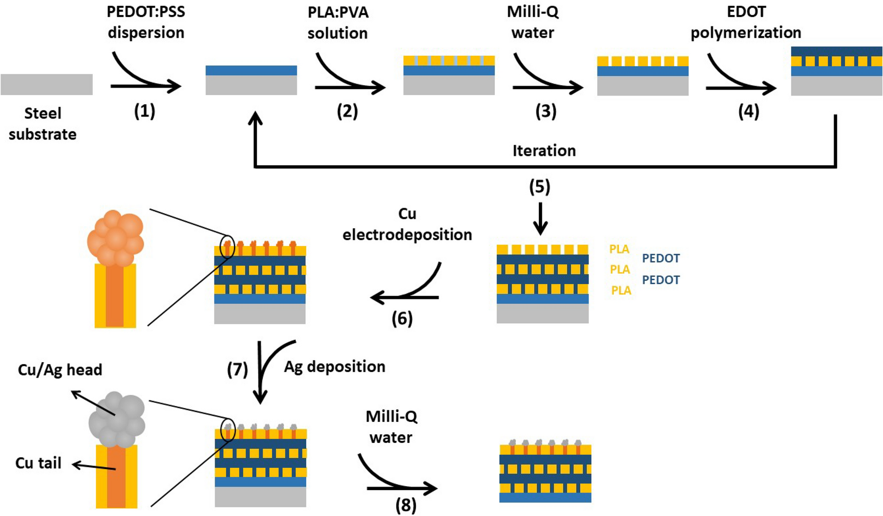
shows a scheme with the different steps followed to obtain the self-supported PLA/PEDOT nanomembranes. First, a sacrificial PEDOT layer was spin coated onto the steel substrate using the PEDOT:PSS aqueous dispersion (1.3 wt%). This sacrificial layer will be removed by immersing it in water since the non-interacting PEDOT:PSS particles will be redispersed, thus allowing peeling the nanomembrane off the substrate. Second, a nanoperforated PLA layer was obtained by spin coating using a PLA/PVA 90:10 v/v solution following the selective PVA etching in water creating the nanoperforations. After that, chronoamperometry was used to obtain a continuous PEDOT coating onto the previous PLA layer. EDOT monomers were polymerized at a constant potential within the nanoperforations that coalesced after emerging the pores forming the PEDOT layer. Unlike PEDOT sacrificial layer, the PEDOT layer obtained by electropolymerization will remain stable even after immersing in water since it is a continuous and covalently linked layer. Steps 2–4 were repeated to finally obtain nanomembranes with five different layers: PLA/PEDOT/PLA/PEDOT/PLA (5-PLA/PEDOT) (Fig. S1). The top PLA layer was used to host the 1D metallic NWs within the nanometric-size pores, aiming to improve their functionality. Thus, copper was incorporated in the pores using electrodeposition (step 6) since it is a very versatile and easy technique for NMs preparation [
30
,
31
] followed by the formation of a silver layer onto Cu nanostructures through a simple redox reaction (step 7). The incorporation of these 1D NMs is expected to improve the sensibility of the biosensors not only due to the higher electrical conductivity of metals (e.g., Cu, Cu/Ag) but also to the higher surface area of the nanostructures. Moreover, the 1D NWs will be oriented and non-aggregated within the membrane, enhancing even more the surface area and therefore the biosensor response (unlike composites where NMs are randomly oriented and aggregated within the matrix).
Schematic illustration showing the fabrication procedure of the 5-PLA/PEDOT, 5-PLA/PEDOT-Cu and 5-PLA/PEDOT-Cu/Ag free-standing nanomembranes. (1) Deposition of the sacrificial PEDOT:PSS layer by spin coating a PEDOT-PSS dispersion. (2) Deposition of a PLA:PVA solution to get a nanosegregated layer. (3) Selective removal of PVA by adding milli-Q water. (4) Growth of a PEDOT layer by EDOT chronoamperometry. (5) The process is repeated until obtaining the PLA/PEDOT/PLA/PEDOT/PLA multilayered nanomembrane (5-PLA/PEDOT). (6) Growth of the 1D cauliflower-shape nanostructures by electrodeposition. (7) Coating Cu nanostructures with a thin Ag layer by a transmetallation reaction. (8) Peeling the membrane off the steel substrate in water to remove the first PEDOT:PSS sacrificial layerFig. 1
Figure
2
shows an image of the fabricated 5-PLA/PEDOT nanomembranes as well as SEM micrographs. As it can be observed, the nanomembranes, with the typical blue color of PEDOT, are continuous, homogenous and self-standing. SEM micrographs also show that the membranes are continuous, homogeneous and flat with perforations homogeneously distributed. The perforations are nanometric (76 ± 40 nm) and the estimated pore density is 1.6 × 10
7
pores cm
−2
. The thickness of the individual layers and the 5-PLA/PEDOT nanomembrane was studied by profilometry. While the individual PLA and PEDOT layer thicknesses were 158 ± 22 nm and 178 ± 24 nm, respectively, the 5-PLA/PEDOT nanomembrane was 731 ± 32 nm thick. The total thickness is lower than the arithmetic sum of the individual layers because of the compression of the lower layers due to the upper ones. On the other hand, the FTIR analysis of the 5-PLA/PEDOT nanomembranes reveal the presence of the main peaks of PLA [1750 cm
−1
(C=O) stretching vibration, 1180 cm
−1
asymmetric C–O stretching, 1085 cm
−1
symmetric C–O stretching] and PEDOT (1527 cm
−1
C=C stretching, 1230 and 1040 cm
−1
C–O–C stretching, 845 cm
−1
C–S stretching) but a bit shifted with respect their positions in the raw PLA and PEDOT:PSS. This shift is indicative of some weak Van der Waals interactions at the interface between the PLA and PEDOT layers (Fig. S2).
A
Optical and
B
,
C
SEM images of the 5-PLA/PEDOT nanomembranesFig. 2

After that, the 5-PLA/PEDOT nanomembranes were modified with the metallic NMs to improve their functionality. First, Cu NWs were first grown by electrodeposition into the perforations aiming to improve electrical conductivity of the nanomembrane. And second, Ag was coated onto previous Cu nanostructures by a transmetallation reaction. Silver confers electrocatalytic properties towards H
2
O
2
, biocompatibility and corrosion resistance as well as high faradaic current density, fast electron transfer rate and high signal-to-noise ratio while keeping the high electrical conductivity [
32
,
33
]. Herein, the nanomembranes are denoted 5-PLA/PEDOT-Cu and 5-PLA/PEDOT-Cu/Ag, respectively. Electrodeposition of Cu was performed by chronopotentiometry at different conditions of current density and time (Figure S3). The SEM analysis revealed that at low current densities no growth of Cu outside the membrane was observed. However, for an applied current density of − 1.4 mA cm
−2
during 40 s, we obtained nanostructures with a cauliflower-shape morphology with nanoparticles (NPs) as the “head” and NWs as the “tail”. Such nanostructures result from the complete filling of the pores and moderate growth outside them (Fig.
3
A, B). For higher times or current densities, these nanostructures grew in size and even coalesced leading to a continuous layer (Figure S4). These conditions are avoided since a nanostructured surface is preferred over a continuous coating to maximize surface area and, therefore, have a higher response towards H
2
O
2
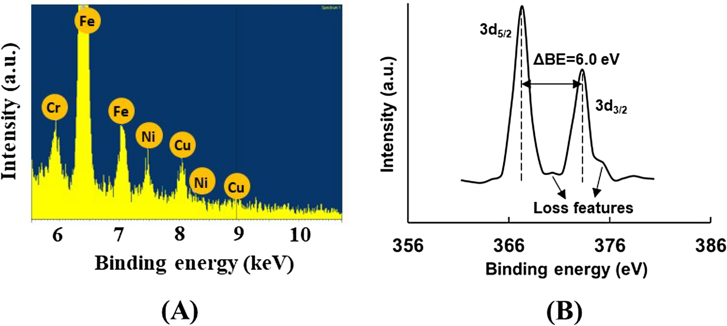
. The presence of Cu is clearly detected by EDX spectroscopy (Fig.
4
A). Other elements (e.g., Fe, Ni, Cr) were also detected coming from the AISI 304 steel substrate used to prepare the nanomembranes. Finally, Ag was coated onto the Cu nanostructures by covering the 5-PLA/PEDOT-Cu nanomembrane’s surface with a 20 mM AgNO
3
solution for 90 s. During this time, the transmetallation reaction between metallic Cu and Ag
+
ions took place leading to an Ag metallic coating onto the Cu nanostructures (Cu + 2Ag
+
→ Cu
2+
+ 2Ag). As we observed by SEM, the presence of the Ag layer did not modify the cauliflower shape of Cu that was probably attributed to the nanometric thickness of the silver layer. XPS analysis confirmed the presence of Ag layer (Fig.
4
B). XPS spectrum shows two peaks at binding energies (BE) of 367 and 373 nm attributed to the spin–orbit components 3
d
5/2
and 3
d
3/2
of Ag, respectively. On the other hand, a calculated full width half maximum (FWHM) of 1.4 eV is in accordance with the reported values for metallic Ag (FWHM = 1.2 eV). Moreover, the following characteristics also corresponds to Ag
0
: (1) the energy distance (ΔBE) between the two peaks is 6.0 eV, (2) the peaks are asymmetric and (3) the presence of loss features observed at the higher BE side of each peak [
34
,
35
].
A
,
B
SEM images of the 5-PLA/PEDOT-Cu nanomembranes after copper electrodeposition at − 1.4 mA cm
−2
during 40 s at different magnifications A
EDX spectrum of the 5-PLA/PEDOT-Cu nanomembrane.
B
XPS spectrum of the 5-PLA/PEDOT-Cu/Ag nanomembraneFig. 3
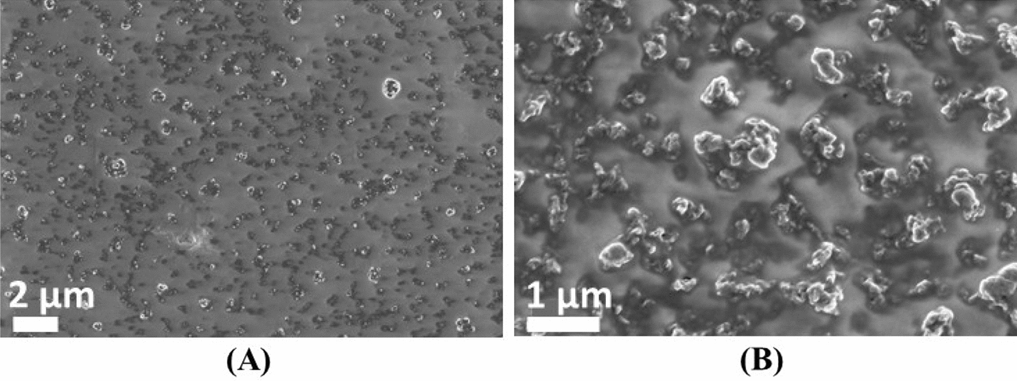
Fig. 4
The membranes were easily peel off from the steel substrate by dipping them in water due to the easy redispersion of the initial PEDOT:PSS sacrificial layer. The free nanomembranes are (1) self-supportive as they keep intact after releasing them from the substrate; (2) flexible/foldable, and (3) conformable on many surfaces. Nanomembrane’s robustness and flexibility can be observed since they can be picked up by a pipette, stored and released without damaging (Videos S1). Moreover, according to the Euler–Bernoulli theory, flexibility is dependent on the inverse of thickness to the third power, meaning that materials are more prone to bent when thickness decreases [ 13 ]. In this sense, we have checked that the fabricated nanomembranes can be bent 180° for a high number of times (> 100) without experience any damage confirming their high flexibility. The bending capacity of the membranes is also observed as they completely adapt onto a curved pipette tip (Video S2) with a bending angle of 180°. Regarding conformability, nanomembranes completely adapt to substrates with different shapes faithfully reproducing their surfaces like a rigid glass slide (planar, rigid substrate), a flexible polyethylene terephthalate (PET) film (planar, flexible substrate), a plastic pipette tip (flexible, rounded substrate), a lemon or a hairy kiwi (curvilinear, rough substrates) or a dynamic finger (curvilinear, rough substrate) (Figure S5, Videos S1–S3). Again, such conformal contact was attributed to the low membrane thickness [ 14 ]. It is also worthy to mention that the nanomembranes were totally stable onto those surfaces for long periods of times without experiencing any damage. Finally, the nanomembranes recovered their planarity after immersing them back in water. Although this means that they do not adhere onto wet surfaces, this is not a drawback since the main applications of the membranes will be onto non-wetted surfaces (e.g., adapted onto skin). A future work would be making these membranes adhesive even onto wet substrates by adding, for example, polydopamine during membrane fabrication.
With these Cu/Ag-modified nanomembranes, we developed a non-enzymatic electrochemical H
2
O
2
biosensor based on the catalytic properties of Ag towards this molecule. The development of easy, fast, flexible and conformable sensors for H
2
O
2
is increasing because this molecule has a key role in many fields (e.g., textile, environmental, food, pharmaceutical) but especially for health monitoring due to its participation in many biological processes like diabetes, cancer, neurodegeneration or aging [
36
]. Although great progress has been made in recent years, so far most of the reported H
2
O
2
electrochemical sensors are in rigid form, which limits their wide application in wearable electronics. First, the electrochemical performance of the modified (5-PLA/PEDOT-Cu, 5-PLA/PEDOT-Cu/Ag) and non-modified (5-PLA/PEDOT) nanomembranes was assessed by cyclic voltammetry in the absence of H
2
O
2
(Fig.
5
A). The current density increases in the following order 5-PLA/PEDOT < 5-PLA/PEDOT-Cu < 5-PLA/PEDOT-Cu/Ag due to the good electrical conductivity of Cu (
σ
= 5.96·10
7
S m
−1
) and the better electrical conductivity of Ag (
σ
= 6.30·10
7
S m
−1
). Later, EIS analyses were conducted on the same samples to assess the influence of Cu and Ag on the electrochemical properties of the nanomembranes. The ion transport was evaluated by measuring the impedance for frequencies ranging from 10
5
to 10
–1
Hz in a 0.5 M NaCl solution. Nyquist plots shows the imaginary impedance, indicative of the capacitive/inductive behaviour, against the real impedance (−
Z
″ vs
Z
′) (Fig.
5
B). In a Nyquist plot, the semi-circular response is attributed to the electron transfer resistance at the higher frequency values (bulk resistance,
R
p
), which determines the electron transfer kinetics of the redox probe at the electrode–electrolyte interface. The incorporation of Cu nanostructures has substantially changed the electrochemical response of the nanomembrane compared to the bare one. As can be observed, a semicircle with a large diameter is only observed for 5-PLA/PEDOT, which is associated with a very large
R
p
(90.5 MΩ) assuming a simplified Randles-like equivalent circuit (Figure S6) in which a CPE (constant phase element) element is used. Meanwhile, the 5-PLA/PEDOT-Cu sample exhibits a semicircle with a much smaller diameter that accounts for a lower
R
p
(683 kΩ) upon adjustment to a Randles-like circuit (Figure S6) indicating the successful incorporation of Cu into the membrane nanoperforations. In the same line, the EIS spectrum of 5-PLA/PEDOT-Cu/Ag nanomembrane shows a semicircle with an even smaller diameter than 5-PLA/PEDOT-Cu leading to the lowest
R
p
(39.7 kΩ), assuming a Randles-like circuit (Figure S6), among the different nanomembranes. These results are in accordance with the CV and clearly show the improvement in the electrical conductivity of the nanomembranes after Cu incorporation and even higher conductivity after its coating with Ag. Figure
5
C shows a magnification of the left part of the Nyquist plot in Fig.
5
B. The pseudo-capacitive behaviour that is noticed in the samples through the presence of the capacitive spike is mainly attributed to the PEDOT/PLA layers. The decreasing slope of the capacitive tail for the 5-PLA/PEDOT-Cu and even more for the 5-PLA/PEDOT-Cu/Ag is due to the presence of Cu or Cu/Ag metals, confirming again a nanomembrane resistance drop.
A
Cyclic voltammetries,
B
Niquist plots and
C
magnified Niquist plots for the 5-PLA/PEDOT, 5-PLA/PEDOT-Cu and 5-PLA/PEDOT-Cu/Ag nanomembranes.
D
Water contact angle of 5-PLA/PEDOT and 5-PLA/PEDOT-Cu/Ag nanomembranesFig. 5

The contact angles ( θ ) measured in water (Fig. 5 D) also provided indirect evidence of the successful modification of 5-PLA/PEDOT with the metallic nanostructures. The wettability of 5-PLA/PEDOT significantly increased upon the incorporation of Cu/Ag as the water contact angle decreased from 70.1° ± 2.9° for 5-PLA/PEDOT to 36.5° ± 2.3° for 5-PLA/PEDOT-Cu/Ag. The increased wettability is due to the metallic nanostructures, characterized by high surface energy which favors hydrophilic behavior and lead water droplets to immediately spread on the surface [ 37 ].
Based on previous results and since the 5-PLA/PEDOT-Cu/Ag nanomembrane showed better properties (e.g., higher current density, lower charge transfer resistance and higher wettability) than the other samples, we evaluated its performance as electrode for H
2
O
2
sensing. Different H
2
O
2
concentrations were added to build the calibration plot and determine the sensitivity as well as the limit of detection (LOD) of the electrochemical sensor. Figure
6
A shows the CVs recorded in different solutions containing H
2
O
2
concentrations in the range 0.35–10 mM. As it can be observed, the current density at negative potentials increases after each addition due to the catalytic reduction of H
2
O
2
due to the presence of silver. From the previous data, the current density-H
2
O
2
concentration calibration plot was obtained showing a linear dependence between the current density and the concentration up to 10 mM (Fig.
6
B). In this range, the precision is very high, as reflected in the R-squared value provided by the linear fit (
R
2
= 0.9971). A good sensitivity of 120.3 ± 8.6 µA cm
−2
mM
−1
was obtained from the absolute value of the slope of the calibration line and a low LOD (7.0 ± 0.4 µM) was calculated as the ratio between three times the standard deviation of the blank solution (without addition H
2
O
2
) and the absolute value of the slope of the calibration curve [
38
]. The low standard deviations for the sensitivity and LOD indicate the good repeatability of the nanomembranes for H
2
O
2
sensing. Table
1
shows a comparison of the performance of the nanomembrane developed in this work with other similar flexible non-enzymatic H
2
O
2
sensors in the literature (Table
1
). Similar sensitivity, linear range and LOD can be observed when compared with reported sensors based only on Ag NMs like our study. For example, Lee et al. developed Ag NW-based films by vacuum filtration. The films, formed by a homogenous and non-oriented Ag NWs network, displayed a LOD of 46 μM [
39
]. Hsiao et al. prepared urchin-like Ag NWs based electrodes by screen printing showing a detection limit of 10 μM [
40
]. Or Kurowska and coworkers fabricated an AgNW array by electrodeposition using an aluminum oxide (AAO) as template to grow the silver nanostructures. After AAO removal, the array showed a LOD of 29.2 μM [
41
]. Thus, the LOD of our 5-PLA/PEDOT-Cu/Ag nanomembranes is much better than the previously reported Ag NWs-based sensors which we attribute either to the random orientation of the NWs [
39
,
40
] or some agglomeration after template removal [
41
]. If we compare with Ag-based composite systems (e.g., Graphene/Ag NPs) [
42
,
43
] or with other materials (e.g., MnO
2
, PtTe
2
) [
29
,
44
,
45
–
46
], the LOD value found in this work is similar or slightly higher than those. For example, the work of Aparicio-Martinez and coauthors showed a LOD of 7.9 μM for a graphene electrode decorated with Ag NPs using laser irradiation [
42
]. Or Gholami and Koivisto quantified a LOD of 0.48 µM for carbon fibers coated with Ag NPs and embedded into nafion [
43
]. Lower LODs have been achieved with electrodes based on MnO
2
(e.g., MnO
2
/Graphene nanosheets) (LOD = 0.19 µM) [
29
] or PtTe
2
(LOD = 1.2 µM) [
46
].
A
Cyclic voltammetries recorded at different H
2
O
2
concentrations (from 0 to 10 mM) using 5-PLA/PEDOT-Cu/Ag nanomembrane as working electrode. Scan rate: 200 mV s
−1
.
B
Calibration plot obtained from graph (
A
).
C
Chronoamperometry curve to evaluate the interference properties of the 5-PLA/PEDOT-Cu/Ag by adding glucose, ascorbic acid and citric acid Performance of different non-enzymatic H
2
O
2
sensors found in the literature Electrode materials Sensitivity Linear range (mM) LOD (mM) References Ag NWs/PET 749 µA mM−1 cm−2 0.2–1.5 46 [ 1.7–3.4 Ag NWs/carbon electrode 4705 µA mM−1 mg−1 cm−2 0.05–10.35 10 [ Ag NWs array/Cu 26.6 μA mM−1 cm−2 0.1–3.1 29.2 [ Ag NPs/graphene 32 µA mM−1 0.1–10 7.9 [ Ag NPs/carbon microfibres/nafion –- 0.1–80 0.48 [ Ag/MnO2/MWCNT/GCE 82.5 μA mM−1 cm−2 0.005–10.4 1.7 [ MnO2/graphite nanofibres 1096 μA mM−1 cm−2 0.1–11 1.25 [ MnO2/graphene nanosheets/GCE 422.1 μA mM−1 cm−2 0.0005–3.5 0.19 [ PtTe2/GCE 10.9 µA mM−1 0.003–0.3 1.2 [ 0.5–1.1Fig. 6
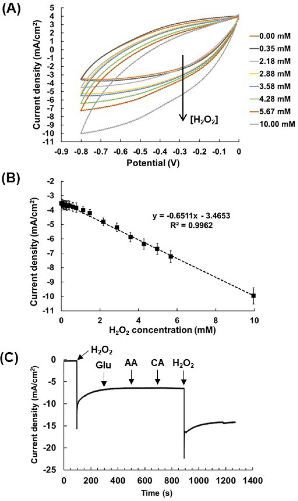
Table 1
Nowadays, there is a controversy about the normal H 2 O 2 levels found in body fluids for healthy people. While some studies report that those levels in plasma should be around 10 µM, other works report values higher than 20 µM [ 47 ]. Regarding other body fluids, concentrations of hydrogen peroxide of 100 µM or higher have been founded in freshly voided human urine and vitreous humor of healthy persons [ 48 ]. However, what it seems clear is that H 2 O 2 concentrations higher than 20 mM triggers inflammatory responses and values higher than 50 µM are cytotoxic to a wide range of cultured animal, plant and bacterial cells [ 48 ]. Moreover, it has been found that levels in the range 30–50 µM are present in certain diseases or during chronic inflammation [ 47 ]. Despite the disparity of values, we believe that the LOD reported in this work for the fabricated nanomembranes allows detecting H 2 O 2 concentrations for healthy and sick people. At this point, we would like to comment that different strategies could be followed to improve the LOD of the nanomembranes like (1) increasing the surface area by increasing pore density in the membrane, reducing pore size or conferring mesoporosity to the Cu nanostructures during electrodeposition, and (2) decorating Cu/Ag nanostructures with transition metal oxides or dichalcogenides as they seem having a better response towards H 2 O 2 .
Finally, the stability and interference properties of the 5-PLA/PEDOT-Cu/Ag nanomembranes were evaluated. CV was employed to quantify the stability of the samples 2 months after their preparation; a 5 mM H 2 O 2 solution showed a 95.5 ± 1.3% retention of the initial current density. The good stability results can be attributed to the noble character of silver as a sensing material. Finally, the anti-interference experiments were performed by chronoamperometry (Fig. 6 C). No significant current changes were observed after adding some common interfering substances like glucose (Glu), ascorbic acid (AA) or citric acid (CA) indicating the good selectivity of the nanomembranes towards H 2 O 2 . Thus, we have demonstrated that 5-PLA/PEDOT-Cu/Ag nanomembranes can be successfully used as electrodes with flexibility and conformability over different substrates with good electrochemical performance (e.g., sensitivity, LOD, linear range) as well as good stability, reproducibility, and selectivity for H 2 O 2 sensing.