skip to main content
Menu
Review Article

The astonishing ultra-small Iron Oxide nanoparticles as positive contrast agents for MR Imaging of cancerous tissues: A review

Authors

Abstract

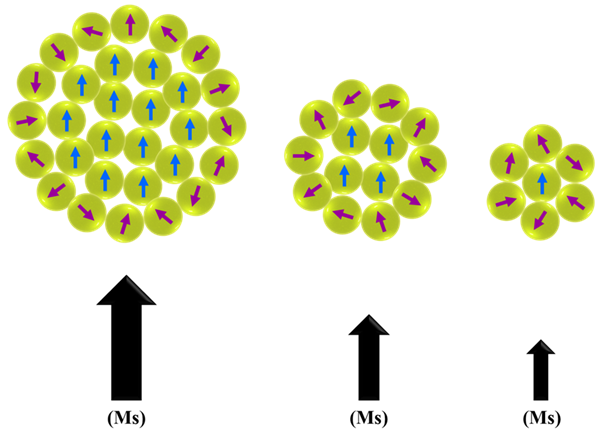
Magnetic nanoparticles, which make up a large part of nanomaterials, have the potential for clinical diagnosis and treatment due to their unique properties such as magnetic and superparamagnetic torque and the power of biological interactions at the cellular and molecular levels. The unique properties of these nanoparticles include super-saturation, superparamagnetic and magnetic susceptibility, which are derived from their inherent magnetic properties. In this review, we investigated the properties of ultra-small iron oxide nanoparticles as a positive contrast agent in MRI. As a result, ultra-small iron oxide nanoparticles have a high potential for use as a T1 weight contrast agent in the clinic. Ultra-small iron oxide nanoparticles have been widely used as a contrast agent in MR imaging. In ultra-small iron oxide nanoparticles, it has been shown that the states change from superparamagnetic to paramagnetic by decreasing in size. Therefore, it can be said that as a result of reducing the size of these nanoparticles, they change from being the T2 contrast agent to a T1 contrast agent. Unlike iron oxide nanoparticles larger than 5 nm, these nanoparticles can create a positive contrast that will facilitate detection. Also, ultra-small iron oxide nanoparticles could solve the problem of gadolinium toxicity and the high magnetic momentum of iron oxide. As a result, this nanoparticle has a high potential for use as a T1 weight contrast agent in the clinic.

Graphical Abstract

Keywords

References

[1] Islami F., Miller K. D., Siegel R. L., Zheng Z., Zhao J., Han X., Ma J., Jemal A., Yabroff, K. R., (2019), National and state estimates of lost earnings from cancer deaths in the united states. JAMA Oncolog. 5: e191460. https://doi.org/10.1001/jamaoncol.2019.1460

[2] Siegel R. L., Miller K. D., Fuchs H. E., Jemal A., (2022), Cancer statistics, CA. Cancer J. Clin. 72: 7-33. https://doi.org/10.3322/caac.21708

[3] Hill D. L., Hawkes D. J., Gleeson M. J., Cox T. C., Strong A. J., Wong W. L., Sofat A., (1994), Accurate frameless registration of MR and CT images of the head: Applications in planning surgery and radiation therapy. Radiol. 191: 447-454. https://doi.org/10.1148/radiology.191.2.8153319

[4] Gu X., Li D. D., Yeoh G. H., Taylor R. A., Timchenko V., (2020), Heat generation in single magnetic nanoparticles under near-infrared irradiation. J. Phys. Chem. Lett. 11: 2182-2187. https://doi.org/10.1021/acs.jpclett.0c00143

[5] Shen Z., Song J., Zhou Z., Yung B. C., Aronova M. A., Li Y., Dai Y., Fan W., Liu Y., Li Z., Ruan H., Leapman R. D., Lin L., Niu G., Chen X., Wu A., (2018), Dotted core–shell nanoparticles for T1‐weighted MRI of tumors. Adv. Mater. 30: e1803163. https://doi.org/10.1002/adma.201803163

[6] Ananta J. S., Godin B., Sethi R., Moriggi L., Liu X., Serda R. E., Krishnamurthy R., Muthupillai R., Bolskar R. D., Helm L., Ferrari M., Wilson L. J., Decuzzi P., (2010), Geometrical confinement of gadolinium-based contrast agents in nanoporous particles enhances T1 contrast. Nat. Nanotechnol. 5: 815–821. https://doi.org/10.1038/nnano.2010.203

[7] Sherwood J. A., Rich M. C., Lovas K., Warram J. M., Bolding M. S., Bao Y., (2017), T1-enhanced MRI-visible nanoclusters for imaging-guided drug delivery. Nanoscale. 9: 11785-11792. https://doi.org/10.1039/c7nr04181k

[8] Sun Y., Kim H. S., Kang S., Piao Y. J., Jon S., Moon W. K., (2018), Magnetic resonance imaging-guided drug delivery to breast cancer stem-like cells. Adv. Healthc. Mater. 7: e1800266. https://doi.org/10.1002/adhm.201800266

[9] Struyfs H., Sima D. M., Wittens M., Ribbens A., Pedrosa de Barros N., Phan T. V., Ferraz Meyer M. I., Claes L., Niemantsverdriet E., Engelborghs S., Van Hecke W., Smeets D., (2020), Automated MRI volumetry as a diagnostic tool for Alzheimer's disease: Validation of icobrain dm. Neuroimage Clin. 26: 102243-102247. https://doi.org/10.1016/j.nicl.2020.102243

[10] Liu K., Dong L., Xu Y., Yan X., Li F., Lu Y., Tao W., Peng H., Wu Y., Su Y., Ling D., He T., Qian H., Yu S., (2018), Stable gadolinium based nanoscale lyophilized injection for enhanced MR angiography with efficient renal clearance. Biomater. 158: 74-85. https://doi.org/10.1016/j.biomaterials.2017.12.023

[11] Ni D., Shen Z., Zhang J., Zhang C., Wu R., Liu J., Bu W., (2016), Integrating anatomic and functional dual-mode magnetic resonance imaging: design and applicability of a bifunctional contrast agent. ACS Nano. 10: 3783-3790.  https://doi.org/10.1021/acsnano.6b00462

[12] Huang G., Li H., Chen J., Zhao Z., Yang L., Chi X., Chen Z., Wang X., Gao J., (2014), Tunable T1 and T2 contrast abilities of manganese-engineered iron oxide nanoparticles through size control. Nanoscale. 17: 10404-10412. https://doi.org/10.1039/c4nr02680b

[13] Mi P., Kokuryo D., Cabral H., Wu H., Terada Y., Saga T., Kataoka K., (2016), A pH-activatable nanoparticle with signal-amplification capabilities for non-invasive imaging of tumour malignancy. Nat. Nanotechnol. 11: 724-730. https://doi.org/10.1038/nnano.2016.72

[14] Zhou H., Guo M., Li J., Qin F., Wang Y., Liu T., Liu Y., (2021), Hypoxia-triggered self-assembly of ultrasmall iron oxide nanoparticles to amplify the imaging signal of a tumor. J. Am. Chem. Soc. 143: 1846-1853. https://doi.org/10.1021/jacs.0c10245

[15] Han Z., Wu X., Roelle S., Chen C., Schiemann W. P., Lu Z., (2017), Targeted gadofullerene for sensitive magnetic resonance imaging and risk-stratification of breast cancer. Nat. Commun. 8: 692-697. https://doi.org/10.1038/s41467-017-00741-y

[16] Li X., Lu S., Xiong Z., Hu Y., Ma D., Lou W., Peng C., Shen M., Shi X., (2019), Light-addressable nanoclusters of ultrasmall Iron Oxide nanoparticles for enhanced and dynamic magnetic resonance imaging of arthritis. Adv. Sci. 6: 1901800. https://doi.org/10.1002/advs.201901800

[17] Cohen D., Mashiach R., Houben L., Galisova A., Addadi Y., Kain D., Bar-Shir A., (2021), Glyconanofluorides as immunotracers with a tunable core composition for sensitive hotspot magnetic resonance imaging of inflammatory activity. ACS Nano. 15: 7563-7574. https://doi.org/10.1021/acsnano.1c01040

[18] Wahsner J., Gale E. M., Rodríguez-Rodríguez A., Caravan P., (2019), Chemistry of MRI contrast agents: Current challenges and new frontiers. Chem. Rev. 119: 957–1057. https://doi.org/10.1021/acs.chemrev.8b00363

[19] Xiao Y., Paudel R., Liu J., Ma C., Zhang Z., Zhou S., (2016), MRI contrast agents: Classification and application (Review). Int. J. Mol. Med. 38: 1319-1326. https://doi.org/10.3892/ijmm.2016.2744

[20] Hasebroock K. M., Serkova N. J., (2009), Toxicity of MRI and CT contrast agents. Expert Opin. Drug Metab. Toxicol. 5: 403-416. https://doi.org/10.1517/17425250902873796

[21] Rasaneh S., Rajabi H., Johari Daha F., (2015), Activity estimation in radioimmunotherapy using magnetic nanoparticles. Chin. J. Cancer Res. 27: 203-208. https://doi.org/10.3978/j.issn.1000-9604.2015.03.06

[22] Rasaneh S., Rajabi H., Babaei M. H., Akhlaghpoor S., (2011), MRI contrast agent for molecular imaging of the HER2/neu receptor using targeted magnetic nanoparticles. J. Nanopart. Res. 13: 2285–2293. 10.1007/s11051-010-9991-5

[23] Rajabi H., Rasaneh S., Salehi S., (2016), Synthesis and biological evaluation of 99mTc-Chitosan nanoparticles as a potential radiopharmaceutical for liver imaging. Synth. React. Inorg. Met. 46: 1450-1454. https://doi.org/10.1080/15533174.2015.1137010

[24] Rasaneh S., Dadras M., (2015), The possibility of using magnetic nanoparticles to increase the therapeutic efficiency of Herceptin antibody. Biomed. Eng-Biomed. Tech. 60: 485-490. https://doi.org/10.1515/bmt-2014-0192

[25] Sadeghi B., (2017), One-Pot synthesis of Ag/Fe3O4 nanocomposite: Preparation and characterization. Adv. Mater. Process. 5: 82-92. https://api.semanticscholar.org/CorpusID:55006948

[26] Szpak A., Fiejdasz S., Prendota W., Strączek T., Kapusta C., Szmyd J. S., Nowakowska M., Zapotoczny S., (2014), T1–T2 dual-modal MRI contrast agents based on superparamagnetic iron oxide nanoparticles with surface attached gadolinium complexes. J. Nanoparticle Res. 16: 1-11. https://doi.org/10.1007/s11051-014-2678-6

[27] Amraee A., Alamzadeh Z., Irajirad R., Sarikhani A., Ghaznavi H., Ghadiri Harvani H., Rabi Mahdavi S., Shirvalilou S., Khoei S., (2023), Theranostic RGD@ Fe3O4-Au/Gd NPs for the targeted radiotherapy and MR imaging of breast cancer. Cancer Nanotechnol. 14: 1-20. https://doi.org/10.1186/s12645-023-00214-6

[28] Torchilin V. P., (2005), Recent advances with liposomes as pharmaceutical carriers. Nat. Rev. Drug Discov. 4: 145-160. https://doi.org/10.1038/nrd1632

[29] Venditto V. J., Szoka F. C., (2013), Cancer nanomedicines: So many papers and so few drugs! Adv. Drug Deliv. Rev. 65: 80-88.  https://doi.org/10.1016/j.addr.2012.09.038

[30] Matsumura Y., Maeda H., (1986), A new concept for macromolecular therapeutics in cancer chemotherapy: Mechanism of tumoritropic accumulation of proteins and the antitumor agent smancs. Cancer Res. 46: 6387-92. https://api.semanticscholar.org/CorpusID:7103917

[31] Lozano-Pérez A. A., Gil A. L., Pérez S. A., Cutillas N., Meyer H., Pedreño M., Ruiz J., (2015), Antitumor properties of platinum (iv) prodrug-loaded silk fibroin nanoparticles. Dalton Trans. 44: 13513-13521. https://doi.org/10.1039/c5dt00378d

[32] Leserman L. D., Weinstein J. N., Blumenthal R., Terry W. D., (1980). Receptor-mediated endocytosis of antibody-opsonized liposomes by tumor cells. Proc. Natl. Acad. Sci. U. S. A. 77: 4089-4093. https://doi.org/10.1073/pnas.77.7.4089

[33] Kamaly N., Xiao Z., Valencia P. M., Radovic-Moreno A. F., Farokhzad O. C., (2012), Targeted polymeric therapeutic nanoparticles: Design, development and clinical translation. Chem. Soc. Rev. 41: 2971-3010.https://doi.org/10.1039/c2cs15344k

[34] Koshkaryev A., Sawant R., Deshpande M., Torchilin V., (2013), Immunoconjugates and long circulating systems: Origins, current state of the art and future directions. Adv. Drug Deliv. Rev. 65: 24-35. https://doi.org/10.1016/j.addr.2012.08.009

[35] Schnorr J., Wagner S., Abramjuk C., Wojner I., Schink T., Kroencke T. J., Schellenberger E., Hamm B., Pilgrimm H., Taupitz M., (2004), Comparison of the iron oxide-based blood-pool contrast medium VSOP-C184 with gadopentetate dimeglumine for first-pass magnetic resonance angiography of the aorta and renal arteries in pigs. Invest. Radiol. 39: 546-553. https://doi.org/10.1097/01.rli.0000133944.30119.cc

[36] Kim B. H., Lee N., Kim H., An K., Park Y. I., Choi Y., Shin K., Lee Y., Kwon S. G., Na H. B., Park J., Ahn T., Kim Y. W., Moon W. K., Choi S. H., Hyeon T., (2011), Large-scale synthesis of uniform and extremely small-sized iron oxide nanoparticles for high-resolution T1 magnetic resonance imaging contrast agents. J. Am. Chem. Soc. 133: 12624-12631. https://doi.org/10.1021/ja203340u

[37] Park E. A., Lee W., So Y. H., Lee Y. S., Jeon B. S., Choi K. S., Kim E. G., Myeong W. J., (2017), Extremely small pseudoparamagnetic Iron Oxide nanoparticle as a novel blood pool T1 magnetic resonance contrast agent for 3 T whole-heart coronary angiography in canines: Comparison with gadoterate meglumine. Invest. Radiol.  52: 128–133. . https://doi.org/10.1097/RLI.0000000000000321

[38] Rui Y., Liang B., Hu F., Xu J., Peng Y., Yin P., Duan Y., Zhang C., Gu H., (2016), Ultra-large-scale production of ultrasmall superparamagnetic iron oxide nanoparticles for T1-weighted MRI. RSC Adv.  6: 22575-22585. https://doi.org/10.1039/C6RA00347H

[39] Shen Z, Chen T, Ma X, Ren W, Zhou Z, Zhu G, Zhang A., Liu Y., Song J., Li Z., Ruan H., Fan W., Lin L., Munasinghe J., Chen X., Wu A., (2017), Multifunctional theranostic nanoparticles based on exceedingly small magnetic iron oxide nanoparticles for T1-weighted magnetic resonance imaging and chemotherapy. ACS nano. 11: 10992-11004. https://doi.org/10.1021/acsnano.7b04924

[40] Vangijzegem, T., Stanicki, D., Boutry, S., Paternoster, Q., Vander Elst, L., Muller, R. N., Laurent, S., (2018), VSION as high field MRI T1 contrast agent: Evidence of their potential as positive contrast agent for magnetic resonance angiography. Nanotechnol. 29: 265103. https://doi.org/10.1088/1361-6528/aabbd0

[41] Wagner M., Wagner S., Schnorr J., Schellenberger E., Kivelitz D., Krug L., Dewey M., Laule M., Hamm B., Taupitz M., (2011), Coronary MR angiography using citrate-coated very small superparamagnetic iron oxide particles as blood-pool contrast agent: Initial experience in humans. J. Magn. Reson. Imaging. 34: 816–823. https://doi.org/10.1002/jmri.22683

[42] Wei H., Bruns O. T., Kaul M. G., Hansen E. C., Barch M., Wiśniowska A., Chen O., Chen Y., Li N., Okada S., Cordero J. M., Heine M., Farrar C. T., Montana D. M., Adam G., Ittrich H., Jasanoff A., Nielsen P., Bawendi M. G., (2017), Exceedingly small iron oxide nanoparticles as positive MRI contrast agents. Proc. Natl. Acad. Sci. U.S.A. 114: 2325–2330. https://doi.org/10.1073/pnas.1620145114

[43] Li F., Liang Z., Liu J., Sun J., Hu X., Zhao M., Liu J., Bai R., Kim D., Sun X., Hyeon T., Ling D., (2019), Dynamically reversible Iron Oxide nanoparticle assemblies for targeted amplification of T1-weighted magnetic resonance imaging of tumors. Nano Lett. 19: 4213–4220. https://doi.org/10.1073/pnas.1620145114

[44] Campbell J. L., Arora J., Cowell S. F., Garg A., Eu P., Bhargava S. K., Bansal V., (2011), Quasi-cubic magnetite/silica core-shell nanoparticles as enhanced MRI contrast agents for cancer imaging. PloS one. 6: e21857. https://doi.org/10.1371/journal.pone.0021857

[45] Reguera J., Jimenez de Aberasturi D., Henriksen-Lacey M., Langer J., Espinosa A., Szczupak B., Wilhelm C., Liz‐Marzán L. M., (2017), Janus plasmonic-magnetic gold-iron oxide nanoparticles as contrast agents for multimodal imaging. Nanoscale. 27: 9467-9480. https://doi.org/10.1039/c7nr01406f

[46] Saraswathy A., Nazeer S. S., Jeevan M., Nimi N., Arumugam S., Harikrishnan V. S., Varma P. R., Jayasree R. S., (2014), Citrate coated iron oxide nanoparticles with enhanced relaxivity for in vivo magnetic resonance imaging of liver fibrosis. Colloids Surf. B. 117: 216–224. https://doi.org/10.1016/j.colsurfb.2014.02.034

[47] Beik J., Alamzadeh Z., Mirrahimi M., Sarikhani A., Ardakani T. S., Asadi M., Irajirad R., Mirrahimi M., Mahabadi V. P., Eslahi N., Bulte J. W. M., Ghaznavi H., Shakeri-Zadeh A., (2021), Multifunctional theranostic Graphene Oxide nanoflakes as MR imaging agents with enhanced photothermal and radiosensitizing properties. ACS Appl. Bio Mater. 4: 4280–4291. https://doi.org/10.1021/acsabm.1c00104

[48] Mekuria S. L., Debele T. A., Tsai H., (2017), Encapsulation of Gadolinium Oxide nanoparticle (Gd2O3) contrasting agents in PAMAM dendrimer templates for enhanced magnetic resonance imaging in vivo. ACS Appl. Mater. Interf. 9: 6782-6795.https://doi.org/10.1021/acsami.6b14075

[49] Ghaghada K. B., Ravoori M., Sabapathy D., Bankson J., Kundra V., Annapragada A., (2009), New dual mode gadolinium nanoparticle contrast agent for magnetic resonance imaging. PloS One. 4: e7628.https://doi.org/10.1371/journal.pone.0007628

[50] Dai Y., Wu C., Wang S., Li Q., Zhang M., Li J., Xu K., (2018), Comparative study on in vivo behavior of PEGylated gadolinium oxide nanoparticles and Magnevist as MRI contrast agent. Nanomedicine: NBM. 14: 547–555. https://doi.org/10.1016/j.nano.2017.12.005

[51] Xu W., Miao X., Oh I. T., Chae K. S., Cha H., Chang Y., Lee G. H., (2016), Dextran‐coated ultrasmall Gd2O3 nanoparticles as potential T1 MRI contrast agent. Chem. Select. 1: 6086-6091. https://doi.org/10.1002/slct.201600832

[52] Zhou Z., Wu C., Liu H., Zhu X., Zhao Z., Wang L., Xu Y., Ai H., Gao J., (2015), Surface and interfacial engineering of iron oxide nanoplates for highly efficient magnetic resonance angiography. ACS Nano. 9: 3012–3022. https://doi.org/10.1021/nn507193f

[53] Zhou Z., Huang D., Bao J., Chen Q., Liu G., Chen Z., Chen X., Gao J., (2012), A synergistically enhanced T(1) -T(2) dual-modal contrast agent. Adv. Mater. 24: 6223–6228.https://doi.org/10.1002/adma.201203169

[54] Zhou Z., Wang L., Chi X., Bao J., Yang L., Zhao W., Chen Z., Wang X., Chen X., Gao J., (2013), Engineered Iron-Oxide-based nanoparticles as enhanced T1 contrast agents for efficient tumor imaging. ACS Nano. 7: 3287–3296. https://doi.org/10.1021/nn305991e

[55] Palani U., Iruson B., Balaraman S., Krishnamoorthy S., Elayaperumal M., (2023), Synthesis and characterization of Iron Oxide, rare earth Erbium Oxide, and Erbium Oxide blended Iron Oxide nanocomposites for biomedical activity application. Int. J. Nano Dimens. 14: 103-114. https://doi.org/10.22034/ijnd.2022.1964912.2170

[56] Yang M., Gao L., Liu K., Luo C., Wang Y., Yu L., Peng H., Zhang W., (2015), Characterization of Fe3O4/SiO2/Gd2O(CO3)2 core/shell/shell nanoparticles as T1 and T2 dual mode MRI contrast agent. Talanta. 131: 661–665. https://doi.org/10.1016/j.talanta.2014.08.042

[57] Choi J. S., Lee J. H., Shin T. H., Song H. T., Kim E. Y., Cheon J., (2010), Self-confirming "AND" logic nanoparticles for fault-free MRI. J. Am. Chem. Soc. 132: 11015–11017. https://doi.org/10.1021/ja104503g

[58] Maeda H., Sawa T., Konno T., (2001), Mechanism of tumor-targeted delivery of macromolecular drugs, including the EPR effect in solid tumor and clinical overview of the prototype polymeric drug SMANCS. J. Control Release. 74: 47-61. https://doi.org/10.1016/s0168-3659(01)00309-1

[59] Tromsdorf U. I., Bigall N. C., Kaul M. G., Bruns O. T., Nikolic M. S., Mollwitz B., Parak W. J., (2007), Size and surface effects on the MRI relaxivity of manganese ferrite nanoparticle contrast agents. Nano lett. 7: 2422-2427. https://doi.org/10.1021/nl071099b

[60] Na H. B., Song I. C., Hyeon T., (2009), Inorganic nanoparticles for MRI contrast agents. Adv. Mater. 21: 2133-2148. https://doi.org/10.1002/adma.200802366

[61] Caravan P., (2006), Strategies for increasing the sensitivity of gadolinium based MRI contrast agents. Chem. Soc. Rev. 35: 512-523. https://doi.org/10.1039/b510982p

[62] Dekkers I. A., Roos R., Molen A. J., (2018), Gadolinium retention after administration of contrast agents based on linear chelators and the recommendations of the European Medicines Agency. Eur. Radiol. 28: 1579-1584. https://doi.org/10.1007/s00330-017-5065-8

[63] Liu, C., Liu, D., B., Long, G., X., Wang, J., F., Mei, Q., Hu, G., Y., Qiu, Hong, (2013), Specific targeting of angiogenesis in lung cancer with RGD-conjugated ultrasmall superparamagnetic iron oxide particles using a 4.7 T magnetic resonance scanner. Chin. Med. J. 126: 2242-2247. https://doi.org/10.3760/cma.j.issn.0366-6999.20130628

 [64] Tao, C., Zheng, Q., An, L., He, M., Lin, J., Tian, Q., Yang, S., (2019), T₁-Weight Magnetic Resonance Imaging Performances of Iron Oxide Nanoparticles Modified with a Natural Protein Macromolecule and an Artificial Macromolecule. J. Nanomater. 9: 170-175. https://doi.org/10.3390/nano9020170

[65] Bai, C., Jia, Z., Song, L., Zhang, W., Chen, Y., Zang, F., Ma, M., Gu, N., Zhang, Y., (2018), Time-Dependent T1–T2 Switchable Magnetic Resonance Imaging Realized by c(RGDyK) Modified Ultrasmall Fe3O4 Nanoprobes. Adv. Funct. Mater. 28: 1802281. https://doi.org/10.1002/adfm.201802281

[66] Du C., Wang J., Liu X., Li H., Geng D., Yu L., Chen Y., Zhang J., (2020), Construction of pepstatin A-conjugated ultrasmall SPIONs for targeted positive MR imaging of epilepsy-overexpressed P-glycoprotein. Biomater. 230:119581. https://doi.org/10.1016/j.biomaterials.2019.119581

[67] Xie M., Wang Z., Lu Q., Nie S., Butch C.J., Wang Y., Dai B., (2020), Ultracompact iron oxide nanoparticles with a monolayer coating of succinylated heparin: a new class of renal-clearable and nontoxic T1 agents for high-field MRI. ACS Appl. Mater. Interf. 12: 53994-54004. https://doi.org/10.1021/acsami.0c12454

[68] Wei R., Cai Z., Ren B.W., Li A., Lin H., Zhang K., Gao J., (2018), Biodegradable and renal-clearable hollow porous iron oxide nanoboxes for in vivo imaging. Chem. Mater. 30: 7950-7961. https://doi.org/10.1021/acs.chemmater.8b03564

[69] Fedorenko S., Stepanov A., Zairov R., Kaman O., Amirov R., Nizameev I., Kholin K., Ismaev I., Voloshina A., Sapunova A., Kadirov M., Mustafina A., (2018), One-pot embedding of iron oxides and Gd(III) complexes into silica nanoparticles—Morphology and aggregation effects on MRI dual contrasting ability. Colloids Surf. A: Physicochem. Eng. 559: 60-67. https://doi.org/10.1016/j.colsurfa.2018.09.044

[70] Luo Y., Yang  J., Yan Y., Li  J., Shen M., Zhang G., Shi X., (2015), RGD-functionalized ultrasmall iron oxide nanoparticles for targeted T1-weighted MR imaging of gliomas. Nanoscale. 7: 14538-14546. https://doi.org/10.1039/c5nr04003e