Department of Biotechnology, Deenbandhu Chhotu Ram University of Science and Technology, Sonipat, Haryana, 131039, IN
Abstract
Infectious diseases have been on rise in the recent past. Early diagnosis plays a role as important as proper treatment and prophylaxis. The current practices of detection are time consuming which may result in unnecessary delays in treatment. Advances in nanodiagnostic approaches have been in focus lately. The rising interest and better understanding of nanoparticles have led to opening up of new frontiers in the concerned area. Optical properties of nanoparticles are being exploited to design detection systems that can provide fast, one-step and reliable results. Based on conserved DNA sequences unique to the target organism, the results offer accuracy comparable to conventional tests. Further, visual or spectrophotometric analysis omits the need of costly apparatus for result interpretation. The present review aims at putting together the information on nanoparticles based DNA conjugate systems for detection of pathogenic microorganisms.
Introduction
Intensive studies have been conducted regarding the synthesis and applications of metal nanoparticles (NPs) in the recent past. Nanomaterials of different shapes and sizes are being synthesized and explored for their applications in various fields of life sciences. Studies are underway to determine their role in disease control in humans and plants. Their unique physicochemical properties make them special when working with modern aspects of biology and medicine [
1
]. However, chemical synthesis of nanoparticles might prove toxic to the environment and also their properties become dependent on the interactions between reducing agents and the metal ions and adsorption of the stabilizing agent onto the NPs [
2
,
3
]. Hence, there is an increasing demand for techniques employing green and non toxic synthesis of metal NPs. Also, the use of biocompatible and non-toxic agents is a preferred choice for materials seeking application in biological fields.
Infectious diseases caused by microorganisms are and have always been a major threat for human lives [
4
,
5
]. One of the major challenges being faced by the medical fraternity is the early and accurate diagnosis of such diseases. Conventional and reliable techniques have been optimized and practiced since decades but suffer with the disadvantages of being time consuming, expensive and may be unsuitable at times [
6
]. Though considerable improvement in the diagnostic techniques has been seen in the recent past, identification of slow growing and fastidious pathogens has not been achieved with much success [
7
]. The time required for conducting conventional laboratory tests can be a major drawback when the disease has reached crucial stage and faster diagnosis is required. Keeping the above mentioned scope for such studies, an attempt has been made to summarize the available information in the form of a review.
Molecular diagnosis
Identification of pathogens based on their nucleic acid detection is very important in current clinical diagnosis. Each microorganism has a fragment of its genomic DNA which is conserved in itself and is preserved over generations without alterations. Detection of this DNA segment can directly confer the presence or absence of the microorganism in the test sample. This portion of DNA forms the basis of molecular detection. Such molecular tests can be broadly categorized as amplification and non-amplification based methods. While non-amplification based methods make use of hybridization strategies with DNA or RNA probes, amplification based methods rely on reactions such as polymerase chain reaction (PCR) to amplify the amount of nucleic acids for better sensitivity [
8
].
Non-amplification based methods
Non-amplification methods are based on direct hybridization reactions between target DNA segments and labeled DNA/RNA probes and are used when the target is in abundance. The labels could be radiolabels, fluorescent labels, enzymatic labels, chemiluminescent labels, etc. The idea behind the analysis is that signal should be emitted only when the probe is bound to the target complementary fragment of nucleic acid. When in free or unbound state, the probe must not emit a signal [
8
].
Amplification-based methods
Amplification-based methods come into picture when the target is present in minute quantities and direct detection cannot be trusted to yield valid results. Hence, the first requirement is to amplify either the target (target amplification) or the signals being generated by the detector molecule used (signal amplification). Signal amplification is particularly used for direct testing of biological materials; target amplification works best on processed samples [
8
].
A number of detection methods are currently available for target amplification systems; normal PCR, real time PCR, transcription mediated amplification (TMA), 16S rRNA amplification and sequencing, species specific gene amplification, etc. to name a few [
8
,
9
].
The nanoparticle based DNA conjugate systems discussed below combines the goods of both the methods and provide a non-amplification based direct detection method, even when the target is present in smaller quantities. Thus, they not only possess good specificity as that of hybridization reactions but also overcome the hurdle of amplification of smaller quantity targets as in, say, PCR based detection.
Nanoparticles in diagnosis
As a result of ultra small size in the range of 1–100 nm [
10
] and high surface-to-volume ratio, NPs are blessed with distinctive physical and chemical properties [
11
]. These unique properties have been under continuous scrutiny since the last few decades with the aim of developing new and improved techniques focused on medical sciences. Gold and silver NPs are being extensively researched for their applications in nanodiagnostic approaches.
Pioneer studies regarding the modification of gold NPs with oligonucleotides were reported way back in 1996 (Alivastos et al. 1996; Mirkin et al. 1996, as cited in [
12
]) and a number of studies have been reported since then concerning gold NP–oligonucleotide conjugate systems aimed at detection of complementary DNA sequences [
13
–
16
]. While gold NPs have been in prime focus, only a few reports are available for silver NPs in such systems. One of the reasons could be that synthesizing silver NPs with repeatable size is a difficult job which in turn affects its optical properties. The first report utilizing silver NPs for detection of target DNA was given by Thompson et al. [
12
] and they emphasized that silver NPs possess 100× greater molar extinction coefficient than gold NPs and can provide increased sensitivity at much lower concentration in both visual and absorption spectrum analysis.
Principle of conjugate synthesis and detection
The basis of conjugate systems is the ability of these NPs to conjugate with biomolecules such as DNA and proteins. Alkanethiolate monolayers onto metallic NPs are quite simple and easy to construct. However, the mechanism behind this adsorption remains unclear. One explanation could be given as oxidative addition of R-S-H to the surface of the metal coupled with the release of hydrogen gas, the equation for which is given below [
17
].
R-S-H+Aun→R-S-Au++Aun-1+1/2H2
Addition of these thiol moieties in nucleic acid molecules can therefore help in their adsorption in the same way.
It is well established that the electrostatic interactions of negatively charged phosphate groups of the DNA backbone and highly polarizable gold NP contribute to the stability of NP suspension against aggregation [
18
]. These forces provide stability to the colloid upon salt addition up to a concentration of 1.5 M NaCl. This can be attributed to the fact that nucleic acid molecules provide buffering action for increasing the ionic strength and improve the stability. Upon increasing the salt concentration further, to 2 M NaCl, NP bound with ssDNA molecules start aggregating. However, if the ssDNA probes are made to hybridize with their complementary targets, they resist aggregation. This improved stability can be accredited to the buffering action in combination with the duplex which keeps the NPs linked to the nucleic acid network and prevent their aggregation [
19
].
One of the basic approaches for detecting hybridization on the surface of the NPs and the resultant aggregation is the surface plasmon resonance (SPR) of the solution. SPR refers to resonant oscillation of free electrons in metals on the boundary of metal surface and the surrounding medium and any change in the particle shape and size is bound to affect the SPR of the material. Aggregation of NP–oligonucleotide conjugates in the presence of high salt concentrations alters the size of NPs and, hence, alters their SPR [
20
].
Synthesis of NP–oligonucleotide conjugates
Synthesis of NP–oligonucleotide conjugates is the first and foremost step for designing such systems since they not only detect the target but also generate result signals. Various authors have stated their protocols for the synthesis of NP–oligonucleotide conjugates [
12
,
13
,
21
–
30
]. These methods offer minor variations, with the basic ideology being the same.
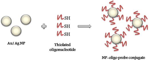
To begin with, the oligonucleotide probes need to be modified with an alkyl thiol group to facilitate their attachment onto the NP surface (Fig.
1
). It was reported that 5′ terminal labeling provided a better efficiency of conjugation as compared to 3′ terminal labeling. This could be attributed to better shielding of NPs from the ionic buffer due to increase in hydrophobicity by the alkyl chain [
12
]. It is also advised to first let the oligonucleotide react with reducing agents such as dithio threitol [
12
,
26
,
29
,
31
] or tris(2-carboxyethyl)phosphine (TCEP) [
32
] so as to reduce any disulphide linkages and ensure full reactivity. If the disulphides are not reduced, active thiol group is lost, adversely affecting the monolayer assembly of oligonucleotides onto the NPs [
33
]. While terminal thiol modification has been conventionally used for functionalizing oligonucleotides, the conjugates synthesized with such probes may suffer from lack of stability. To overcome this problem, oligonucleotides modified with triple cyclic disulphide moieties, multiple thiols and thioctic acid have also been investigated for conjugate formation [
28
,
34
,
35
]. It was reported that presence of multiple thiol groups resulted in increased stability of the conjugates [
35
] because of the fact that polydentate ligands offer more stable metal–ligand complexes [
36
].
Fig. 1
Conjugation of thiolated oligonucleotides with metallic nanoparticles
Once the oligonucleotides are modified and reduced, they are added to nanoparticle suspension and the resulting mixture is incubated overnight followed by the addition of phosphate buffered saline and surfactant such as sodium dodecyl sulphate. While surfactant helps in preventing aggregation [
26
], addition of PBS prior to adding oligonucleotide suspension can lead to irreversible aggregation of the nanoparticles by disrupting the stabilizing citrate layer on the surface as stated by Thompson and others [
12
]. Finally, a salting buffer is added over a period of 2 days and the solution is allowed to equilibrate overnight. Addition of salt must be gradual as charge repulsion is the main force behind maintaining the colloidal repulsion and sudden change in salt concentration could lead to irreversible aggregation. Salt, on one hand, helps in overcoming the nucleotide–metal interactions and, on the other hand, favours the adsorption of thiols onto the NP surface with steady increase in concentration. Similar methods of conjugate formation can be used for both AuNPs and AgNPs but salt increments used for AuNP reactions would lead to irreversible aggregation if used for AgNPs [
31
] and hence, the process needs to be carefully monitored.
A comparison of amount of DNA that could be conjugated onto gold and silver NPs showed that silver NPs offer a better coverage (22.6 ± 2.4 pmol/cm
2
for 30 nm silver NP) than gold NPs (15 ± 4 pmol/cm
2
for 13 nm gold NP) [
12
,
23
].
Characterization
Properly functionalized conjugates retain the same colour as unmodified AuNPs with no visible aggregates and can be stored in this state for 1 month [
26
]. NP suspension shows a shift in the surface plasmon resonance after functionalization. The absorption maxima for AuNPs was reported to shift from 518 to 523 nm upon oligonucleotide conjugation by Nicewarner-Pena et al. [
37
] and for AgNPs from 396 to 400 nm as stated by Vidal et al. [
31
]. Generation of UV–visible spectrograms at timely intervals along with visual examination can help access the stability of these conjugates. A single extinction peak ensures that no aggregation in the NP suspension resulted during the conjugation with oligonucleotides [
32
]. Moreover, electron microscope studies can also be undertaken to ensure oligonucleotide conjugation with NPs.
Hybridization detection strategy
Ultrasensitive DNA and RNA detection systems have been in investigation for long and these emerging techniques hold the promise of detecting even smaller and minute concentrations of biological materials omitting the need for amplification reactions such as PCR [
14
,
19
,
38
–
43
].
In case of nucleic acid based detection, DNA probes specific to conserved regions of pathogenic DNA can be coupled with NPs to formulate biosensors. NPs based assays can provide direct detection of genomic DNA sequence based on optical scattering properties of NPs [
14
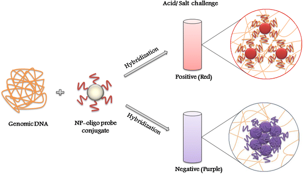
]. When these conjugates encounter genomic DNA of the target organism, the probe immobilized onto the NP hybridizes with the complementary sequences in the genome. This is performed under stringent conditions such that hybridization does not occur unless the sequences are perfectly complementary to each other. On subjecting to acid/salt challenge, conjugates which successfully hybridize with the complementary DNA do not undergo acid induced aggregation, maintain their red colour while unhybridized conjugates aggregate which in turn leads to colour change from red to purple [
19
,
38
,
39
,
42
,
43
] (Fig.
2
). This aggregation is also accompanied by an absorbance peak shift towards longer wavelength due to change in surface plasmon resonance of the NPs [
38
,
39
]. Absorbance peak of hybridized gold NP-probe solution is observed at approximately 520 nm which shifts to around 575 nm in the absence of hybridization [
43
]. Hence, the effect of acid/salt induced aggregation of gold NP-probe conjugates into bigger clusters can be easily observed by visible colour change and spectrophotometric analysis of the test solutions before and after the completion of the aggregation reaction.
Fig. 2
Schematic for gold NP–oligonucleotide conjugates based detection system
Diagnostic applications
DNA based
Non-specific detection of DNA sequences have been reported by a number of workers using both gold [
13
,
32
,
44
] and silver NPs [
12
,
27
]. Where species specific DNA detection is concerned, studies have been published using NP–oligonucleotide conjugates for a few pathogens, the results of which are summarized in Table
1
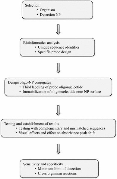
. The stated methods make use of detection of unamplified DNA using oligonucleotide probes as well as detection using NP–oligonucleotide conjugates combined with PCR. A schematic plan of basic strategy followed for designing NP based detection systems is given in Fig.
3
.
Table 1
Gold NP based detection systems for pathogenic organisms
Target organism
Target gene
Probe sequences
Detection limit
References
Methicillin-resistant Staphylococcus aureus
mecA
ATGGCATGAGTAACGAAGAATA
TTCCAGATTACAACTTCACCA
AAAGAACCTCTGCTCAACAAG
GCACTTGTAAGCACACCTTCAT
66 pg/µl
[14]
23S rRNA mecA
GAGAGACAACATTTTCGACT
GGAACGAAGGTATCATCTTG
500 ng amplicon
[45]
Mycobacterium tuberculosis
RNA polymerase β-subunit
GGACGTGGAGGCGATC
0.75 µg
[38]
Mycobacterium avium subsp. paratuberculosis
16–23 s ITS DNA
CAGTCCGTGTGGTGTCCCT
18.75 ng/µl
[39]
Salmonella enteritidis
insertion element (Iel)
AATATGCTGCCTACTGCCCTACGCTT
1 ng/µl
[40]
Salmonella typhimurium
–
CCAAAGGCTACGCGTTAACGTGGTGTTGG
105 cfu/ml
[41]
Escherichia coli O157:H7
–
CCGGACGCTTATGCCTTGCCATCTACAGAGCAGGTGTGACGG
105 cfu/ml
[41]
Escherichia coli
malB
ACAAAGGGAGAAGGGCATG
11.4 ng
[42]
Leishmania spp.
kinetoplast minicircle DNA
GTTAGCCGATGGTGGTCTTG
ACGGGTGTCTTTGATGATGC
TAGTCTGGTGGGATGCTTCG
GTGCCTTTGATGTGGGTGTT
11.5 ng/µl
[43]
Plasmodium falciparum
Lactate dehydrogenase
CTGGGCGGTAGAACCATAGTGACCCAGCCGTCTAC
57 pg/μl
[46]
Fig. 3
Basic strategy for designing nanoparticle–oligonucleotide based detection system
Colorimetric assays based on NP–oligonucleotide conjugates have been developed for the detection of various pathogenic microorganisms. Probe sequences from conserved genomic regions were selected based on their uniqueness for the organism in question. They were tested so as to ensure that they do not share homology with non-target organisms to prevent non-specific associations and hence, false positive results. These probe sequences were then conjugated with AuNPs and used for hybridization studies. Successful assays have been developed for methicillin-resistant
S. aureus, M. tuberculosis, M. avium
subsp.
paratuberculosis, E. coli, S. enteritidis, S. typhimurium,Leishmania
spp. and
P. falciparum
[
14
,
38
–
43
,
45
,
46
] (Table
1
). These systems were tested using various clinical samples and results were observed as visible colour change of the NP suspension or with the help of spectrophotometry which depicted an absorbance peak shift in case of negative hybridization.
An interesting approach involving catalytic DNA molecules, known as DNAzymes was reported by Zagorovsky and Chan. They conjugated two sets of AuNPs, A and B, with two fragments of the DNAzyme, specific to one single gene. These fragments shared complementarities with the ends of a linker DNA, the central part of which is the target for active DNAzyme. In the presence of target gene in the test sample, the two DNAzyme fragments come in close proximity and form a complex with the target gene and the linker DNA. This interaction leads to cleaving of the linker from its central part. The cleaved linker fragments cannot link the AuNPs together and the solution remains red. In the absence of target, the linker DNA remains intact and cross links AuNP-A and AuNP-B together leading to aggregation and colour change from red to purple. Different sets of AuNPs were prepared for
N. gonorrhoeae, T. pallidum, P. falciparum
and hepatitis B virus for rapid detection of gonorrhea, syphilis, malaria and hepatitis B infection. Multiplexed reaction was also observed and multiple targets were successfully detected in one experiment [
47
].
Antibody based
In addition to the above stated system, various other diagnostic systems have been postulated using NPs. Conjugation of NPs with antibodies in the form of immune-chromatographic strip (ICS) has already been commercialized. Antibodies offer an advantage in detection as they can be targeted to not only the target microorganism but also to the toxins secreted and the antibodies generated by immune response [
6
,
7
]. Antibody coupled NPs have been designed for the detection of
S. aureus
in clinical samples,
E. coli
O157:H7 in food samples, multiplex detection of
E. coli, S. typhimurium
and
S. aureus
, detection and photo thermal lysis of
S. typhimurium
and detection of various others [
48
–
53
].
Conclusion and future prospects
Currently used detection methods rely on culturing bacterial isolates, which need an incubation period of at least 24 h, and then testing to establish their identity, which is a time consuming work. Other systems such as enzyme immunoassays work only when the target pathogen is present in abundance [
54
,
55
]. More specific molecular techniques such as PCR can work efficiently with lesser amount of samples but require costly apparatus.
The above stated systems can directly detect target DNA and can circumvent the need for culturing of pathogens and amplification of target DNA in the specimens. In addition of providing better detection and reliable results, these methods also allow for even easier result interpretations which can be analyzed by visible colour change and determining the shift in the absorbance spectra peak of the conjugates before and after the addition of target DNA using UV–visible spectroscopy. With both of these being fast methods, the system will not only result in faster diagnosis than conventional methods but will also provide authentic result interpretation. Also, as no special equipments or tedious protocols are required for analysis, these systems can be used as routine screening tool in clinical pathology laboratories.
However, work still needs to be done to establish the behavior of such systems when they interact with biological fluids in raw form and whether reliable results can be obtained in unprocessed samples. Also, there is a need to improve the sensitivity of these systems for better detection of pathogens present in even smaller quantities. With such advances, diagnosis of slow growing and difficult to culture fastidious microorganisms could also be done routinely with little hesitation.
Acknowledgments
The author PJ is thankful to Assured Opportunity for Research Careers (AORC), Department of Science and Technology (DST), Ministry of Science and Technology, New Delhi for awarding INSPIRE fellowship.
Author’s contributions
This work was carried out in collaboration between all authors. PJ conceptualized and drafted the manuscript and the figures. PK coordinated the design and literature search. JSR guided technical writing and critically revised the manuscript.
References
Jo et al. (2009) Antifungal activity of silver ions and nanoparticles on phytopathogenic fungi 93(10) (pp. 1037-1043) 10.1094/PDIS-93-10-1037
Knoll and Keilmann (1999) Near-field probing of vibrational absorption for chemical microscopy (pp. 134-137) 10.1038/20154
Sengupta et al. (2005) Temporal targeting of tumour cells and neovasculature with a nanoscale delivery system (pp. 568-572) 10.1038/nature03794
Fauci (2001) Infectious diseases: considerations for the 21st century 32(5) (pp. 675-685) 10.1086/319235
Houpikian and Raoult (2002) Traditional and molecular techniques for the study of emerging bacterial diseases: one laboratory’s perspective 8(2) (pp. 122-131) 10.3201/eid0802.010141
Kaittanis et al. (2010) Emerging nanotechnology-based strategies for the identification of microbial pathogenesis 62(4–5) (pp. 408-423) 10.1016/j.addr.2009.11.013
Thompson et al. (2008) Ultrasensitive DNA detection using oligonucleotide-silver nanoparticle conjugates 80(8) (pp. 2805-2810) 10.1021/ac702403w
Storhoff et al. (1998) One-pot colorimetric differentiation of polynucleotides with single base imperfections using gold nanoparticle probes (pp. 1959-1964) 10.1021/ja972332i
Storhoff et al. (2004) Homogeneous detection of unamplified genomic DNA sequences based on colorimetric scatter of gold nanoparticle probes 22(7) (pp. 883-887) 10.1038/nbt977
Chen et al. (2004) Gold and silver nanoparticles functionalized with known numbers of oligonucleotides per particle for DNA detection (pp. 2804-2805) 10.1039/b411181h
Cai et al. (2010) One-pot polymerase chain reaction with gold nanoparticles for rapid and ultrasensitive DNA detection (pp. 557-563) 10.1007/s12274-010-0016-6
Pradeep (2007) (pp. 128-155) Tata McGraw-Hill Publishing Company Ltd.
Sandström et al. (2003) Nonspecific and thiol-specific binding of DNA to gold nanoparticles (pp. 7537-7543) 10.1021/la034348u
Baptista et al. (2005) Colorimetric detection of eukaryotic gene expression with DNA-derivatized goldnanoparticles (pp. 111-117) 10.1016/j.jbiotec.2005.02.019
Zeng et al. (2014) Nanomaterials enhanced surface plasmon resonance for biological and chemical sensing applications (pp. 3426-3452) 10.1039/c3cs60479a
Mirkin et al. (1996) A DNA-based method for rationally assembling nanoparticles into macroscopic materials (pp. 607-609) 10.1038/382607a0
Reynolds et al. (2000) Homogeneous, nanoparticle-based quantitative colorimetric detection of oligonucleotides (pp. 3795-3796) 10.1021/ja000133k
Demers et al. (2000) A fluorescence-based method for determining the surface coverage and hybridization efficiency of thiol-capped oligonucleotides bound to gold thin films and nanoparticles (pp. 5535-5541) 10.1021/ac0006627
Storhoff et al. (2002) Sequence-dependent stability of DNA-modified gold nanoparticles (pp. 6666-6670) 10.1021/la0202428
Sato et al. (2003) Rapid aggregation of gold nanoparticles induced by non-cross-linking DNA hybridization 125(27) (pp. 8102-8103) 10.1021/ja034876s
Hill and Mirkin (2006) The bio-barcode assay for the detection of protein and nucleic acid targets using DTT-induced ligand exchange 1(1) (pp. 324-336) 10.1038/nprot.2006.51
Liu et al. (2006) Silver nanoparticle-based ultrasensitive chemiluminescent detection of DNA hybridization and single-nucleotide polymorphisms 78(11) (pp. 3738-3744) 10.1021/ac0522409
Lee et al. (2007) Silver nanoparticle–oligonucleotide conjugates based on DNA with triple cyclic disulfide moieties 7(7) (pp. 2112-2115) 10.1021/nl071108g
Thompson et al. (2010) Precise control of the assembly of dye-coded oligonucleotide silver nanoparticle conjugates with single base mismatch discrimination using surface enhanced resonance raman scattering 114(16) (pp. 7384-7389) 10.1021/jp909639b
Kim et al. (2011) The affinity ratio—its pivotal role in gold nanoparticle-based competitive colorimetric aptasensor (pp. 4058-4063) 10.1016/j.bios.2011.03.030
Vidal et al. (2005) Stability and hybridization-driven aggregation of silver nanoparticle–oligonucleotide conjugates (pp. 812-816) 10.1039/b417683a
Cordray et al. (2012) Gold nanoparticle aggregation for quantification of oligonucleotides: optimization and increased dynamic range 431(2) (pp. 99-105) 10.1016/j.ab.2012.09.013
Zhang et al. (2009) Fluorescent bio-barcode DNA assay for the detection of Salmonella entericaSerovar enteritidis (pp. 1377-1381) 10.1016/j.bios.2008.07.081
Dougan et al. (2007) Enhanced oligonucleotide-nanoparticle conjugate stability using thioctic acid modified oligonucleotides 35(11) (pp. 3668-3675) 10.1093/nar/gkm237
Li et al. (2002) Multiple thiol-anchor capped DNA-gold nanoparticle conjugates 30(7) (pp. 1558-1562) 10.1093/nar/30.7.1558
Cotton et al. (1999) (pp. 27-29) Wiley
Nicewarner-Pena et al. (2002) Hybridization and enzymatic extension of au nanoparticle-bound oligonucleotides 124(25) (pp. 7314-7323) 10.1021/ja0177915
Baptista et al. (2006) Gold nanoparticle-probe-based assay for rapid and direct detection of Mycobacterium tuberculosis DNA in clinical samples 52(7) (pp. 1433-1434) 10.1373/clinchem.2005.065391
Liandris et al. (2009) Direct detection of unamplified DNA from pathogenic mycobacteria using DNA-derivatized gold nanoparticles 78(3) (pp. 260-264) 10.1016/j.mimet.2009.06.009
Vetrone et al. (2012) Detection of non-PCR amplified S. enteritidis genomic DNA from food matrices using a gold-nanoparticle DNA biosensor: a proof-of-concept study 12(8) (pp. 10487-10499) 10.3390/s120810487
Wu et al. (2012) Aptasensors for rapid detection of Escherichia coli O157:H7 and Salmonella typhimurium 7(1) (pp. 658-664) 10.1186/1556-276X-7-658
Bakthavathsalam et al. (2012) A direct detection of Escherichia coli genomic DNA using gold nanoprobes (pp. 8-17) 10.1186/1477-3155-10-8
Andreadou et al. (2014) A novel non-amplification assay for the detection of Leishmania spp. in clinical samples using gold nanoparticles (pp. 56-61) 10.1016/j.mimet.2013.10.011
Fujita et al. (2012) Structural study on gold nanoparticle functionalized with DNA and its non-cross-linking aggregation 368(1) (pp. 629-635) 10.1016/j.jcis.2011.11.021
Chan et al. (2014) Detection of methicillin-resistant Staphylococcus aureus using a gold nanoparticle-based colourimetric polymerase chain reaction assay (pp. 105-111) 10.1016/j.bios.2013.09.027
Cheung et al. (2013) Structural basis for discriminatory recognition of Plasmodium lactate dehydrogenase by a DNA aptamer 110(40) (pp. 15967-15972) 10.1073/pnas.1309538110
Zagorovsky and Chan (2013) A plasmonic DNAzyme strategy for point-of-care genetic detection of infectious pathogens (pp. 3168-3171) 10.1002/anie.201208715
Huang (2006) Gold nanoparticle-based immunochromatographic test for identification of Staphylococcus aureus from clinical specimens (pp. 139-143) 10.1016/j.cca.2006.05.026
Huang et al. (2009) Single-domain antibody-conjugated nanoaggregate-embedded beads for targeted detection of pathogenic bacteria (pp. 9330-9334) 10.1002/chem.200901397
Varshney and Li (2007) Interdigitated array microelectrode based impedance biosensor coupled with magnetic nanoparticle–antibody conjugates for detection of Escherichia coli O157:H7 in food samples (pp. 2408-2414) 10.1016/j.bios.2006.08.030
Wang et al. (2007) Fluorescent nanoparticles for multiplexed bacteria monitoring 18(2) (pp. 297-301) 10.1021/bc060255n
Wang et al. (2010) Rapid colorimetric identification and targeted photothermal lysis of Salmonella bacteria by using bioconjugated oval-shaped gold nanoparticles (pp. 5600-5606) 10.1002/chem.201000176
Phillips et al. (2008) Rapid and efficient identification of bacteria using gold-nanoparticle–poly(para-phenyleneethynylene) constructs (pp. 2590-2594) 10.1002/anie.200703369
Bennett et al. (1996) The isolation and detection of Escherichia coli O157 by use of immunomagnetic separation and immunoassay procedures 22(3) (pp. 237-243) 10.1111/j.1472-765X.1996.tb01151.x
Chapman et al. (2001) A comparison of immunomagnetic separation and culture, Reveal™ and VIP™ for detection of E. coli O157 in enrichment cultures of naturally contaminated raw beef, lamb and mixed meat products 32(3) (pp. 171-175) 10.1046/j.1472-765x.2001.00883.x