Results and discussion
XRD
An X-ray diffraction (XRD) pattern of the sample was done at room temperature on D8 Advance Bruker diffractometer with a Cu Kα radiation (
λ
= 0.154 nm). From Fig.
1
, it can be observed that the diffraction peaks exhibit a phase face centred cubic structure and was in good agreement with the JCPDS [space group Fd3m (227), JCPDS #89-4319]. Using Scherrer formula [
31
,
32
], average crystallite sizes were estimated. It was found that the size of nanoparticles were 33–40 nm from the X-ray line broadening.
XRD patterns of Fe
3
O
4Fig. 1
Where
λ
is the X-ray wavelength (1.54 Å) for copper Kα.
θ
is the Bragg’s angle.
β
is full width half maximum value. Dislocation density (
δ
) is calculated with the crystalline size.
From the calculated
d
spacing value, the lattice constants are calculated as follows: with the below formulae.
Micro strain arises due to the lattice misfit which varies on the deposition conditions and thus it is calculated by the formula.
We have observed that dislocation density has decreased with the increase in the crystallite size. Similarly, the micro strain has increased with the decrease in the crystallite size. These results are shown in the Table
1
.
The values of standard ‘
d
’, observed ‘
d
’, absolute ‘
a
’ difference, percentage of lattice contraction, crystalline size, dislocation density, strain and
h
,
k
,
l
of Fe
3
O
4 Observed 2 Standard d (Å) Observed d (Å) Absolute ‘ % of lattice contraction Crystalline size Dislocation density ( Strain ( 30.34 2.968 2.94541 0.02259 2.259 42.71 5.48 8.11 2 2 0 35.72 2.531 2.51185 0.01915 1.915 36.54 7.48 9.48 3 1 1 43.4 2.098 2.08272 0.01528 1.528 37.89 6.96 9.91 4 0 0 53.92 1.713 1.70053 0.01247 1247 17.68 21.9 19.58 4 2 2 57.39 1.615 1.60328 0.01172 1.172 34.6 834 10.01 3 3 3 63.01 1.484 1.47271 0.01129 1.129 33.54 8.88 103 4 4 0Table 1
TG/DTA
In the differential thermal analysis/thermogravimetric analysis (DTA/TG), we have observed both exothermic and endothermic graphs. There is a gradual weight loss in the TG graph as the temperature increases. At 100 °C, there was a sudden fall in the graph indicating that there was weight loss due to the loss of moisture in the sample. There was gradual weight loss due to the loss of carbon at 500 °C. Correspondingly, in Fig.
2
, there was DTA graph showing the endothermic peak at 350 °C indicating that maximum heat was absorbed into the sample.
TG/DTA of Fe
3
O
4Fig. 2
Particle size analyzer
The particle size was calculated at various cumulative factors using particle analyser. In ethyl alcohol at room temperature and at low concentration the sample was suspended and ultra sonicated for 10 min and subjected to laser of 245 nm wavelength in the particle analyser instrument. Thus in Fig.
3
average particle size for sample was shown with histogram.
Particle analyzer of Fe
3
O
4Fig. 3
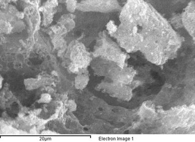
SEM, TEM and EDAX
The morphological studies were done by using SEM. In Fig.
4
, we have observed that the sample has many pores on the surface of the sample indicating that the obtained sample has porous nature.
SEM image of Fe
3
O
4Fig. 4
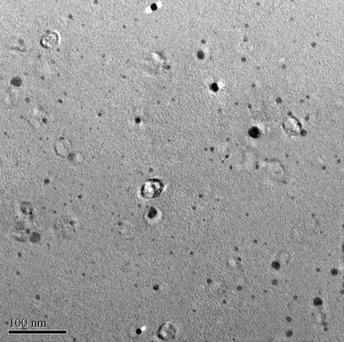
The main benefit of a TEM is that it can simultaneously give evidence in real space (in the imaging mode) and reciprocal space (in the diffraction mode). Using the TEM image, both the size and shape of the obtained nanoparticles were observed. They were spherical in shape and porous surface as seen in the Fig.
5
. The crystallite size obtained by the Scherrer’s formula and the size from the TEM confirm each other.
TEM image of Fe
3
O
4Fig. 5
The energy-dispersive X-ray spectroscopy results are shown in the Fig.
6
. It shows the presence of oxygen and iron. This confirms the existence of oxygen and iron in the sample (Fig.
6
).
EDAX of Fe
3
O
4 Antimicrobial activity of Fe
3
O
4
at control levelFig. 6
Fig. 7
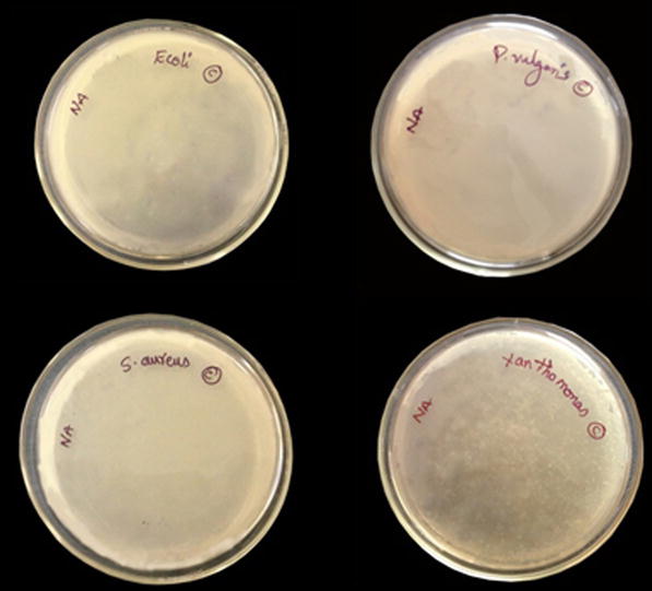
Antibacterial activity
Fe
3
O
4
showed antibacterial effect against gram-positive as well as gram-negative bacteria which clearly indicates that these nanoparticles are effective antibacterial agents. In Figs.
7
,
8
and
9
the control, low and high concentrations of Fe
3
O
4
were shown, respectively. Many antibacterial studies were made using different nanoparticles. The reason for the bactericidal activity is due to the presence of reactive oxygen species (ROS) generated by different nanoparticles [
33
]. Chemical interaction between hydrogen peroxide and membrane proteins or between the chemical produced in the presence of Fe
3
O
4
nanoparticles and the outer bilayer of bacteria could be the reason for the antibacterial activity of Fe
3
O
4
. The hydrogen peroxide produced enters the cell membrane of bacteria and kills them. It is also noted that nanoparticles continue to be in interaction with dead bacteria once the hydrogen peroxide is generated; thus foiling further bacterial action and continue to produce and release hydrogen peroxide to the medium [
34
]. In Figs.
8
and
9
we clearly see the antibacterial activity in brown and yellow colours indicating that the bacteria is completely destroyed and antibacterial activity is still active, respectively.
Antimicrobial activity of different extracts with Fe
3
O
4
at low concentration Antimicrobial activity of different extracts with Fe
3
O
4
at high concentration Activation index against various microorganismsFig. 8
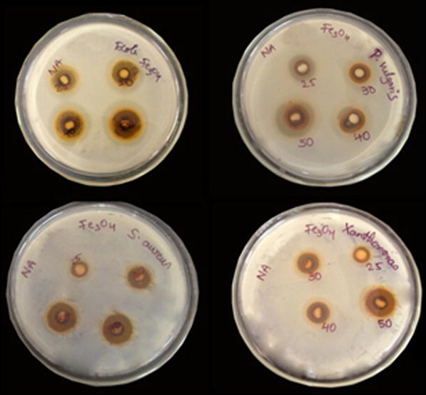
Fig. 9
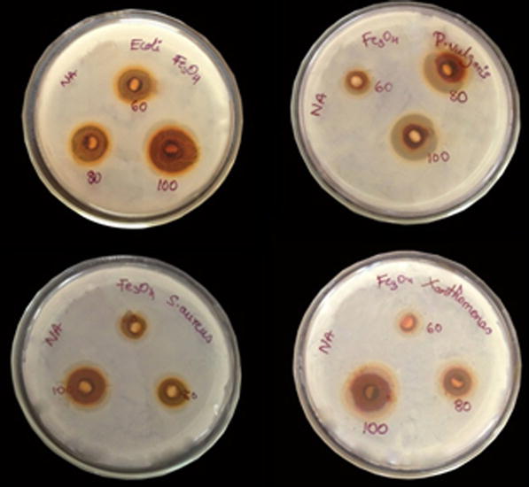
Fig. 10
The possible mechanism of action is that the metal nanoparticles are carrying the positive charges and the microbes are having the negative charges which create the electromagnetic attraction between the nanoparticles and the microbes. When the attraction is made, the microbes get oxidized and die instantly [ 35 ]. Generally, the nanomaterials release ions, which react with the thiol groups (–SH) of the proteins present on the bacterial cell surface which leads to cell lysis [ 36 ].
The central mechanism that caused the antibacterial activity by the particles might be through oxidative stress caused by ROS [
37
,
38
]. ROS includes radicals like superoxide radicals (
The nanoparticles can also produce bactericidal effects as verified by a few authors have verified. A few authors like Lee et al. [ 40 ] stated that the iron nanoparticles caused the inactivation of E. coli by zero-valent and the diffusion of the small particles ranging from 10 to 80 nm into E. coli membranes. Nano scale zero valent iron could interact with intracellular oxygen thus generating oxidative stress and ultimately triggering the interference of the cell membrane. Nanoparticles of ZnO and MgO also have revealed that with a decrease in particle size, antibacterial activity increases [ 41 , 42 ]. Likewise, Taylor and Webster also made a study studies on iron oxide nanoparticles and its bactericidal effects of on S. epidermidis [ 43 ]. They also described that bacterial inhibition depends on concentration. We need to note that iron oxide nanoparticles do not negatively impact all cells but with an appropriate magnetic field of iron oxide, nanoparticles may be engaged to destroy bacteria.
The results revealed that the microorganisms are sensitive to the test samples in varying magnitudes. The Antibacterial activity of Fe 3 O 4 nanoparticles on 4 bacterial strains is summarized in Fig. 10 . The Fe 3 O 4 Nanoparticle showed a good antibacterial activity on E. coli and P. vulgaris than the S. aureus bacterial strains. The gram-negative bacteria are more sensitive when compared to gram-positive bacteria. Earlier studies also indicate that gram-negative bacteria are less sensitive than gram-positive bacteria. A strong bactericidal activity was observed against E. coli and P. vulgaris .