Results and discussion
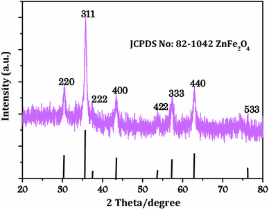
XRD patterns of all samples show very broad peaks, indicating poor crystallinity and ultrafine nature of the particles. The crystalline structure of the as-synthesized ZnFe
2
O
4
hollow sphere was characterized by powder XRD. As shown in Fig.
1
, the diffraction peaks match well with the standard patterns of the cubic structure of spinel-phase Zn ferrite (JCPDS file no. 82–1042), where the diffraction peaks at 2
θ
values of 30.0°, 35.3°, 42.9°, 53.2°, 56.7°, 62.3°, and 76.6° can be attributed to the reflection of (220), (311), (400), (422), (333), (440), and (533) planes of the spinel ZnFe
2
O
4
, respectively. No additional peak of the impurity phase was observed in the XRD patterns, showing that the prepared ferrites are pure.
XRD patterns of the as-synthesized porous ZnFe
2
O
4
hollow microspheresFig. 1
The general morphology of the as-prepared ZnFe
2
O
4
hollow sphere products observed by field emission SEM (FE-SEM) and typical images at different magnifications are shown in Fig.
2
a–c, it was observed that the major morphological feature is a regular spherical shape with an average particle diameter of about 100–210 nm. The high-magnification FE-SEM image (Fig.
2
c) reveals that each micro-hollow sphere is very similar to hollow spheres and it is actually composed of aggregates of more primary particles.
SEM (
a
–
c
) and EDS images of the as-synthesized porous ZnFe
2
O
4
hollow microspheresFig. 2

EDX results from hollow spheres showed that it was mainly composed of Zn, Fe, and O (Fig. 2 d). Quantification of the EDX spectrum showed that the ratio of Zn, Fe, and O was about 1:2:4, suggesting that the porous hollow spheres had a chemical formula of ZnFe 2 O 4 .
On the basis of the above experimental results and observations, a formation mechanism of the ZnFe
2
O
4
hollow spheres is designed, schematically illustrated in Fig.
3
. The reaction begins with the mixture of FeCl
3
·6H
2
O, ZnCl
2
·4H
2
O, Sodium acetate (CH
3
COONa 3H
2
O), EG, and PEG. In our system, the Zn
2+
and Fe
3+
ions were nucleated under hydrothermal conditions with the water generated from metal precursors to form nanosized crystalline ZnFe
2
O
4
. PEG serves as a structure-directing template, as it can easily self-assemble to form spherical grains. The Ostwald ripening process also plays an important role in the formation of nanocrystals. According to the Ostwald ripening mechanism, crystalline particles will grow into crystalline nuclei, which aggregate isotropically to form spherical grains in ethanol solution and further to microspherical crystallites. During growth, the smaller, less crystalline particles will be dissolved gradually, while the larger, more crystalline particles will grow bigger. Eventually, the core can grow gradually to form a solid sphere. As the crystal grows, the porous ZnFe
2
O
4
microsphere was formed at last. Finally, the massy porous ZnFe
2
O
4
microspheres are formed, as evidenced by XRD (Fig.
1
), SEM (Fig.
2
), TGA (Fig.
4
), and FTIR (Fig.
6
).
The proposed preparation scheme of the porous ZnFe
2
O
4
hollow microspheres TG curve of the as-synthesized porous ZnFe
2
O
4
hollow microspheresFig. 3

Fig. 4
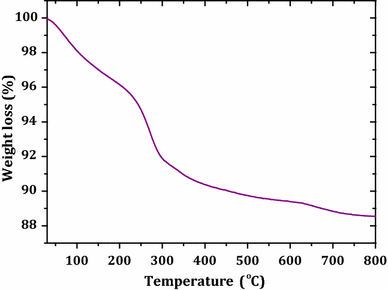
As shown in Fig. 4 , the TG curve shows that porous ZnFe 2 O 4 hollow spheres have three weight loss steps from room temperature to 800 °C under air atmosphere. The first weight loss of about 3.7 % in the range 25–220 °C was due to the loss of residual water in ZnFe 2 O 4 hollow spheres. The combustion of carbon was complete at a relatively low temperature (<400 °C). From the weight change between 150 and 400 °C, the organic matter content (carbon and oxygen containing surface groups) was determined to be 40 wt%.
The nitrogen sorption isotherm of resultant ZnFe
2
O
4
hollow spheres and their corresponding pore-size distribution curve are exhibited in Fig.
5
. The nitrogen adsorption desorption isotherm belongs to type IV category according to international union of pure and applied chemistry classification, suggesting the presence of mesopores. The specific surface area was thus estimated, by BET equation [
25
], to be 44 m
2
g
−1
. In addition, the sorption exhibits type IV isotherm, and the pore analysis has revealed that the pore sizes in the porous microspheres mainly fall into 3–9 nm, as seen in the inset of Fig.
5
. Hysteresis loops can be observed in the curve, attest to the existence of a mesoporous structure. The high surface area and pore volume further confirm that the hollow microspheres have porous structure which will broaden the application of the product.
N
2
sorption isotherm of the porous ZnFe
2
O
4
hollow microspheres. The
inset
shows the corresponding BJH pore-size distributionFig. 5

Theoretically, all MFe 2 O 4 either normal or inverse spinel oxides of transition metals have four infrared active modes. These vibrations occur at frequencies of ν 1 (650–550 cm −1 ), ν 2 (525–390 cm −1 ), ν 3 (380–335 cm −1 ), and ν 4 (300–200 cm −1 ) [ 26 ].
The ν 1 and ν 2 bands are generated due to intrinsic vibrations of tetrahedral and octahedral coordination compounds. Absorption of ν 1 happens due to the bond stretching of tetrahedral metal ions and oxygen, while ν 2 vibration is observed due to the vibration of oxygen in the direction perpendicular to the axis joining the tetrahedral ions and oxygen. The ν 3 mode is obtained from the Fe 3+ –O complexes at octahedral sites [ 27 ]. The frequency of ν 4 vibration depends on the mass of tetrahedral metal ion complexes, which gives information about the vibration of ions occupying at tetrahedral site.
In order to make sure of its chemical compositions, the Fourier transform infrared spectrometry (FTIR) spectrum of the ZnFe
2
O
4
hollow sphere product was verified. As shown in Fig.
6
, the two bands at 582 and 435 cm
−1
represented the characteristic peaks of tetrahedral and octahedral Fe–O stretching for ZnFe
2
O
4
. The appearance of bands at 1,081 and 2,925 cm
−1
is assigned to the stretching of ether groups and the characteristic absorptions of alkyl (R-CH
2
) stretching modes. In addition, the bands at 3,432 and 1,650 cm
−1
are attributed to the surface hydroxyl and the adsorbed water molecules, respectively [
28
]. Hydroxyl group contribution was observed at 3,432 cm
−1
. The appearance of these peaks in the spectrum confirmed the presence of PEG and adsorbed EG on the surface of particles. The above FTIR analysis matches well with the XRD result, which confirms that the as-obtained products are pure-phase spinel Zn ferrite.
FTIR spectra of the as-synthesized porous ZnFe
2
O
4
hollow microspheresFig. 6

Such porous oriented ZnFe
2
O
4
hollow spheres exhibited good magnetic property [
29
]. Magnetic measurements of the samples were carried out at room temperature using a PPMS with a peak field of 20 kOe. The hysteresis loops for porous zinc ferrite hollow microspheres are shown in Fig.
7
. As clearly shown, the variation of magnetization as a function of the applied field presents a narrow cycle, and the observed hysteresis loops are a characteristic behavior of soft magnetic materials. The saturation magnetization (
M
s
) of the zinc ferrite hollow spheres is about 32 emu g
−1
, which is close to the value of bulk ZnFe
2
O
4
(36 emu g
−1
) [
30
]. The low saturation magnetization of the ferrite NPs is generally believed to be due to the decreased particle size and the presence of an anti-ferromagnetic layer on the surface. In the close-up view (not shown in Figure), the curve presents a very small hysteresis loop with a remnant magnetization (
M
r
) of 1.3 emu g
−1
and a coercivity (
H
c
) of 16 Oe, denoting the ferromagnetic behavior of the sample. Table
1
shows the previously reported saturation magnetization values for zinc ferrite nanocrystallines synthesized by various methods [
31
–
37
], from which we could conclude that our present
M
s
value at ambient temperature is obviously higher than that of most of the reported uniform ZnFe
2
O
4
nanocrystallines through the facial hydrothermal synthesis.
Magnetization hysteresis loop of the porous ZnFe
2
O
4
hollow microspheres Comparison of the saturation magnetization (
M
s
) values for ZnFe
2
O
4
hollow spheres with literature values Number Method of synthesis Temp (K) Size (nm) Ref 1 Hydrothermal method 300 210 32 This study 2 Hydrothermal in ammonia solution 300 80 61.87 [ 3 Hydrothermal in supercritical methanol 5–20 4.2 38 [ 4 Oil-in-water micelles 3.7 3 70 [ 5 Co-precipitation at 373 K 55 4.2 46.9 [ 6 Sol–gel method 30 300 7.8 [ 7 Ultrasound-assisted emulsion 12 5 25 [ 8 Hydrothermal method 8 250 17.5 [Fig. 7

Table 1