Department of Chemistry, Science and Research Branch, Islamic Azad University, Tehran, IR
Department of Chemistry, Payame Noor University, Zanjan, IR
Abstract
Magnetic nanoparticles iron oxide with average sizes of 6 nm were synthesized by a chemical coprecipitation method from mixtures of FeCl
2
·4H
2
O and FeCl
3
·6H
2
O. For preparation, multi-walled carbon nanotubes (MWCNTS) with outer diameter of 50 nm, wall thickness from 1 to 2 nm and length from 500–2,000 nm were used. Characterization of the MWCNT–Fe
3
O
4
by X-ray powder diffraction (XRD), Fourier transform infrared (FTIR) spectroscopy, scanning electron microscope (SEM), transmission electron microscope (TEM), thermo-gravimetric analysis (TGA) and magnetic characterization was conducted on a vibrating sample magnetometer (VSM).
Introduction
Since discovery of their structures in 1991 [
1
], carbon nanotubes (CNTs) have attracted considerable interdisciplinary interest because of their unique physical and chemical properties; however, the hydrophobicity of CNTs may limit their application [
2
,
3
]. Therefore, surface modification of CNTs with functional groups and/or nanoparticles so as to disperse them into aqueous solutions becomes a key step for their applications [
4
].
Magnetic nanoparticles are gaining importance as they can be used as highly effective, efficient and economically-viable adsorbents, with the additional advantage of their easy separation under a magnetic field for reuse [
5
].
Herein, we describe a very simple, direct, and effective approach for the decoration of multi-walled carbon nanotubes (MWCNTs) by Fe
3
O
4
magnetic nanoparticles. Our results suggest a novel and simple approach for controlling the size and size distribution of Fe
3
O
4
on the surface of MWCNTs, which leads to a further development of CNT-based nanomaterials.
Experimental
Material
MWCNTs were purchased from NanoAmor Nanostructured & Amorphous Materials, Inc, USA [Purity >95 %, outer >50 nm, length 500–2,000 nm, surface area ~40m
2
/g, and manufacturing method catalytic chemical vapor deposition (CVD)]. Other chemicals were purchased from Merck Inc, USA.
Instrumentation
X-ray powder diffraction (XRD) analysis was conducted on a Rigaku Smart Lab Diffractometer operated at 40 kV and 35 mA using Cu Kα radiation.
The characteristics of the MWCNT-Fe
3
O
4
were analyzed by attenuated total reflection-Fourier transform infrared spectrometer (100 spectra accumulation, 2 cm
−1
resolution, BOMEM, Canada). FTIR samples were prepared by grinding dried MWCNTs and MWCNT-Fe
3
O
4
together with potassium bromide (KBr) to make a pellet that was dried in an oven for 8 h before the test.
A morphological analysis was carried out using a scanning electron microscope (SEM), (TESCAN, VEGA 3, USA).
Transmission electron microscopy (TEM) analysis was performed using a JEOL JEM microscope (TEM, JEOL 2010, Japan) operating at 200 kV by depositing sample onto the lacey carbon-coated copper grids.
Thermo-gravimetric analysis (TGA) of all samples was done using SDT Q600 instrument from TA instrument. The temperature ranged from 25 to 1,000 °C and the heating rate was 10 °C/min.
Magnetic characterization was conducted on a vibrating sample magnetometer (VSM) (PPMS VSM, Model 6000).
Synthesis of MWCNT-Fe3O4
Iron oxide nanoparticles were firstly synthesized by chemical coprecipitation according to the method reported with a minor modification [
6
,
7
].
Typically, FeCl
2
·4H
2
O and FeCl
3
·6H
2
O with a molar ratio of 1:2 were mixed in deionized water. Afterwards, 10 mL of 2 mol/L HCl was added. The solution was heated to 30 °C, under nitrogen protection with vigorous stirring. The as-formed solution was stirred at 30 °C at a constant pH value of 9(NH
3
·H
2
O was used to adjust pH value) for 1 h. The obtained precipitates were continuously mixed under constant heating at 50 °C for 30 min. After cooling to room temperature, the obtained magnetic nanoparticles (denoted as 6 nm Fe
3
O
4
) were several times washed with water and ethanol, ultrasonicated for 30 min, and then dried under vacuum at 60 °C overnight.
The typical procedure for the preparation of MWCNT–Fe
3
O
4
was as follows: 3 g MWCNTs was dispersed into 150 mL of concentrated HNO
3
(37 % by mass fraction), followed by oil bath heating at 110 °C for 5 h [
8
,
9
]. The solution was diluted by deionized water until the pH value of the filtrate was around 7. Such treatment decorated –OH and –COOH groups onto the surface of MWCNT, which was confirmed by infrared spectrum analysis. After further rinsing and drying, 200 mg acid-purified nanotubes and 50 mg Fe
3
O
4
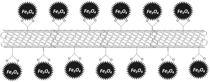
nanoparticles were dispersed in 40 mL solution of deionized water/ethanol (1:1, volume ratio). The mixture was ultrasonicated for 1 h and then stirred at room temperature for 96 h. The solution was separated from the residue using 200 nm filter membrane, followed by vacuum drying at 50 °C for 16 h. A simplified scheme of MWCNT–Fe
3
O
4
is illustrated in Fig.
1
.
Fig. 1
Illustration of MWCNT–Fe3O
4
Results and discussion
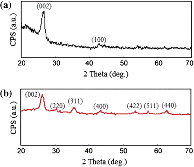
XRD analysis
XRD powder patterns of MWCNT–COOH and MWCN–Fe
3
O
4
hybrid are shown in Fig.
2
a and b respectively. The diffraction peaks at 2theta of 26,088 and 43,088 are attributed to the graphite structure (002) and (100) planes of the MWCNTs [
10
] in Fig.
2
a, b showing that the hybrid is composed of two phases: cubic Fe
3
O
4
and MWCNTs. No obvious peaks from other phases were observed [
11
]. The main peaks of Fe
3
O
4
in the XRD pattern are broadened, indicating the smaller crystallite size of Fe
3
O
4
NPs [
10
]. The mean size of the crystallites of Fe
3
O
4
in the MWCNT–Fe
3
O
4
(assuming spherical morphology) was estimated as 9 ± 3 nm from the diffraction pattern by X-ray line profile fitting [
11
].
Fig. 2
XRD powder pattern of
a
MWCNT–COOH and
b
MWCNT–Fe
3
O
4
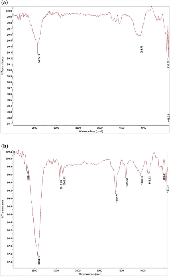
FTIR analysis
The FTIR spectra of the samples are shown in Fig.
3
. The band observed near 1,580 cm
−1
in all samples shows the presence of the cylinder like carbon structure (rolled graphene sheet). According to Jishi et al. [
12
], several infrared active modes may have a wavenumber near 1,580 cm
−1
, while the wavenumbers depend on the geometry of the CNT. Besides, infrared wavenumbers are dependent on the nanotube diameter. The broadness of the band observed at 1,580 cm
−1
for SWCNT samples can be explained by the polydispersity in the geometry of nanotubes. The stretching C=O vibration of the carboxyl (COOH) group is visible in the spectra of the modified carbon nanotubes. It was also observed at 1,713 cm
−1
for MWCNT–COOH, and at 1,724 cm
−1
for MWCNT–Fe
3
O
4
. Compared to MWCNT–COOH, this band reduced in the spectrum of MWCNT–Fe
3
O
4
. This reduction was ascribed to the linkage between magnetite particles and nanotubes, which was formed by a reaction of carboxyl groups with the surface of magnetite particles [
13
]. In the spectra of MWCNT–Fe
3
O
4
, the broadband around ca. 585 cm
−1
showed the presence of iron oxide, primarily magnetite [
13
].
Fig. 3
FT-IR spectrum of
a
MWCNT–COOH and
b
MWCNT–Fe
3
O
4
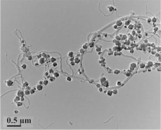
TEM analysis
As shown in Fig.
4
, the morphologies of the samples were observed by TEM.
Fig. 4
Representative TEM images of MWCNT–Fe
3
O
4
composites
Figure
4
depicts an entangled network of acidified MWCNTs, which are attached to clusters of iron oxides. The iron oxide nanoparticle aggregates were composed of even smaller subunits of about 10 nm [
14
], implying that large particles may be formed via precipitation followed by a step-like aggregation process. In Fig.
4
, MWCNTs are covered with the carboxylic acid (COOH) component in which iron oxide nanoparticles were uniformly dispersed without obvious aggregation. The carboxylic acid component grafted on the surface of the MWCNT was not destroyed in the process of loading iron oxides. Carboxylic acid can form complexes with metal ions because of their high number of coordinating functional groups [
15
]. This Figure shows representative TEM images of the MWCNT–Fe
3
O
4
nanocomposites.
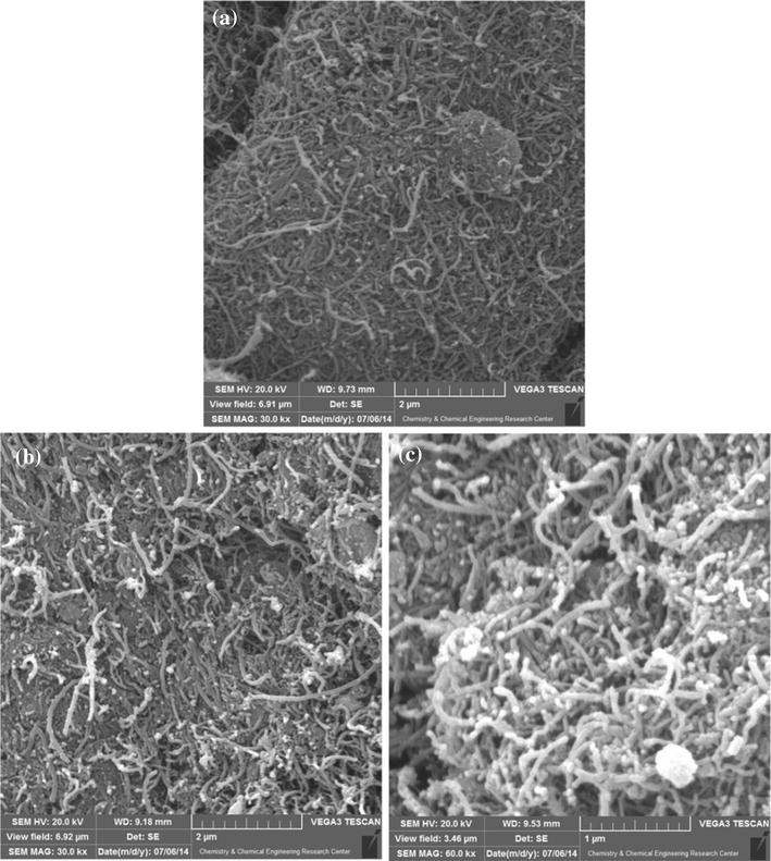
SEM analysis
Scanning electron microscopy shown in Fig.
5
is the typical morphology of the MWCNT–COOH (Fig.
5
a), and MWCNT–Fe
3
O
4
(Fig.
5
b, c). SEM image (Fig.
5
b, c) of the composites depicts an entangled network of oxidized MWCNTs (MWCNT–COOH) with clusters of iron oxides attached to them. The Surface area of the prepared composite was measured using BET method. The specific surface area of MWCNT–Fe
3
O
4
composite was 92 m
2
/g. Under the reaction conditions employed, four iron oxides are commonly formed. These are Fe
3
O
4
(magnetite), γ-Fe
2
O
3
(maghemite), α-Fe
2
O
3
(hematite) and α-FeO(OH) (goethite). Among them, two magnetite and maghemite are magnetic [
16
]. Most of the nanoparticles formed showed a tendency to congregate. Similar results were reported by Fan and Li [
13
].
Fig. 5
SEM images of
a
the MWCNT–COOH,
b
,
c
MWCNT–Fe
3
O
4
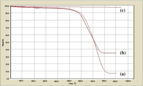
TGA
TGA was used to characterize the iron oxide content in the as-obtained composites. For the iron oxide sample (Fig.
6
c), the weight increase at the broad temperature is attributable to the oxidation of the Fe
3
O
4
into Fe
2
O
3
. The TGA curve of MWCNTs (Fig.
6
a) shows a small mass loss at lower temperature because of the removal of absorbed water and the functional groups. An obvious weight loss in the temperature range of 635–730 °C is caused by the oxidization of the nanotubes [
17
]. Compared with the pristine MWCNTs, the MWCNT–Fe
3
O
4
composites lost their weight at a lower temperature range of 490–635 °C (Fig.
6
b), leaving iron oxide residue weight of around 26.65 % for 6 nm Fe
3
O
4
/MWCNT a. This is attributed to the catalytic role of metal oxide nanoparticles in the oxidation of carbon materials [
18
].
Fig. 6
TGA curves of
a
MWCNTs,
b
6 nm MWCNT–Fe
3
O
4
composite, and
c
Fe
3
O
4
VSM analysis
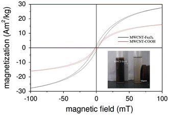
The magnetic property of the MWCNT–Fe
3
O
4
nanocomposite was investigated by VSM. Figure
7
shows the magnetization curves measured at 25 °C for the sample fabricated at 200 °C for 12 h. Magnetization increased with an increase in the magnetic field. MWCNT–COOH possessed good magnetic properties, although the saturation magnetization was slightly lower than that of MWCNT–Fe
3
O
4
. Both compounds exhibited an extremely small hysteresis loop and low coercivity, as typically characteristic of superparamagnetic particles [
19
]. It is worth mentioning, this analysis shows that the saturation magnetization of the magnetic MWCNT composites was different and is significantly higher at functionalized samples.
Fig. 7
Magnetization curves of MWCNT–Fe
3
O
4
composite and MWCNT–COOH at room temperature
Conclusions
A simple, effective and reproducible method has been carried out for synthesizing MWCNT–Fe
3
O
4
magnetic nanocomposites. Magnetic measurements proved that MWCNT–Fe
3
O
4
composite had superparamagnetic characteristics at room temperature. This study would help to develop a new composite material with excellent properties employed in large-scale fabrication of magnetic CNT hybrid materials.
Acknowledgments
HS, RS and MK would like to thank Islamic Azad University for all supports.
Ouyang et al. (2002) One-dimensional energy dispersion of single-walled carbon nanotubes by resonant electron scattering 88(6) 10.1103/PhysRevLett.88.066804
Thostenson et al. (2001) Advances in the science and technology of carbon nanotubes and their composites: a review 61(13) (pp. 1899-1912) 10.1016/S0266-3538(01)00094-X
Bahr et al. (2001) Dissolution of small diameter single-wall carbon nanotubes in organic solvents? (pp. 193-194) 10.1039/b008042j
Singh et al. (2013) Functional oxide nanomaterials and nanocomposites for the removal of heavy metals and dyes
Giersig and Hilgendorff (2005) Magnetic nanoparticle superstructures (pp. 3571-3583) 10.1002/ejic.200500497
Unknown ()
Chen et al. (2007) Tuning of redox properties of iron and iron oxides via encapsulation within carbon nanotubes 129(23) (pp. 7421-7426) 10.1021/ja0713072
Fan and Li (2012) Preparation and magnetic properties of multiwalled carbon nanotubes decorated Fe3O4 nanoparticles 50(10) 10.1016/j.carbon.2012.04.015
He et al. (2010) The attachment of Fe3O4 nanoparticles to graphene oxide by covalent bonding 48(11) (pp. 3139-3144) 10.1016/j.carbon.2010.04.052
Gupta et al. (2011) Chromium removal by combining the magnetic properties of iron oxide with adsorption properties of carbon nanotubes 45(6) (pp. 2207-2212) 10.1016/j.watres.2011.01.012
Fan and Li (2012) Preparation and magnetic property of multiwalled carbon nanotubes decorated by Fe3O4 nanoparticles 27(2) (pp. 111-116) 10.1016/S1872-5805(12)60007-9
Chang et al. (2011) Characterization of magnetic soluble starch-functionalized carbon nanotubes and its application for the adsorption of the dyes 186(2) (pp. 2144-2150) 10.1016/j.jhazmat.2010.12.119
Yao et al. (2003) Polymerization from the surface of single-walled carbon nanotubes-preparation and characterization of nanocomposites 125(51) (pp. 16015-16024) 10.1021/ja037564y
Perales Perez et al. (1998) Precipitation and densification of magnetic iron compounds from aqueous solutions at room temperature 50(3) (pp. 223-242) 10.1016/S0304-386X(98)00054-1
Zhang et al. (2003) Thermogravimetric analysis for the array of C60 molecules formed in single-wall carbon nanotube 369(5) (pp. 680-683) 10.1016/S0009-2614(03)00036-8
Li et al. (2007) Preparation of nanocomposites of metals, metal oxides, and carbon nanotubes via self-assembly 129(30) (pp. 9401-9409) 10.1021/ja071122v
Liu et al. (2008) Fiberlike Fe2O3 macroporous nanomaterials fabricated by calcinating regenerate cellulose composite fibers 20(11) (pp. 3623-3628) 10.1021/cm703623v