Results and discussion
Characterization of thioglycolic acid-capped CdS quantum dots
Synthesized thioglycolic acid-capped CdS QDs (TGA-capped CdS QDs) are optically characterized by UV–vis absorption spectroscopy and fluorometry. The TGA-capped CdS QDs show long emission wavelengths and a high separation between the excitation and emission wavelengths (ca. 125 nm) simplifying fluorescence measurements. The absorption spectrum shows well-defined excitonic absorption peak at wavelength of 427 nm, and a sharp band edge emission peak (
λ
= 555 nm), which was independent of the excitation wavelength, was obtained in the FL spectrum (Fig.
1
).
a
Absorption,
b
Fluorescence spectra and
c
TEM image of synthesized aqueous TGA-capped CdS QDsFig. 1
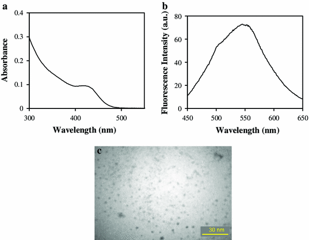
Also TEM (transmission electron microscopy) image of Synthesized QDs is shown in Fig. 1 c.
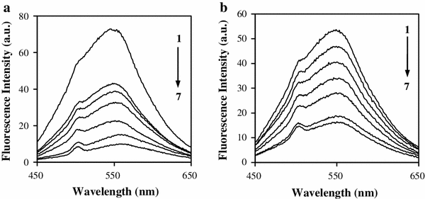
Effect of cations on the fluorescence of QDs
The fluorescence intensity (
λ
ex
= 427 nm) of quantum dots drastically decreases with some cations in a certain condition. It is observed that synthesized QDs have high tendency for Pb
2+
and Cr
3+
. The more the Pb
2+
or Cr
+3
concentration, the lower was the FL intensity obtained. This decrease was proportional to each cation concentration (Fig.
2
).
Fluorescence spectra of aqueous TGA-capped CdS QDs in the presence of different concentrations of
a
Cr
3+
and
b
Pb
2+
.
a
QDs 75 µL, phosphate buffer 0.01 M, pH = 6 (1–7: 0, 25, 50, 100, 200, 300, 400 ng mL
−1
).
b
QDs 100 µL, acetate buffer 0.01 M, pH = 5 (1–7: 0, 1, 2.5, 5, 10, 50, 75 ng mL
−1
)Fig. 2
Optimization of condition
It is clear that pH influences the fluorescence intensity of the complex significantly. To investigate the effects of pH on determinations, the pH ranges between 4 and 9. At lower pHs, there is no fluorescence intensity because of the protonation of the thiol groups and aggregation of the QDs [
35
]. The maximum value of
F
0
/
F
was obtained when pH was 6 and 5 for Cr
3+
and Pb
2+
, respectively (Fig.
3
). (
F
0
and
F
are the fluorescence intensity of the aqueous CdS QDs without and at a given analyte concentration). Therefore, these optimal pH values were chosen to be applied in further experiments.
Effects of pH on the
F
0
/
F
for
a
Cr
3+
10 ng mL
−1
(QDs = 100 µL, phosphate buffer 0.05 M) and
b
Pb
2+
5 ng mL
−1
(QDs = 75 µL, acetate buffer 0.05 M)Fig. 3
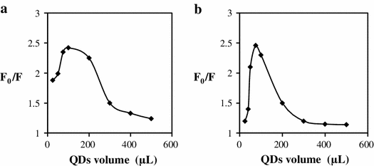
It was found that the concentration of QDs affected not only the fluorescent intensity but also the sensitivity of the assay. As shown in Fig.
4
, high concentration of aqueous QDs decreases the sensitivity and causes self-quenching of the QDs fluorescence. Also, very low concentration of QDs gives very weak fluorescence intensity, which may result in narrow linear range. Thus, 100 and 75 µL of QDs was chosen for Cr
3+
and Pb
2+
determinations, respectively. In selected optimal values of the quantum dots, wide linear range and low detection limit concentrations are achieved.
Effects of QDs amount on the
F
0
/
F
for
a
Cr
3+
10 ng mL
−1
(phosphate buffer 0.05 M, pH = 6) and
b
Pb
2+
5 ng mL
−1
(acetate buffer 0.05 M, pH = 5)Fig. 4
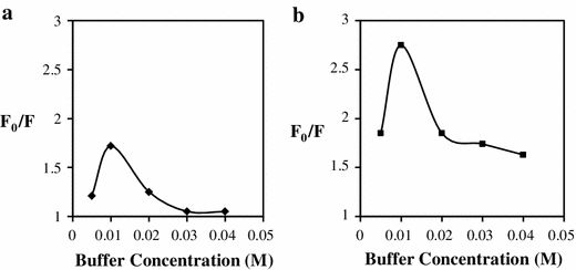
The effect of buffer solution on fluorescence intensity system was discussed. To do so, different buffer solutions such as phosphate, tris and acetate were investigated. The results showed that phosphate and acetate have the greatest effect on the fluorescence quenching of TGA-capped CdS QDs in the presence of Cr
3+
and Pb
2+
, respectively. Hence, these buffers were selected in all experiments. Optimization of buffer concentration was carried out and the result is showed in Fig.
5
. Maximum signal was obtained in 0.01 M for both states.
Effects of buffer concentration on the
F
0
/
F
for
a
Cr
3+
10 ng mL
−1
(QDs = 100 µL, phosphate buffer pH = 6) and
b
Pb
2+
5 ng mL
−1
(QDs = 75 µL, acetate buffer pH = 5)Fig. 5
In results, the best optimal conditions for the measurement of Cr 3+ are: QDs = 100 µL, phosphate buffer 0.01 M, pH = 6, and for Pb 2+ are: QDs = 75 µL, acetate buffer 0.01 M, pH = 5.
Quantitative characteristics
Under optimal condition, the ratio of FL intensity (
F
0
/
F
) in the absence (
F
0
) and presence of analyte (
F
) was plotted versus the concentration of each analyte in ng mL
−1
. Calibration graphs and corresponding FL spectra are shown in Fig.
6
. Obtained analytical characteristics are indicated in Table
1
.
Calibration curve of the
a
Cr
3+
and
b
Pb
2+
in optimum condition Analytical characteristics of the method for the determination of Cr
3+
and Pb
2+ Parameter Cr3+ Pb2+ Linear range (ng mL−1) 3–400 0.8–100 Slope 0.0024 0.0098 Intercept 1.065 1.057 Correlation coefficient ( 0.9993 0.9979 Number of points 10 10 LOD (ng mL−1) 1.2 0.8 RSD% ( 3.2 (8 ng mL−1) 2.1 (5 ng mL−1) RSD% ( 3.0 (50 ng mL−1) 3.9 (10 ng mL−1) RSD% ( 1.4 (300 ng mL−1) 2.4 (50 ng mL−1)Fig. 6
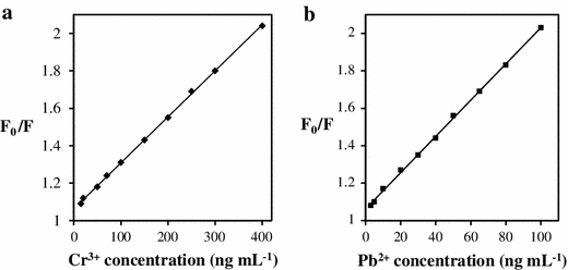
Table 1
The quenching ability of Cr 3+ and Pb 2+ can be expressed by Stern–Vollmer equation: where F and F 0 are fluorescence quantum yield or fluorescence intensity of quantum dots with ( F ) and without ( F 0 ) extinguishing or analyte. K sv is the Stern–Vollmer constant. Linear equations between concentration and relative fluorescence intensity obtained for Cr 3+ and Pb 2+ ( y = 0.0024 x + 1.065 and y = 0.0098 x + 1.057, respectively) are consistent with Stern–Vollmer equation.
Interference of coexisting foreign substances
In order to investigate the effect of coexisting substances on Cr
3+
and Pb
2+
determination in real samples, various concentrations of interfering ions are added to determination solution (1 μg mL
−1
analyte) and,
F
/
F
0
value is measured versus interference ion concentration. Interference limit is considered 5 % deviation from the initial
F
/
F
0
value (Table
2
). The results show that the method for analysis of Cr
3+
and Pb
2+
is free from any interference.
Interfering effects of some coexisting ions on Cr
3+
and Pb
2+
determination Coexisting ions Interference ratio for Cr3+ Interference ratio for Pb2+ Hg2+, Fe3+ 800 800 Cd2+ 300 300 Zn2+ 500 300 Na+ 2,000 2,000 Co2+ 150 150 Cu2+ 200 250 Li+, Ni2+, K+, NO3− 500 500 5,000 4,000 4,000 400 Cl− 7,000 600Table 2
Analysis of real samples
The represented method was successfully applied to the quantitative determination of Cr
3+
and Pb
2+
in the real samples of various compositions such as Nahand dam, Karkaj and Azarshahr well, tab and mineral waters. The real samples were spiked with standard solution of each analyte, and then were analyzed by standard addition method. In Tables
3
and
4
, the obtained recovery values in real samples are listed.
Results for the determination of Pb
2+
in the real samples Sample (Pb2+) Add (ng mL−1) Founda (ng mL−1) purposed method Recovery % Founda (ng mL−1) standard method Karkaj well water 0 4.22 ± 0.2 – 4.51 ± 0.3 5 9.28 ± 0.1 101 ± 2.0 – 10 14.40 ± 0.3 102 ± 2.9 – Nahand dam water 0 0 – Not detect 5 4.85 ± 0.11 97.2 ± 2.2 – 10 10.38 ± 0.21 104 ± 2.3 – Azarshahr well water 0 Not detect – Not detect 5 5.12 ± 0.1 102 ± 2.1 – 10 10.14 ± 0.2 101 ± 1.9 – Mineral water 0 Not detect – Not detect 5 5.21 ± 0.1 104 ± 1.8 – 10 10.38 ± 0.3 104 ± 2.9 – Tabriz tab water 0 Not detect – Not detect 5 5.11 ± 0.1 102 ± 1.7 – 10 10.09 ± 0.3 101 ± 2.5 – Results for the determination of Cr
3+
in the real samples Sample (Cr3+) Add (ng mL−1) Founda (ng mL−1) purposed method Recovery % Founda (ng mL−1) standard method Karkaj well water 0 5.81 ± 0.12 – 5.48 ± 0.31 5 10.72 ± 0.18 97.2 ± 3.6 – 10 15.58 ± 0.25 97.7 ± 2.4 – Nahand dam water 0 12.50 ± 0.20 – 4.51 ± 0.14 5 17.70 ± 0.15 104 ± 2.8 – 10 22.29 ± 0.32 97.9 ± 3.3 – Azarshahr well water 0 9.16 ± 0.11 – 4.51 ± 0.22 5 14.37 ± 0.15 104 ± 3.1 – 10 19.00 ± 0.30 98.4 ± 3.2 – Mineral water 0 Not detect – Not detect 5 5.13 ± 0.14 103 ± 2.8 – 10 10.05 ± 0.12 101 ± 1.1 – Tabriz tab water 0 Not detect – Not detect 5 4.88 ± 0.19 97.6 ± 3.7 – 10 9.72 ± 0.30 97.2 ± 2.9 –Table 3
Table 4
The recoveries were between 97 and 104 %, showing that the proposed procedure is applicable to the determination of Cr 3+ and Pb 2+ in environmental samples.
On the other hand, the results were compared with those obtained by a standard method using electrothermal atomic absorption spectroscopy. Statistical analysis using Student t test showed that there are no significant differences between the results of two methods.