Review
Introduction
At present lung cancer accounts for 23% of total cancer-related mortality, outnumbering breast cancer, colon cancer, and prostate cancer combined together [ 1 , 2 ]. The extreme lethality of lung cancer is ascribed to the lack of early diagnostic strategies as in almost 50% of the cases the disease is confirmed in stage IV, leaving low chance of survival [ 3 ]. The inaccessibility to the deeper portions of the lung for conventional therapy further adds up to the complication in the treatment process [ 4 ].
The incidence of lung cancer can be broadly classified into two major types on the basis of histologic appearance, one being small cell lung cancer (SCLC) and the other being non-small cell lung cancer (NSCLC). SCLC is less prominent and more aggressive with mean survival of 4 months if left untreated [ 5 ]. Its extreme lethality roots from rapid growth rate, early metastasis, and fast metabolism. SCLC originates from neuroendocrine tumors and is thus studded with neurosecretory vesicles and neurofilaments [ 6 ]. It accounts for almost 80% to 85% of the lung cancers and is not susceptible to conventional chemotherapy and radiation therapy. NSCLC can be further subclassified into epidermoid, large cell, broncho-alveolar, adenocarcinoma, and squamous cell carcinoma [ 7 ]. Each of these NSCLC histological subtypes is distinct and responds in diverse means to specific therapies.
Tobacco smoking has been identified as the major cause of both types of lung cancers (i.e., SCLC and NSCLC) owing to exposure of pulmonary system to aromatic mutagenic agents present in inhaled smoke [ 8 ]. The only subclass of lung cancer that is not associated with smoking is adenocarcinoma which arises due to occupational and environmental exposure to carcinogenic agents such as radon, asbestos, and other types of radiation. Apart from this, factors such as familial predisposition to lung cancer, genetic alteration ( alk , met , ros1 genes, etc.), and Helicobacter pylori infection form a minor class of lung cancer instigators [ 9 , 10 ].
Current therapeutic strategies such as chemotherapy and radiation therapy is only effective in the initial stages of treatment of SCLC, whereas NSCLC are less sensitive to such treatment modalities, which leaves surgery (only in stages I, II, and some of IIIA) and gene therapy as other possible alternative to tackle NSCLC [ 11 ] and lung cancer stem cells. Thus, the complete eradication of lung cancer requires a new approach such as utility of nanoscale materials. It is by the virtue of nanoscale dimension of lung cancer therapeutic and/or diagnostic system that they are capable of effectively transcending bronchial epithelium barrier and accumulating in deep lung regions. Some of such nanoscale formulations that have given promising results include nanogels or nano-sprays which are intratracheally administered into the lungs, and the results have confirmed that intratracheal means of drug delivery for lung cancer therapy are much better than the parenteral route. In one such approach, inhalable Ex4-C16-loaded DOCA-GC nanogels were synthesized for treatment of hyperglycemia. The therapeutic efficacy of this nanogel formulation was monitored in type 2 diabetic C57BL/6 db/db mice, and the cytotoxicity associated with them was established by using A549 and Calu-3 cell lines. The use of chitosan-based nanogels for pulmonary delivery did not instigate any immune response and prolonged hyperglycemic effect even at lower concentration of drug. This work establishes the possibility of using such nanogel-based pulmonary delivery system for delivery of anticancer drugs specifically to lung cancer cells [ 12 ].
As a prerequisite to device a nanoscale therapeutic system, its therapeutic, diagnostic, and the delivery system should be of nanoscale dimension. The nanoscale lung cancer therapeutic agents encompass nanoscale formulations of metal nanoparticles, chemotherapeutic drugs, and herbal extract, whereas nanoscale delivery system for lung cancer includes metal nanoparticles, polymeric nanoparticles, liposomes, and protein cages.
The extensive research in the field of nanotechnology has opened up a whole new range of nanomaterials for cancer therapy and diagnosis [
13
,
14
]. The applications of these nanoparticles in cancer therapies has been effective to a great extent owing to their inherent small dimensions which enables them to specifically accumulate in tumor cells as they permeate through the leaky vasculature in the vicinity of tumor cell mass (enhanced permeability and retention effect) [
15
]. The poorly developed lymphatic drainage also contributes indirectly to nanoparticle accumulation at the site of the tumor. Another advantage of nanoscale system is that they are capable of effectively overcoming clearance by the kidney and thereby provide good blood circulation time for the drugs they carry. Apart from these benefits, the most favorable property of such a system is its ability to support high loading capacity of therapeutic and imaging agents owing to high surface-area-to-volume ratios of nanoparticles [
16
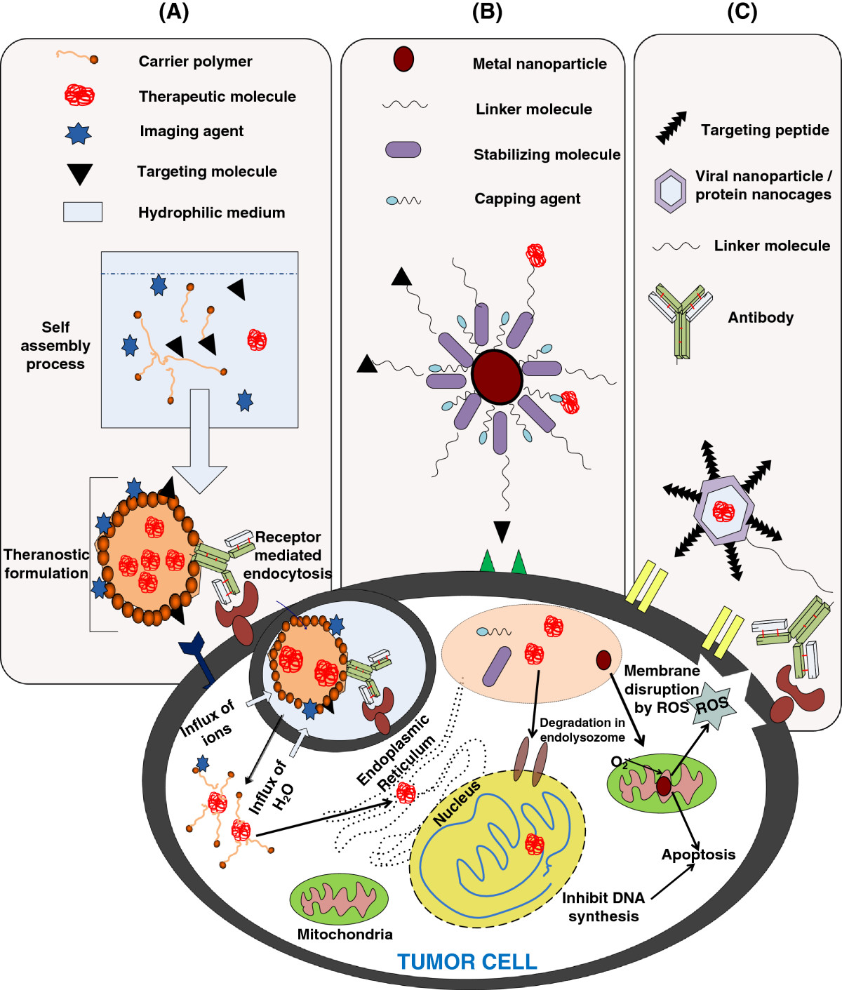
]. Further functionalization with specific targeting molecules and stabilizing agent (e.g., PEG (poly(ethylene glycol)) can result in fabrication of targeted nanotheranostic agent against lung cancer cells. The area of nanomedicine is too broad to cover all the aspects in a single review article. Thus, here we emphasize on nanomaterials that have shown great promise for applications in lung cancer diagnosis and therapy. This review is broadly divided into three sections: (1) polymeric nanoparticles-based approaches (2) metal nanoparticles-based approaches, and (3) bio-nanoparticles-based approaches. The schematic representation of these approaches is depicted in Figure
1
.
Schematic representation of different approaches for cancer therapy and diagnosis.
(A)
Polymeric nanoparticle-based approach,
(B)
metal nanoparticle-based approach, and
(C)
bio-nanoparticle-based approach.Figure 1
Polymeric nanoparticles-based approaches
Polymeric nanoparticles provide a common platform for inclusion of a drug of therapeutic potential, an imaging agent, and an appropriate targeting moiety to end up with a perfect nanotheranostic drug delivery system. The versatility in physiochemical modification of polymer properties enables it to be tuned to the requirements for drug encapsulation. Apart from being biodegradable and biocompatible, these polymeric systems are capable of giving rise to sustained-release profile of the drugs encapsulated. In addition to chemotherapeutic drugs, the polymer systems have been fabricated to carry nucleic acids and proteins to effectuate their therapeutic potential over target tumor cells. The most commonly used polymer systems for lung cancer therapeutics includes poly(ϵ-caprolactone) (PCL), polylactic acid (PLA), poly(lactide-
co
-glycolide) (PLGA), alginic acid, gelatin, and chitosan. The biodegradability and toxicity of carrier polymers are monitored closely in case of pulmonary application, as remnant polymers can interact with the bio-surfactants present in the alveoli which can further lead to a cascade of events eventually leading to severe inconvenience in breathing. Few of such polymer-based pulmonary therapeutic approaches have been enlisted in Table
1
.
Polymeric formulations for pulmonary delivery of therapeutic or imaging agents Carrier molecule Therapeutic/imaging agent Model system under study References PLGA Paclitaxel HeLa and NMRI mice [ 9-Nitro-camptothecin [ pDrive-sh AnxA2 plasmid DNA Preclinical (mice) [ PEI PEI pCMV Luc DNA Intravenous injection in mice [ PEG-PEI copolymer A549, Calu-3 cells, and preclinical (mice) [ PEI p53 Plasmid Intravenous injection and aerosol inhalation in mice [ B16-F10 tumor-bearing mice [ PEG PEG-PLGA NF-κB decoy Explanted lungs from patients with PAH and rat models [ Poly- Genomic DNA of Injected into mice through intranasal route [ PEG-substituted PLL Firefly luciferase Injected into C57BL/6 mice through intranasal route [ PEGylated gelatin nanoparticle pCMV β-gal Intravenously and intratumorally administered to LLC-bearing female C57BL/6 J mice [ Chitosan Chitosan/tripolyphosphate nanoparticles Model therapeutic protein insulin Lysozyme in PBS [ Chitosan oligomer polyplexes FITC-labeled pCMV-Luc HEK 293 cells [ Liposomes flt-1 gene-encoding tyrosine kinase Pulmonary arterial endothelial cells of rabbits [ Recombinant human superoxide dismutases Intravenously injected into anesthetized pigs [ Solid lipid nanoparticles Cyclosporine A, calcitonin, and somatostatin Administered by parenteral routes or by oral, nasal, and pulmonary routes in rats [ Branched polyester 5(6)-Carboxyfluorescein Rabbit lung model [ Others Glycerol and poloxamer-188 T cell-specific surface antigen Human bronchial cell line, Calu-3 cells [Table 1
In addition, encapsulation of such nanotheranostic systems within polymer alters bio-distribution by making the uptake and distribution properties primarily those of the carrier, rather than those of the neat therapeutic, thereby increasing circulating half-life, avoiding degradation of therapeutic in transit to the delivery site.
Poly(d,l-lactide-co-glycolide)
PLGA is among the most successful Food and Drug Administration (FDA)-approved biodegradable polymers used for formulation of a nanoscale drug delivery system. Apart from drugs, PLGA can be used for delivery of proteins and various other bio-macromolecules such as RNA, DNA, and peptides [ 35 – 37 ].
Considering the specific case of lung cancer, the polymer PLGA has proved to be a prospective carrier molecule. In one such attempt by Wu et al. in 2001 [ 38 ], endostatin-loaded PLGA microspheres were fabricated by emulsification-evaporation technique. This system could attain the desired therapeutic effect at lower concentration of drug thus avoiding predisposition of normal healthy cells to cytotoxic drugs.
In an approach to effectively eliminate NSCLC, simultaneous administration of cytotoxic and antiangiogenic drugs was carried out to exploit their synergistic effects. In order to accommodate such combinations in a single delivery formulation, a research group headed by Sengupta et al. in 2005 [ 39 ] fabricated bi-phospholipid-coated PLGA core nanoparticles wherein doxorubicin (doxo) is conjugated to PLGA while comberstatin is mixed with phospholipid and encapsulated in the outer lipid bilayer.
In order to employ PLGA for delivery of nucleic acids to lung cancer cells, it is necessary to introduce positive groups to form stable polyionic complex with nucleic acids. In a recent study to deliver nucleic acid, PLGA coupled to the diamine derivatives of PVA (polyvinyl alcohol), (DEAPA (3-(diethylamino)propylamine)-PVA- g -PLGA) was used as delivery vehicles for siRNA into H1299-EGFP cells (lung cancer cells expressing green fluorescent protein). These lung cancer cells exhibited energy-dependent and clathrin-mediated cellular uptake of drug-loaded microspheres for initial 2 h. The extent of cellular uptake of these particles was further improved by addition of lung surfactant to the carrier molecules [ 40 ]. In another attempt by Nguyen et al. in 2008, human lung epithelial (H1299 luc) cell lines were successfully transfected using tertiary-amine-modified PVA grafted over PLGA as siRNA delivery construct [ 41 ].
A drawback of PLGA which limits its application as a drug carrier for lung cancer therapy is its rapid clearance from the circulatory system. In order to address this issue, PEG-modified PLGA was adapted as and the inclusion of PEG moiety effectively increased the blood circulation time of the carrier molecule [ 42 ]. PLGA-PEG copolymeric nanoparticles were employed as a common platform for coupling imaging agent superparamagnetic iron oxide nanoparticle (SPION) and an anticancer drug molecule, doxo hydrochloride, as an effective theranostic strategy against lung cancer. Until recent past, there has not been any significant report of inclusion of tumor-targeting peptide into such PLGA-based carrier for lung cancer; thus, work related to this is under way in our laboratory.
Chitosan
Chitosan bears number of ionisable amino group which could be scaled easily to serve the need for the delivery of the therapeutic agent. Its versatile nature along with its non-toxic, biodegradable, and bio-adhesive nature has led to its wide-scale application in drug delivery. Owing to the cationic nature of chitosan, it is generally employed for the delivery of nucleic acids to lung cancer cells. Chitosan in dry powder form was evaluated as a carrier for intratracheal delivery of pCMV-Muβ-encoding murine interferon-β in mice pre-infused with appropriated doses of CT26 cells [ 43 ]. The system attained therapeutic levels at the target site at lower dosage level and improved the mean survival time of mice to a significant extent. In another similar study, Okamoto et al. in 2003 used low molecular weight chitosan as a vector for delivering pCMV-Luc gene into the lung cancer cells through nasal administration into the mice model [ 44 ]. This system exhibited high transfection rate and higher expression of the luciferase protein in the cells lining the walls of the bronchioles.
Though chitosan has produced significant results in delivery of siRNA into H1299 lung cancer cell lines, its in vivo application is limited because of its interaction with extracellular molecules such as hyaluronic acids. To avoid such non-specific interactions, Varkouhi et al. in 2010 introduced thiol groups in trimethyl chitosan (TMC) [ 45 ]. The siRNA/thiolated chitosan polyplex system attained 60% to 80% gene silencing activity in H1299 human lung cancer cells and has been carried over for in vivo evaluations.
While considering chitosan as carriers for pulmonary delivery of chemotherapeutic drugs, a derivative of chitosan with groups capable of encapsulating the drug is to be introduced. The most common anticancer drug administered for NSCLC is paclitaxel (PTX). A chitosan derivative, i.e., N -((2-hydroxy-3-trimethylammonium)propyl)chitosan chloride (HTCC) was investigated as carrier for PTX by Lv et al. in 2011 [ 46 ]. The nanocarrier synthesized had a diameter of 130 nm (roughly) with high PTX loading efficiency. These PTX-loaded HTCC nanoparticles (HTCC-NP:PTX) were assessed for in vitro cytotoxicity and they exhibited preferential accumulation in subcutaneous tumor tissues as a result of enhanced permeability and retention (EPR) effect.
A second generation drug, gemcitabine, has been effective in NSCLC treatment; and in order to attain effective delivery of gemcitabine to NSCLC cells, Ventura et al. in 2011 came up with chitosan-dextran-based delivery system [ 47 ]. In this approach gemcitabine was encapsulated in chitosan microspheres with different amount of dextran sulfate by spray-drying technique. The nanoparticle construct was found to have porous surface morphology with a size range of 1 to 5 μm. The addition of dextran sulfate improved the release profile to a prolonged duration of 30% over 4 days from 70% in 30 min. The cytotoxicity of the construct was carried out in vitro on human lung cancer cell line A549.
Dendrimers
Dendrimers has its own significance in the field of cancer therapeutics and diagnosis [
48
]. The word
dendrimer
has its origin from two Greek words, the word
dendron
, meaning tree, and
meros
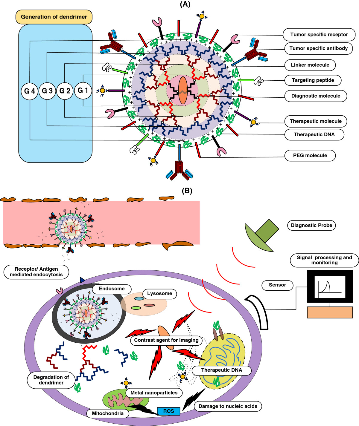
, meaning part. It is by virtue of its unique branched, multivalent, globular architectural design that it has extensive medical applications such as drug delivery, gene transfection, tumor therapy, and diagnostics. The feasibility of extensive range of surface functionalization of dendrimers with targeting, therapeutic and diagnostic molecules provides scope for effective therapy and diagnosis of lung cancer. A schematic representation of dendrimer-based theranostic system is depicted in Figure
2
.
Schematic representation of (A) dendrimer structure and (B) mode of action of dendrimer-based theranostic system.Figure 2
The dynamics of cellular entry and ibuprofen delivery by poly(amidoamine) (PAMAM) dendrimers and hyperbranched polyol polymers has been studied in A549 human lung epithelial carcinoma cells. It was confirmed from the study that PAMAM dendrimer was rapidly taken by the lung carcinoma cells (A549) as compared to that of hyperbranched polyol. Encapsulation of drug inside these polymers led to considerably lower inflammatory response and resulted in increased cellular uptake by the cells [ 49 ].
It came to news in recent past that a pharmaceutical company named Starpharma Holdings Ltd has come up with dendrimer-doxorubicin formulation which on intratracheal administration to rats appears to yield substantially higher efficacy in overcoming lung metastases as compared to that of the drug alone [ 50 ].
A new class of biocompatible polyester dendrimer called as PGLSA (poly(glycerol-succinic acid)) has also been investigated as a carrier for water-insoluble drugs such as camptothecin, 10-hydroxycamptothecin and 7-butyl-10-aminocamptothecin in four different lung cancer cells. The therapeutic potential of such system was validated against human colorectal adenocarcinoma, breast adenocarcinoma, non-small cell lung carcinoma, and glioblastoma cells. An improved cellular uptake and retention of these anticancer compounds were observed in cancer cells [ 51 ].
A new approach to deliver nucleic acids by means of dendrimers came into existence when a research group reported enhanced penetration efficiency and improved stability of small interfering RNA (siRNA) by utilizing surface-engineered poly(propyleneimine) (PPI) dendrimers. The siRNA nanoparticles were covered by a dithiol containing cross-linker molecules, followed by a layer of PEG polymer in order to confer lateral and steric stability to the delivery system. In addition to these, a synthetic analog of luteinizing hormone-releasing hormone (LHRH) peptide was attached at the distal end of PEG polymer which aided in cancer cell-specific delivery of siRNA. The high specificity and efficacy of these nanoparticles were further reinforced by in vivo experiments [ 52 ].
Apart from polycationic polymer such as poly(ethyleneimine)(PEI) and chitosan, PAMAM is one another emerging polycationic polymer used for interference RNA (iRNA) delivery. The additional edge that reducible hyperbranched (rHB) PAMAM could provide over PEI is that it carries variable ratios of reducible and non-reducible disulfide linkages. A group led by Rahbek in 2010 successfully transfected H199 human lung cancer cell line with pre-miRNA EGFP by a similar rHB-based formulation [ 53 ].
In recent past, Liu et al. in 2010 have successfully conjugated lung cancer-targeting peptide (LCTP) and fluorescent-labeled molecule (FITC) on the surface of acetylated derivative of PAMAM (4G) dendrimer [ 54 ]. This system demonstrated time- and concentration-dependent cellular uptake under in vitro conditions and in athymic mice, it was thus established as a promising drug carrier for targeted cancer nanotheranostics.
Poly(N-2-hydroxyethyl)-d,l-aspartamide
The need for delivery of combinational drugs to overcome lung carcinoma has introduced new class of polymers such as poly( N -2-hydroxyethyl)- d , l -aspartamide (PHEA). A research group headed by Licciardi et al. in 2012 synthesized PHEA copolymer carrier in a two-step synthesis mechanism [ 55 ]. The spherical PHEA microparticles had an average diameter of 1 to 3 μm which were loaded with beclomethasone dipropionate (BDP) and flutamide. The system was investigated under in vitro conditions for studying its release profile, extent of mucoadhesion and enzymatic degradation over bronchial epithelial cells (16HBE) which further showed a considerable extent of success as compared to conventional carrier molecules.
Poly(ethyleneimine)
In regard with gene therapy for cancer cells, the polymer under extensive use for this purpose is PEI due its ability to form highly stable polyplexes with nucleic acids. In order to improve the hydrophobicity of PEI-based delivery system and thereby enable its easier transit across the membrane, cholesterol molecule has been linked to PEI. The lung cancer cell line, A549 was successfully transfected with green fluorescent protein by this PEI-Cho l/DNA complex. This gene delivery system could overcome interaction with plasma proteins which further contributes to the improvement of its efficacy. Owing to mucoadhesive nature of PEI, a research group investigated PEI-derived aerosol system for topical gene delivery (p53) to the lungs of B16-F10 murine melanoma mice model [ 23 ]. An increment of 50% in mean length of survival of the in vivo model was observed. The system was found to transfect mainly epithelial cell lining in the airways, with diffuse transfection in alveolar lining cells and the tumor foci.
The most effective PEI-based gene delivery strategy would be to target the lung cancer stem cells, the reason being the fact that they are responsible for frequent recurrence of lung cancer after chemotherapy or radiotherapy. Such targeted delivery of microRNAs (miR145) to CD133 marker screened lung adenocarcinoma stem cells was reported by Chiou et al. in 2012. They adapted polyurethane-short branch-polyethylenimine (PU-PEI) as favorable carrier for microRNAs. The delivered miR145 specifically suppressed the stem cell-like properties and render them susceptible to chemotherapy or radiotherapy [ 56 ].
A PEI-based carrier for delivery of therapeutic gene which suppresses the expression of metastatic signals by lung cancer cells was investigated by Zhou et al. in 2011. They used heparin-conjugated PEI for the delivery of therapeutic gene pIL15 (encoding interleukin-15) in murine models of lung metastasis. The post-treatment therapeutic assessment indicated apoptosis and inhibition of cell proliferation in lung tumor foci, which could curb the growth of cancer cell mass to a great extent [ 57 ].
Though PEI-based gene delivery systems exhibit high transfection efficiency in lung cancer models, they have been associated with toxicity which limits their in vivo application. In search of an alternative, Hong et al. in 2012 developed glycerol triacrylate-spermine (GT-SPE), a polyspermine as a nanosized gene carrier for transfection of lung cancer cells with small hairpin Akt1 (shAkt1) RNA. The delivery of shAkt1 in a K-ras (LA1) lung cancer mice model was found to induce apoptosis in target lung cancer cells [ 58 ].
Poly(ethylene glycol)
PEG is a biocompatible hydrophilic polymer, which is inculcated in polymeric drug carriers to prolong their residence time in body to decrease their susceptibility to metabolic enzymes and lower their immunogenicity. Only in rare instances, it has been used as such for delivery of therapeutic drugs to pulmonary cells, whereas most of the time it forms a component of a copolymeric carrier molecules.
In a recent study by Guthi et al. in 2010, a multifunctional PEG- b -PDLLA(poly( d , l -lactide) micelle system grafted with LCTP was loaded with SPIONs and doxo [ 59 ]. The formulation exhibited α v β 6 -dependent cell targeting towards H2009 lung cancer cells with very good specificity. The integrated multifunctional micelle (MFM) theranostic design enables image-guided targeted delivery of therapeutic agents to lung cancer. Considering the pitfall such as stability of such micellar systems, Tan et al. in 2012 used diblock copolymers of PEG and PE to encapsulate hydrophobic drug molecules such as quercetin. The stand out aspect of such a system from other conventional ones is that it is sensitive to overexpression of lactose dehydrogenase enzymes which is a characteristic feature of human lung cancer cell lines (A549). The incorporation efficiency of the drug quercetin was estimated to be around 89% in the nanomicelles. The other significant aspect of this micellar nanoparticle formulation is its unique stability at both highly acidic pH (1.2) and at pH of 7.4 which further channelizes the drug specifically to lung cancer cells [ 60 ].
PEGylated phospholipid-polyaminoacid conjugate copolymer has also been used for efficient delivery of Beclomethasone dipropionate (BDP) to lung carcinoma cells. The amphiphilic nature of this polymer enabled it to form micelles in an aqueous solution with BDP once the polymer concentration attains critical micelle concentration of 1.23 × 10 −7 M. The formulation with drug (3.0 wt.%) loaded within it was evaluated on human bronchial epithelium (16HBE) for its cytotoxicity and drug release profile. In another similar study, cross-linked PEG thiol with 1,6-hexane-bis-vinylsulfone (HBVS) was verified as a stable nanogel for pulmonary cancer cell-targeted therapy. The construct was validated with a fluorescent dye HiLyte Fluor 750 (AnaSpec Inc., Fremont, CA, USA) and was confirmed by a suitable imaging system [ 61 ].
PEG-based copolymeric systems for lung cancer therapy
Of the few PEG-based copolymeric drug delivery systems, the most successful ones have been PEG-PCL and PEG-PEI. A marked improvement in the transfection efficiency of PEI-based gene delivery polymers against the lung tumor cells was attained by Kleemann et al. in 2005 when they conjugated protein transduction domain, i.e., HIV-1 TAT over PEI through heterobifunctional PEG spacer molecules [ 62 ]. The efficacy of TAT-PEG-PEI composite was tested by the level of expression of luciferase in A549 cells and in mice after intratracheal instillation. The in vivo study provided significant expression of reporter genes in bronchial and alveolar tissues. A novel biodegradable polymeric carrier molecule consisting of PEI-PEG copolymer was employed for Akt1 shRNA delivery in lung cancer cells by Dhananjay et al. in 2008. The Akt1 shRNA-mediated silencing of oncoprotein Akt1 induced specific apoptosis in lung cancer cells. It was established from their study that the new system under investigation demonstrated nearly 1.5 times higher level of transfection as compared to that of standard PEI [ 63 ].
A customizable polymer carrier based on PEG and PCL microparticles was initially studied for its ability to be tuned to deliver a wide range of drug molecules. A biocompatible side chain is grafted on this PEG-PCL core microparticle depending upon the nature of drug to be incorporated. The addition of stearic acid to the construct enabled sustained and prolonged delivery of 10-hydroxycamptothecin to A549 cell line. In the same manner, a PEG-PCL copolymer micelle with norcantharidin entrapped within was fabricated by self-assembly and was supplemented to A549 cell line. The same construct was infused into mice bearing S180 sarcoma and was found to have high efficacy [ 64 ]. At times when PCL alone would serve the purpose, a PCL loaded with zinc phthalocyanine (ZnPc) nanoparticles of 187.4 ± 2.1 nm diameter was fabricated by Fadel et al. in 2010 with a purpose to evaluate its photodynamic therapy against human lung adenocarcinoma. The carrier was investigated for this efficacy against A549 cells and demonstrated encapsulation efficiency of 67.1% ± 0.9%. Exposure of the treated cells to red light (600 nm) for a time period of 24 h eliminated about 95.9% ± 1.8% of A549 cells [ 65 ].
Paclimer
A standard formulation called paclimer (Guilford Pharmaceuticals Inc., Baltimore, MD, USA) was developed to provide gradual and sustained systemic levels of the PTX for a prolonged period. In this formulation, polilactofate polymer was loaded with PTX (10%) drug (Paclimer). This formulation was developed by Harper et al. in 1999 who later assessed its efficacy in treatment of NSCLC. The two critical factors about paclimer microspheres which has endowed them with distinct recognition are their nanoscale dimension (in the range of 20 to 200 nm) and the other is their slow and sustained-release profile (approximately 1% to 2% per day for around 90 days) [ 66 ].
Another such standard nanoparticle formulation specific for lung cancer cells, called expansile, was developed by Griset et al. in 2009. It was validated against Lewis lung carcinoma cells in murine models. It was enabled with a unique potential to release drug payload in response to highly acidic pH present in the vicinity of cancer cells [ 67 ]. Once the nanoparticle arrives at endosomes following uptake by the cells, the acidic conditions that prevail therein degrade the acid-labile hydrophobic protecting groups on the polymer, which leads to swelling of the polymeric nanoparticle and release of its payload. This system thus attained effective reduction in bystander effects of drugs. In another similar work by Zubris et al. in 2012, a pH-responsive hydrogel loaded with PTX expansile was synthesized and was concluded to be a promising system for targeted delivery to pulmonary lung adenocarcinoma cell lines (A549) [ 68 ].
Metal nanoparticles-based approaches
In the present world, we regularly come in contact with metal nanoparticles through various means, such as water, food, cosmetics, and medicine, as they are widely used in a variety of everyday appliances. Some of these nanoparticles have showed cytotoxic effects on lung cells. However, their cytotoxicity depends on various factors, including size, concentration, and time of exposure. A precise control over these parameters can enable their application in lung cancer therapy and diagnosis. Some of the commonly used metal nanoparticles in lung cancer therapy and diagnosis are as follows (Table
2
):
Metal-based nanoparticles for pulmonary delivery of therapeutic or imaging agents Carrier molecule Therapeutic/imaging agent Model system under study Ref CNT CNT-gold hybrid Doxorubicin-HCl A549 cell line [ SWCNT HDL-stabilized semiconducting SWCNT (photodynamic and photothermal effect) NCI-H460 cell line [ DEX-MWCNTs Doxorubicin-HCl A549 lung epithelial cancer cell line [ SWCNT-graphene oxide Paclitaxel A549 and NCI-H460 cell lines [ Amino-functionalized MWCNT siRNA Human lung xenograft model [ Calcium phosphate Dicalcium phosphate dihydrate (DCPD) Magnetic dicalcium phosphate dihydrate (hyperthermia cancer therapy) A549 and HFL1 (human lung fibroblast) cell lines [ PeGylated calcium phosphate nanoparticles siRNA, doxorubicin Human small airway epithelial cells (SAEC), A549, H520, H292, and SKLU-1 (human NSCLC) cell lines [ Lipid/calcium/phosphate nanoparticle platform Gemcitabine triphosphate H460 (human NSCLC cells) and female nude mice [ Lipid-coated calcium phosphate nanoparticle siRNA NCI-H-460 human lung cancer cells line, Female athymic nude. [ B16F10 melanoma cells, C57BL/6 mice [ DOPA-coated calcium phosphate nanoparticle siRNA Human H460 lung cancer cells [ Magnetic nanoparticles Fe3O4 Mice osteoblast and He99 lung cancer cell line [ Thermally cross-linked superparamagnetic iron oxide nanoparticles (TCL-SPIONs) Doxorubicin, Cy 5.5 Tumor-bearing mice [Table 2
Gold nanoparticles
Among all nanoparticles, gold nanoparticles (Au NPs) have been extensively studied for lung cancer therapy and diagnosis. Au NPs either alone or in conjugation with other molecules are widely used in medicines, biomedical applications, bioimaging, and photothermal therapy.
A photothermal therapeutic agent has been developed using hollow Au/Ag nanostructures with a dendritic morphology for the destruction of A549 lung cancer cells [ 82 ]. Similarly, studies had been done to find out the comparative efficiency of Au-based nanomaterials (silica@Au nanoshells conjugated with antibody, Au/Ag hollow nanospheres, and Au nanorods) for the photothermal destruction of various tumor cells including A549 lung cancer cells using a continuous-wave near-infrared laser [ 83 ]. Moreover, Au NPs in conjugation with methotrexate, an analog of folic acid, also produced a cytotoxic effect in LL2 (Lewis lung carcinoma) [ 84 ].
Most of the conventional diagnostic strategies available for lung cancer are expensive and less accurate. So a novel technique has been developed for the diagnosis of lung cancer from exhaled breath sample by using an array of sensors based on Au NPs. The composition of volatile organic compound in exhaled breath is different in healthy human being as compared to lung cancer patient. About 42 volatile organic compounds have been identified, which are used as lung cancer biomarkers [ 85 ]. Similarly, hollow gold nanospheres (HGNs) have been used to develop a highly sensitive and fast immunoassay technique for the lung cancer detection which is 100 to 1,000 times more sensitive than enzyme-linked immunosorbent assay having a limit of detection 1 to 10 pg/mL. This surface-enhanced Raman scattering (SERS)-based immunoassay technique utilizes the HGNs for the immunoanalysis of lung cancer marker, carcinoembryonic antigen, while magnetic beads are used as an immunocomplex-supporting substrate [ 86 ].
An electrochemical-based immune sensor technique has been developed to quantitatively test human lung cancer-associated antigen by using a (alpha-enolase) ENO1 antibody conjugated to Au NPs for lung cancer diagnosis [ 87 ]. Similarly, based on electrochemical and contact angle measurements, a highly sensitive and rapidly identifying method has been demonstrated for detection of different cancer cells including lung cancer [ 88 ]. Moreover, Medley et al. in 2008 utilized the Au NPs - conjugated aptamer for the calorimetric assay for the direct visualization/detection of cancerous cell including lung cancer cell [ 89 ].
Recently, Barash et al. in 2012 proposed a nanodevice based on gold NPs sensors that classify the lung cancer histology by detecting the lung cancer-specific patterns of volatile organic compound profiles. It is capable to differentiate between healthy and lung cancer cell, small and non-small cell lung cancer and subtypes of NSCLC [ 90 ].
Silica nanoparticles
Silica nanoparticles are widely used in various biomedical applications, such as biosensors for biomolecular assay, biomarkers for tumor identification, and drug/DNA delivery agents in cancer therapy because of its biocompatibility and rapid renal clearance. It has been reported that the A549 lung cancer cells specifically taken up the multifunctional magnetic nanoparticles such as cobalt ferrite, encapsulated inside silica shell along with imaging agent, i.e., organic dye (FITC), and a tumor-targeting antibody (Ab CD-10) [ 91 ]. Similarly, Zhang et al. in 2010 developed a molecular imaging agent to detect a single miRNA in lung cancer cells. In this work, Ru(bpy) 3 2+ fluorescent metal complexes were encapsulated in silica sphere with thin silver shell to enhance emission intensity and photostability of the complex [ 92 ].
Inorganic layered metal hydroxide nanoparticles
Layered metal hydroxide (LMH) nanoparticles having diameter of 200 nm is made up of anionic clay coated with positively charged metal hydroxide. It can be used as an efficient drug/gene delivery system in tumor therapy because of its biocompatibility and efficient cellular uptake via clathrin-mediated endocytosis and EPR. These LMH nanoparticles below 250 μg/mL concentration for a time duration of 48 h is more cytotoxic to lung cancer cells as compared to normal lung cells, whereas at higher concentration of LMH, i.e., 250 to 500 μg/mL for a span of 72 h, tumor cells were observed to suffer from oxidative stress and membrane damage [ 93 ].
Neodymium oxide nanoparticles (rare earth elements)
Neodymium, one of the rare earth elements, is found to be cytotoxic against cancer cells [ 94 ]. The micromolar concentration of this nanosized neodymium oxide (nano Nd 2 O 3 ) has been found to induce extensive autophagy and massive vacuolization in NSCLC cells (NCI-H460). Apart from this, it also arrests cell cycle in S phase by perturbing the mitochondrial membrane potential and ceasing the activity of proteasome [ 95 ].
Silver nanoparticles
The cytotoxicity of silver nanoparticles (Ag NPs) to various cell lines is effectuated by apoptosis and necrosis mechanisms, which are in turn fostered by altering membrane structure and up-regulating apoptotic signaling molecules [ 96 , 97 ]. The cytotoxicity of these nanoparticles depends on their shape, size, surface chemistry, etc., as spherical silver nanoparticles and microparticles are almost non-toxic to human alveolar epithelial cells, while silver wires shows strong cytotoxicity against it [ 98 ].
The only drawback that withholds extensive application of silver nanoparticle is its poor biocompatibility to the in vivo system. In one recent work to overcome this problem, a significant improvement in biocompatibility of silver NPs was observed when they were organically modified by capping them with stem latex from medicinal plant, Euphorbia nivulia . These NPs are found to be cytotoxic against human lung carcinoma cells (A549) in a dose-dependent manner [ 99 ]. The peptide and terpenoid contents of the latex help in the synthesis of latex-capped silver nanoparticles (LAg NPs), which transverse the cell membrane and can be used as a biocompatible carrier for the NPs.
Nanodiamond
Nanodiamond (ND), a carbon nanomaterial, is non-toxic and biocompatible as it does not induce cytotoxicity in lung cells and can be used in biomedical application such as labeling and tracking of cancer cells [ 100 , 101 ]. These NPs get conjugated with various chemicals, biomolecules, and anticancer drugs via covalent or non-covalent bonds. ND is used in lung cancer therapy, by covalently conjugating it with the PTX. This complex, when infused into xenograft of severe combined immunodeficiency mice, inhibited tumor growth and lung cancer cell formation by inducing mitotic arrest and apoptosis [ 102 ].
Iron oxide nanoparticles
Supermagnetic iron oxide is widely used as a MRI contrasting agent, which, if combined with a suitable carrier and targeting agents, can be used for cancer theranostic applications. In an attempt to develop such a theranostic system for lung cancer, these NPs, along with the anticancer drug doxo, were encapsulated within MFM system. In order to achieve lung cancer cell specific delivery of these micellar complex, LCTP was grafted onto its surface [ 59 ]. Similarly, a biocompatible and water-soluble theranostic anticancer drug delivery carrier has been prepared by conjugating fluorescent polymer chain (polymethacrylic acid) and folic acid with magnetic silica/iron oxide nanocomposites. The folic acid introduced into the system aids in targeted delivery of drugs, whereas the polymethacrylic acid serves as imaging agent [ 103 ].
Other metal-based anticancer drugs
Cisplatin is a transition metal complex containing platinum metal ion in its center. This metal complex is widely used as an effective anticancer drug [ 104 , 105 ]. The anticancer drug cis-diamminedichloroplatinum(II) (DDP, cisplatin) is used against different types of tumors, but its use is limited as it lacks tumor-specific targeting and leads to severe side effects in post-administration phase. Peng et al. in 2011 reported the synthesis of biocompatible epidermal growth factor receptor (EGFR)-targeted heparin-DDP nanoparticles by conjugating single-chain variable fragment anti-EGFR antibody (ScFvEGFR) to it as targeting ligand for lung cancer [ 106 ]. Moreover, it had been found that the anti-microtubule agents, noscapine (nos), synergistically enhance the anticancer activity of cisplatin for the treatment of A549 and H460 lung cancer cells and in vivo in murine xenograft model by increasing the expression of apoptotic-related proteins, which suggests its application for lung cancer therapy [ 107 ].
Recently, ruthenium complexes have emerged as a new class of metal-based anticancer drugs because of their low toxicity and more effectiveness than platinum-based drugs. A small number of such ruthenium-based anticancer drugs have passed phase I clinical trials. In one such instance, hexanuclear self-assembled arene-ruthenium nano-prismatic cages were synthesized which showed cytotoxicity against A549 cell line by interfering the cell cycle regulatory pathways via apoptosis [ 108 ].
Bio-nanoparticles-based approaches
Though metal-based nanotherapeutic system has been the major subject of research, when it comes to their
in vivo
application for lung cancer treatment, its toxicity and biocompatibility remain a concern to be addressed. In order to overcome these two issues, current researchers have shifted their focus towards utilizing the bio-nanotechnology-based therapeutic system, wherein a pre-existing biological system/component is integrated to the therapeutic nanoparticles. Inclusion of such biological system/component renders the system with improved stability and biocompatibility. In the recent past, such systems have been successfully devised and targeted specifically to lung cancer cells, few of those which deserve mention are as follows (Table
3
):
Bio-nanoparticles-based carriers for pulmonary delivery of therapeutic or imaging agents Carrier molecule Therapeutic/imaging agent Model system under study Ref Albumin Albumin nanoparticles Paclitaxel Phase I/II trials on patients with stage IV or recurrent NSCLC [ ABI-007 (albumin-bound) Paclitaxel Phase I and pharmaco-kinetic study on patients [ Abraxane (albumin-bound) Paclitaxel Patients having NSCLC [ Hematoporphyrin-linked albumin nanoparticles (HP-ANP) Gamma-emitting nuclides (99mTc) A549 (human alveolar epithelial cancer cell line) [ Albumin nanoparticles Paclitaxel, carboplatin, and bevacizumab Phase II trial, patients with advanced (stage IIIB or IV) non-squamous NSCLC [ Albumin nanoparticles Paclitaxel, carboplatin Patients with stage IIIB to IV NSCLC [ Elderly patients with advance NSCLC [ Other RGD-functionalized apoferritin Green fluorescent protein (GFP), ferrimagnetic iron oxide nanoparticles Human lung adenocarcinoma A549 cells [ Gelatin nanoparticles (GPs) Biotinylated epithelial growth factor (EGF) molecules A549 and HFL1, CB-17/lcrCrl-scid-bg mice [ Cholesterol (attached with cell penetrating peptide TAT(48–60), and penetratin) siRNA against p38 MAP kinase mRNA Mouse fibroblast L929 cell line, male BALB/c mice [ Tail-less T4 viral nanoparticles heads Cy3 and Alexa Fluor 546 A549 lung cancer epithelial cells [ Dimerized HIV-1 TAT peptide-based nanoparticle vector (dTAT NP) Luciferase or angiotensin II type 2 receptor (AT2R), plasmid DNA (pDNA) Lewis lung carcinoma (LLC) cells cultured [ Dual lectins-based system Serum from lung cancer patient and normal healthy person [ Lactose-based spray-dried powders GPs Fine particle fraction (FPF) and mass median aerodynamic diameter (MMAD) studies [Table 3
Apoferritin
Ferritin is a protein nanocage composed of self-assembling 24 polypeptide subunits having internal and external diameters of 8 and 12 nm, respectively. When the iron core is removed, the hollow protein cage left is called as apoferritin which undergo assembly and disassembly with the change in pH. This property is further exploited for its use as a template for the synthesis of variety of nanoparticles which would be used for various cancer theranostic applications. These apoferritin-encapsulated nanoparticles enter into target tumor cell by receptor-mediated endocytosis [ 123 ], clathrin-mediated endocytosis, and macropinocytosis process [ 124 ]. In course with such findings, Li et al. in 2012 constructed a ferritin-based multifunctional nanostructure that would be used for the diagnosis of human lung adenocarcinoma A549 cells by fluorescence and MR imaging [ 116 ].
The antioxidant enzymes present inside the human body, such as superoxide dismutase (SOD), are not capable in protecting the cells from sudden oxidative damage. So, in recent years research has been focused in the development of artificial antioxidants that can be used to reduce oxidative stress and can be utilized for lung cancer therapy. In the recent past, Liu et al. in 2012 reported that apoferritin-CeO 2 nano-truffle can be utilized as artificial redox enzyme as it mimics the SOD activity [ 124 ]. Similar results were obtained by using apoferritin-encapsulated Pt nanoparticles that can act as artificial antioxidant as they mimic the biological enzymes such as catalase, peroxidase, and SOD that can be exploited in fighting against the ROS-mediated disease by scavenging hydrogen peroxide and superoxide [ 125 , 126 ].
Viral nanoparticles
Viral nanoparticles (VNPs) emerged as an interesting topic of research in the field of biomedical applications specifically for drug delivery owing to their biocompatible nature, wide range of shapes and sizes, and ease in supporting surface modification by a variety of functional moieties [ 127 , 128 ]. VNPs obtained from different sources such as plant viruses, animal viruses, and bacteriophages have been used in variety of biomedical applications ranging from biosensing, bioimaging, to drugs/gene delivery system and also in vaccine development [ 127 – 130 ].
Lung cancer developed an intrinsic and acquired drug resistance for most of the current small molecule-based anticancer drugs. This has shifted the focus of current researchers to employ conventional therapies in tandem with immunotherapeutic approaches. Such multi-faceted therapeutic approaches have significantly reduced the chances of developing drug resistance. One such attempt was made by Veljanski et al. in 2012, wherein they used conventional chemotherapeutic drug along with genetically modified oncolytic viruses (OVs) for lung cancer therapy. The inability of chemotherapeutic agents to kill cancer stem cells is well complemented by OVs-mediated gene therapy [ 131 ].
In course with similar approach, a research group headed by Robertson in 2011 has demonstrated the use of engineered T4 viral nanoparticles as a molecular probe and has used the same to study uptake mechanism in lung cancer cell (A549) [ 132 ]. They have also demonstrated its cellular imaging and flow cytometric applications, by bioconjugating the fluorescent dyes (Cy3 and Alexa Fluor 546) with the 100 nm-sized head of the T4 bacteriophage. The inclusion of T4 bacteriophage provided larger surface area for the accumulation of about 19,000 dyes/viral nanoparticles that lead to the enhancement in the fluorescence of about 90% in the case of Cy3 dye.
Protein-based nanoparticles
Protein nanoparticles have been used for the drug delivery purposes either alone or in combination with biodegradable polymers. These nanoparticles are basically prepared from naturally occurring protein, such as albumin, gelatin, gliadin, and legumin [ 133 , 134 ]. It had been demonstrated that the protein-based nanoparticles (porcine gelatin, human serum albumin) can be used as a suitable drug and gene delivery carrier because of their biocompatibility, high cellular uptake efficiency, and lack of inflammation in human bronchial epithelial cells [ 135 ].
Wiley et al. in 2009 developed a immunoprophylactic strategies by utilizing protein cage nanoparticles (PCN) obtained from small heat-shock protein (sHsp 16.5) of hyperthermophilic archaeon Methanococcus jannaschii [ 136 ].The exposure of pulmonary cell with these PCN enhances the protective immune responses by increasing the formation of inducible bronchus-associated lymphoid tissue (iBALT) against the primary viral infection of the lung caused by various respiratory viruses and also restricts the pulmonary damage caused due to these immune responses.
Liposomes
Oral drug delivery to pulmonary system has been hampered because of low bioavailability of drugs. To overcome this problem, a layer-by-layer assembly of polyelectrolytes over liposomes was designed by Jain et al. in 2012 for the administration of PTX. The PTX-coupled stearyl amine formed the core of the nanoparticle which was further overlaid with subsequent layers of anionic poly(acrylic acid) (PAA) and then cationic poly(allylamine hydrochloride) (PAH). Lung adenocarcinoma cells (A549) were used to verify the efficacy of the designed system for lung cancer treatment [ 58 ]. Another PTX-based liposomal system was devised by solid lipid nanoparticles (composed of glycerol palmitostearate and 50% ( w / w ) polysorbate 80 to target tumors in a murine lung cancer model. This system enabled attainment of high PTX concentration in the target lung cancer cells with reduced systemic toxicity and increased therapeutic index. PTX and doxo are the most effective drugs against lung cancer, and a range of carrier molecules has evolved around them for effective targeted delivery [ 59 ]. Liposome-mediated drug delivery is rendered with inherent ability to transit across membrane barriers more efficiently. In one such attempt to fabricate liposome-based drug delivery system for lung cancer therapy, a group led by Zhao L in 2011 fabricated Tween-80/HSPC/cholesterol liposomes of 501.60 ± 15.43 nm diameter loaded with PTX. This system exhibited 15-fold higher concentration of PTX as compared to when PTX alone was administered intravenously [ 60 ].
Gene delivery to lung cancer cells has been successful with DOTAP/cholesterol-based lipoplexes; but due to the presence of hydrophobic moieties in such systems, they have been associated with extensive interaction with blood components, which leads to lower efficacy in lung cancer treatment. In order to overcome such drawbacks, they were grafted with PEG which stabilized and shielded them from the blood components. The study carried out by Gjetting et al. in 2010 established PEG-modified DOTAP/cholesterol lipoplexes as the much better gene delivery system as compared to that of non-PEGylated counterpart [ 61 ].
Solid lipid nanoparticles
Solid lipid nanoparticles (SLNs) are natural or synthetic lipid-based drug delivery system of submicron size (50 to 1,000 nm) [ 137 ]. Some common solid lipids used to make SLNs include triglycerides (e.g., Compritol 888 ATO and Dynasan 112), carnauba wax, beeswax, cetyl alcohol, emulsifying wax, cholesterol, and cholesterol butyrate [ 138 , 139 ]. In realizing the promises and scope of SLNs in the field of drug delivery, a detailed review article has been published by Mehnert and Muller et al. on SLN syntheses and characterization [ 140 , 141 ]. Owing to the inherent ability of SLN to render improved bioavailability for water insoluble drugs, they have been successfully designed as carrier for delivery of various anticancer drugs, such as doxorubicin, idarubicin, paclitaxel, camptothecins, and etoposide [ 142 ].
The lungs offer a high surface area by avoiding first-pass effects. It also facilitates rapid drug absorption of aerosolized drugs (in the 1 to 3 μm size range) as the walls of alveoli in the deep lung are extremely thin [ 143 , 144 ]. Apart from the delivery of anticancer drug by SLNs, they have been used as efficient gene delivery system in in vitro lung cancer cells (A549). In this work cationic SLN was formed by mixing tricaprin (TC), 3β-[ N -( N , N -dimethylaminoethane)-carbamoyl] cholesterol (DC-chol), dioleoylphosphatidylethanolamine (DOPE) and Tween 80. The fabricated SLNs were loaded with anti-microRNA for suppression of microRNA-21 functions in human lung cancer cells. In the recent past antitumor efficacy of SLNs-encapsulated phospho-sulindac was examined in human lung cancer xenograft models. The solid lipid particle (SLP) used in this work was fabricated by variable proportions of stearic acid, lecithin and phosphatidylserine [ 145 ].
SLNs have also been used to deliver radioactive contrast agents to diagnose any abnormality in lungs. A group headed by Videira has synthesized 99m Tc radiolabeled SLP aerosols which were administered to adult male Wistar rats. The radiation emitted by 99m Tc was acquired and quantized by gamma camera which was further analyzed to arrive at the extent of 99m Tc bio-distribution. The results confirmed the feasibility of SLP as colloidal carriers for lymphoscintigraphy or therapy upon pulmonary delivery [ 146 ].
The feasibility of SLN as nanocarriers for delivery of therapeutic drug and diagnostic contrast agents has been well complemented by a study of cytotoxic effect of SLNs on A549 cells. It has been estimated from a study that SLN of homogenized triglycerides and phospholipids on repeated inhalation exposure to BALB/c mice were safe at concentrations lower than a 200 μg [ 147 ].