Results and discussion
Isolation and screening of fungi for nanoparticle synthesis
In order to access the biosynthesis potentials of Zn, Mg and Ti nanoparticles, more than one hundred fungi were isolated from the soil of agricultural fields of CAZRI, Jodhpur. Initially all the isolated fungi were screened for synthesis of nanoparticles using various precursor salts. Out of which, only 14 fungal isolates (Table
1
) were found to be proficient in breaking down of macroscale precursor salt particles in the range of nanoscale particles at least at one dimension.
Molecular characterization and extra cellular enzymatic protein profile of nanoparticle producing fungi Fungus Isolate NCBI GenBank Accession No. Nanoparticle produced TFR-1 JN 194185 Zn, Mg CZR-2 JF 681301 Zn, Mg, Ti TFR-7 JQ 675308 Ti TFR-10 JQ 675293 Zn, Mg, Ti TFR-11 JQ 675294 Zn, Mg, Ti TFR-12 JQ 675295 Zn, Mg, Ti CZR-1 JF 681300 Zn, Mg, Ti TFR-2 JN 194186 Zn, Ti TFR-3 JN 126255 Mg TFR-5 JQ 675306 Mg TFR-4 JQ 675305 Zn, Mg, Ti TFR-6 JQ 675307 Zn TFR-8 JQ 675291 Mg, Zn TFR-9 JQ 675292 Zn, Mg, TiTable 1
Molecular identification of fungi
The genomic DNA of all the 14 fungal isolates grown on potato dextrose broth (PDB) for 7 days was successfully isolated. The purified genomic DNA of each fungal isolate was subjected to PCR amplification of 5.8S rRNA gene region using ITS 1 and ITS 4 primers. Upon gel electrophoresis a single prominent band was obtained. The polymerase chain reaction (PCR) amplified products were subjected to DNA sequencing. The nucleotide sequences were subjected to Basic Local Alignment Search Tool (BLAST) search of National Center for Biotechnology Information (NCBI), USA and each fungal isolate was designated up to species level based on the maximum similarity with the GenBank reference sequences. All the 14 novel gene sequences were submitted to NCBI databases and assigned GenBank accession number (Table 1 ). Out of 14 fungal isolates, six were identified as Aspergillus flavus, two each as A. terreus and A. tubingensis , and one each as A. niger, R. bataticola, A. fumigatus and A. oryzae.
Biosynthesis of Zn, Mg and Ti nanoparticles
Cell-free filtrate collected from fungal mycelia was exposed to precursor compound of Zn, Mg and Ti, and the mixture was allowed to react for a period of 72 h. Initially the particle size distribution of Zn, Mg and Ti biosynthesized nanoparticles using DLS analysis was ascertained for intensity distribution (Table
2
). The results clearly exhibited that, isolates CZR1, TFR2, TFR3, TFR5, TFR8 and TFR12 synthesized nanoparticles and obtained intensity distribution shows average particle size in close vicinity of 50 nm. Although all the selected 14 isolate of fungi showed the intensity distribution of nanoparticles below 100 nm sizes for targeted nanoparticles, the fungal isolate CZR1 was found the best among all the isolates for its potential synthesis of entire targeted nanoparticles, therefore isolate CZR1 was used for further experimentation.
Intensity distribution of bio-transformed precursor compound after 72 h of incubation with fungus extracellular protein Fungal isolate Nanoparticles Zn Mg Ti CZR1 51 ± 0.5a 49 ± 0.8 43 ± 0.3 CZR2 84 ± 0.3 96 ± 0.5 91 ± 0.8 TFR1 88 ± 0.6 98 ± 0.3 NF TFR2 55 ± 0.4 NF 53 ± 0.3 TFR3 NF 48 ± 0.5 NF TFR4 89 ± 0.4 82 ± 0.3 74 ± 0.8 TFR5 NF 52 ± 0.5 NF TFR6 92 ± 0.3 NF NF TFR7 NF NF 95 ± 0.5 TFR8 56 ± 0.3 65 ± 0.3 NF TFR9 94 ± 0.5 73 ± 0.6 84 ± 0.3 TFR10 98 ± 0.8 67 ± 0.2 73 ± 0.6 TFR11 79 ± 0.2 83 ± 0.3 89 ± 0.4 TFR12 88 ± 0.3 64 ± 0.8 56 ± 0.5Table 2
Response to various precursor compounds for biosynthesis of Zn, Mg and Ti nanoparticles
The results of performance of various precursor compounds for biosynthesis of Zn, Mg and Ti nanoparticles with regards to average size of nanoparticles, polydispersity index (PDI), and percent nanoparticle yield are presented in Table
3
. Four zinc based precursor compounds were tested and the smallest average nanoparticle size of 30 nm was recorded upon utilization of ZnO by the extracellular enzymes secreted by the fungus with PDI of 0.119 and 100 % conversion of macro precursor compound into nanoparticles. Whereas out of four, magnesium based precursor compound MgO, resulted in production of the smallest nanoparticle of 10 nm with the PDI of 0.236 and 100 % conversion of macro precursor compound into nanoparticles. With regards to two compounds of titanium, TiO
2
(anatase) was found more efficient as compared with TiO
2
(rutile) for obtaining relatively smaller size of nanoparticles.
Response to various precursor compounds for biosynthesis of Zn, Mg and Ti nanoparticles Metal Precursor compound Avg. nanoparticle size (nm) PDIa % Nanoparticle yield Zn Zinc nitrate 46 ± 1.4 0.122 100.0 Zinc chloride 74 ± 0.8 0.784 6.5 Zinc sulfate 88 ± 0.7 1.114 4.1 Zinc oxide 30 ± 0.6 0.119 100.0 Mg Magnesium nitrate 15 ± 0.3 0.228 100.0 Magnesium chloride 69 ± 1.6 0.987 3.8 Magnesium sulfate 96 ± 2.7 1.418 1.2 Magnesium oxide 10 ± 0.8 0.236 100.0 Ti TiO2 Rutile 17 ± 0.7 0.274 100.0 TiO2 Anatase 13 ± 0.4 0.261 100.0Table 3
Optimization of salt concentration for nanoparticle production
The results of optimization of salt concentration for biosynthesis of Zn, Mg and Ti nanoparticle (Table
4
) exhibited that the formation of nanoparticle below 100 nm increased, in general, with the decrease in concentration of precursor compounds from 1 M to 0.01 mM. The least average nanoparticle size was obtained from oxides of all the three metal precursor compounds. Although the least nanoparticle size was biosynthesized at 0.01 mM concentration for production of Zn nanoparticles from ZnO, Mg nanoparticles from MgO and Ti nanoparticles from TiO
2
(anatase).
Optimization of salt concentration for biosynthesis of Zn, Mg and Ti nanoparticles Metal Precursor compound 1 M 0.5 M 0.1 M 1 mM 0.5 mM 0.1 mM 0.01 mM Zn ZnO 100 ± 0.2a 100 ± 0.3 95 ± 0.3 88 ± 0.2 71 ± 0.2 30 ± 0.1 26 ± 0.2 ZnNO3 100 ± 0.3 100 ± 0.5 100 ± 0.2 97 ± 0.1 76 ± 0.3 46 ± 0.3 43 ± 0.3 ZnSO4 100 ± 0.8 100 ± 0.3 100 ± 0.3 100 ± 0.8 100 ± 0.5 92 ± 0.2 88 ± 0.5 ZnCl2 100 ± 0.5 100 ± 0.5 100 ± 0.8 100 ± 0.3 98 ± 0.2 89 ± 0.8 74 ± 0.3 Mg MgNO3 100 ± 0.4 100 ± 0.5 92 ± 0.4 83 ± 0.8 54 ± 0.2 15 ± 0.3 12 ± 0.3 MgO 100 ± 0.8 100 ± 0.2 100 ± 0.5 92 ± 0.2 62 ± 0.2 10 ± 0.1 8 ± 0.4 MgSO4 100 ± 0.8 100 ± 0.5 100 ± 0.3 100 ± 0.5 100 ± 0.3 100 ± 0.8 96 ± 2.7 MgCl2 100 ± 0.3 100 ± 0.3 100 ± 0.8 100 ± 0.5 96 ± 0.8 82 ± 0.5 69 ± 1.6 Ti TiO2 Rutile 100 ± 0.4 100 ± 0.3 98 ± 0.6 84 ± 0.3 48 ± 0.1 15 ± 0.1 13 ± 0.3 TiO2 Anatase 100 ± 0.3 100 ± 0.4 100 ± 0.7 93 ± 0.2 52 ± 0.3 13 ± 0.2 12 ± 0.5Table 4
Optimization of reaction period for nanoparticle biosynthesis
Based on the previous experimentation on performance of precursor compounds and salt concentration two compounds each of Zn, Mg and Ti metals were exposed to fungal extracellular enzyme secrets. A perusal of data (Table
5
) revealed that none of the precursor compounds produced nanoparticles up to 24 h of incubation period. The biosynthesis of nanoparticle was detected only after 36 h of incubation onwards, and the reactions in various compound combinations stabilized the size of nanoparticles around 72 h. It was also observed that further incubation beyond 72 h, up to 120 h neither reduced the size of nanoparticles nor was economical to harvest appreciable yields. The results clearly indicated that all the potential precursor compounds of three metals, resulted in best yields of nanoparticles with reduced size at 72 h of incubations. Therefore, for further experimentation, the incubation period was kept at 72 h.
Optimization of reaction period for biosynthesis of Zn, Mg and Ti nanoparticles Metal Precursor compound Time (h) 36 48 60 72 84 96 120 Zn ZnO 82 ± 0.1a 64 ± 0.4 44 ± 0.3 30 ± 0.2 29 ± 0.1 29 ± 0.1 29 ± 0.1 ZnNO3 90 ± 0.3 72 ± 0.3 41 ± 0.3 46 ± 0.1 46 ± 0.2 46 ± 0.2 46 ± 0.3 Mg MgNO3 84 ± 0.4 58 ± 0.8 38 ± 0.4 15 ± 0.7 15 ± 0.4 15 ± 0.4 14 ± 0.3 MgO 91 ± 0.4 61 ± 0.4 36 ± 0.3 10 ± 0.4 10 ± 0.4 10 ± 0.7 10 ± 0.4 Ti TiO2 Rutile 84 ± 0.3 54 ± 0.4 29 ± 0.3 15 ± 0.3 15 ± 0.3 15 ± 0.4 15 ± 0.4 TiO2 Anatase 89 ± 0.5 59 ± 0.5 31 ± 0.4 13 ± 0.4 13 ± 0.4 12 ± 0.4 12 ± 0.3Table 5
Standardization of reaction medium pH for nanoparticle biosynthesis
In order to standardize the pH of the reaction mixture of precursor compounds with extracellular enzyme secrets only the best precursor compound of each metal (ZnO for Zn; MgO for Mg; TiO
2
anatase for Ti) was tested at various pH levels ranging between 4 and 8 with an increment of 0.5. The average nanoparticle size was reduced with increase in pH from 4.0 to 5.5, which was further enhanced up to pH 8.0 (Table
6
). In general, the pH 5.5 was found the most suitable not only for accelerating the rate of reaction but also to substantially reduce nanoparticle size as compared to other pH levels. The least nanoparticle size of 8 nm was recorded using MgO for magnesium nanoparticles followed by 11 nm by TiO
2
(anatase) for Titanium and 26 nm by ZnO for zinc metal nanoparticles at 5.5 pH.
Average distribution of Zn, Mg and Ti nanoparticle at different pH of the reaction medium Precursor compound pH 4.0 4.5 5.0 5.5 6.0 6.5 7.0 7.5 8.0 ZnO 45 ± 0.3a 30 ± 0.4 28 ± 0.2 26 ± 0.1 30 ± 0.2 38 ± 0.2 59 ± 0.3 63 ± 0.1 98 ± 0.8 MgO 35 ± 0.3 30 ± 0.2 12 ± 0.1 8 ± 0.1 14 ± 0.2 25 ± 0.3 35 ± 0.4 54 ± 0.7 72 ± 0.7 TiO2 (Anatase) 39 ± 0.4 21 ± 0.2 12 ± 0.3 11 ± 0.1 24 ± 0.1 37 ± 0.2 62 ± 0.2 83 ± 0.6 98 ± 0.9Table 6
Optimization of temperature for nanoparticle biosynthesis
To standardize the temperature for optimum production of different nanoparticles, various temperatures between 20 and 40 °C were tested. The maximum production of Zn, Mg and Ti nanoparticles was recorded at 28 °C temperature (Table
7
). The average distribution with regards to size of Zn, Mg and Ti nanoparticles increased when temperature deviated from 28 °C.
Optimization of temperature for biosynthesis of metal nanoparticles Precursor compound Temperature (°C) 20 23 25 28 30 32 34 38 40 ZnO 63 ± 0.4a 48 ± 0.3 34 ± 0.8 26 ± 0.2 28 ± 0.7 28 ± 0.4 29 ± 0.3 38 ± 0.5 52 ± 0.4 MgO 58 ± 0.5 37 ± 0.9 19 ± 0.3 8 ± 0.4 11 ± 0.4 12 ± 0.7 12 ± 0.3 19 ± 0.6 36 ± 0.7 TiO2 57 ± 0.3 34 ± 0.4 21 ± 0.8 12 ± 0.6 14 ± 0.4 14 ± 0.3 17 ± 0.2 29 ± 0.4 43 ± 0.3Table 7
Effect of storage time on nanoparticle stability
Once the nanoparticle is synthesized in solution, agglomeration is a recurrent problem with storage and adversely affects field applications. Therefore, effect of storage time on stability of biosynthesized nanoparticles was tested from zero to 125 days. The results presented in Table
8
, exhibited that the size of the nanoparticles remains unchanged from 0 to 7 days and increased thereafter up to 125 days. The results of the effect of storage on stability of biosynthesized nanoparticles suggest that the best result of their application in field can be effectively used up to 90 days for Zn and Ti nanoparticles and up to 105 days for biosynthesized Mg nanoparticles.
Effect of storage time on stability of biosynthesized Zn, Mg and Ti nanoparticles Type of nanoparticle Time (days) 0 1 3 7 15 30 45 60 75 90 105 125 Zn 26.4 ± 0.1a 26.4 ± 0.1 26.8 ± 0.8 26.8 ± 0.4 27.1 ± 0.3 28.9 ± 0.8 36.8 ± 0.1 66.4 ± 0.8 83.4 ± 0.4 96.3 ± 0.4 148.6 ± 0.4 219.6 ± 0.8 Mg 8.2 ± 0.5 8.2 ± 0.4 8.5 ± 0.7 8.8 ± 0.4 9.2 ± 0.6 14.6 ± 0.7 22.6 ± 0.4 56.3 ± 0.4 68.4 ± 0.3 84.5 ± 0.8 98.3 ± 0.3 138.6 ± 0.8 Ti 12.2 ± 0.4 12.2 ± 1.0 12.5 ± 0.8 12.9 ± 0.9 13.4 ± 0.7 15.2 ± 0.6 23.4 ± 0.8 52.1 ± 0.4 62.3 ± 0.8 85.8 ± 0.6 102.8 ± 0.4 142.9 ± 0.8Table 8
Characterization of biosynthesized Zn, Mg and Ti nanoparticles
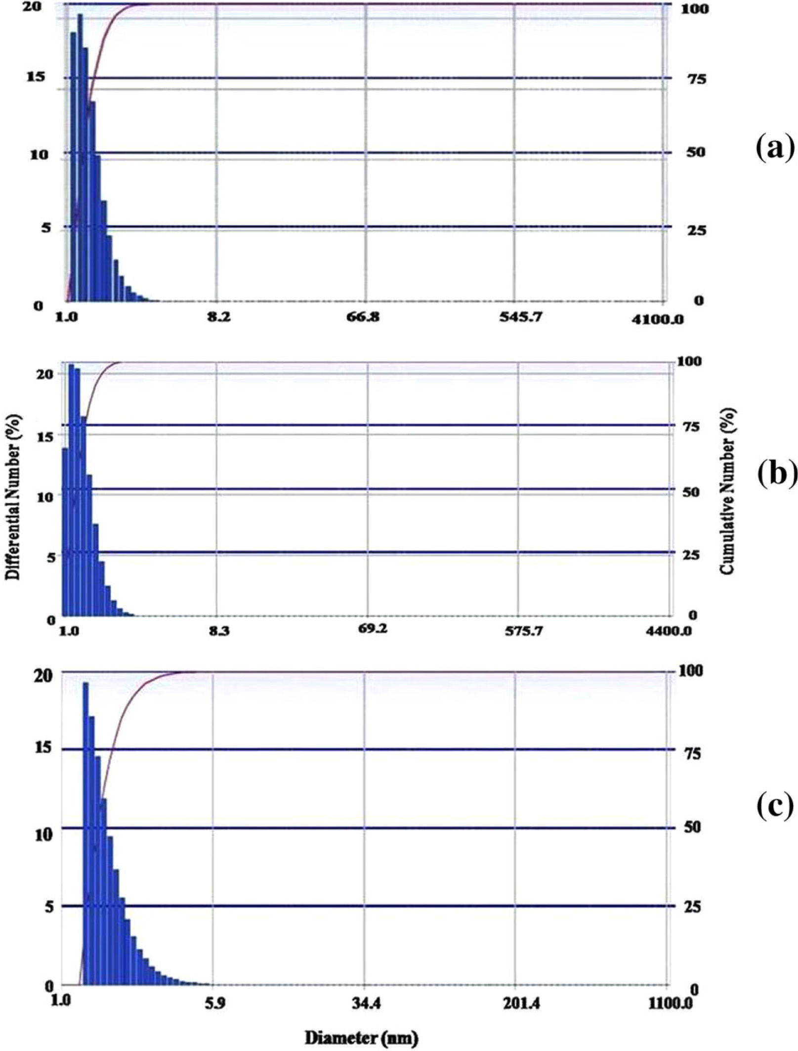
The size of bio-transformed Zn, Mg and Ti nanoparticles was initially measured by DLS technique which analyzes particle size distribution in the solution phase. The histograms for biosynthesized nanoparticles of Zn, Mg and Ti metal (Fig.
1
a, c) clearly exhibit that mean hydrodynamic diameter of size distribution for Zn nanoparticles was below 10 nm and Mg nanoparticles size was less than 7.8 nm, whereas, Ti nanoparticles showing size lower than 6 nm on the basis of number distribution in the solution. To measure the exact shape and size of synthesized nanoparticles, TEM examination was performed.
Histogram obtained from DLS technique showing number distribution of biologically synthesized
a
Zn,
b
Mg,
c
Ti nanoparticlesFig. 1
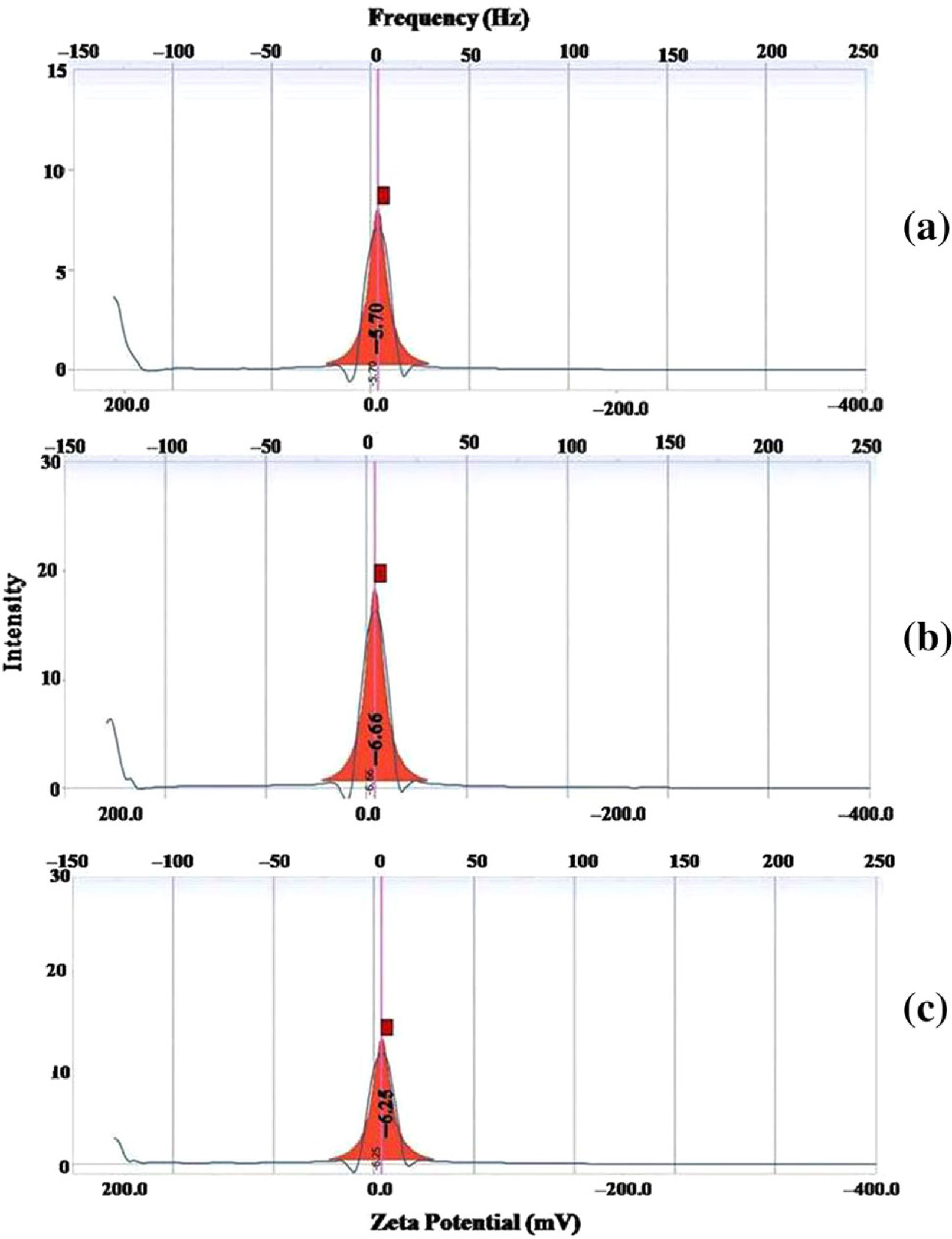
The surface charge of the nanoparticles plays a crucial role during interaction with molecule of other biological system such as plant, which should normally be in the range of −30 to +30 mV. The surface charge of biosynthesized Zn nanoparticle was measured as zeta potential of −5.70 mV, whereas, −6.66 and −6.25 mV of Mg and Ti, respectively (Fig.
2
a, c).
Zeta potential of biologically synthesized
a
Zn,
b
Mg,
c
Ti nanoparticlesFig. 2
The transmission electron microscopic (TEM) images of biosynthesized Zn, Mg and Ti nanoparticles were shown in Fig.
3
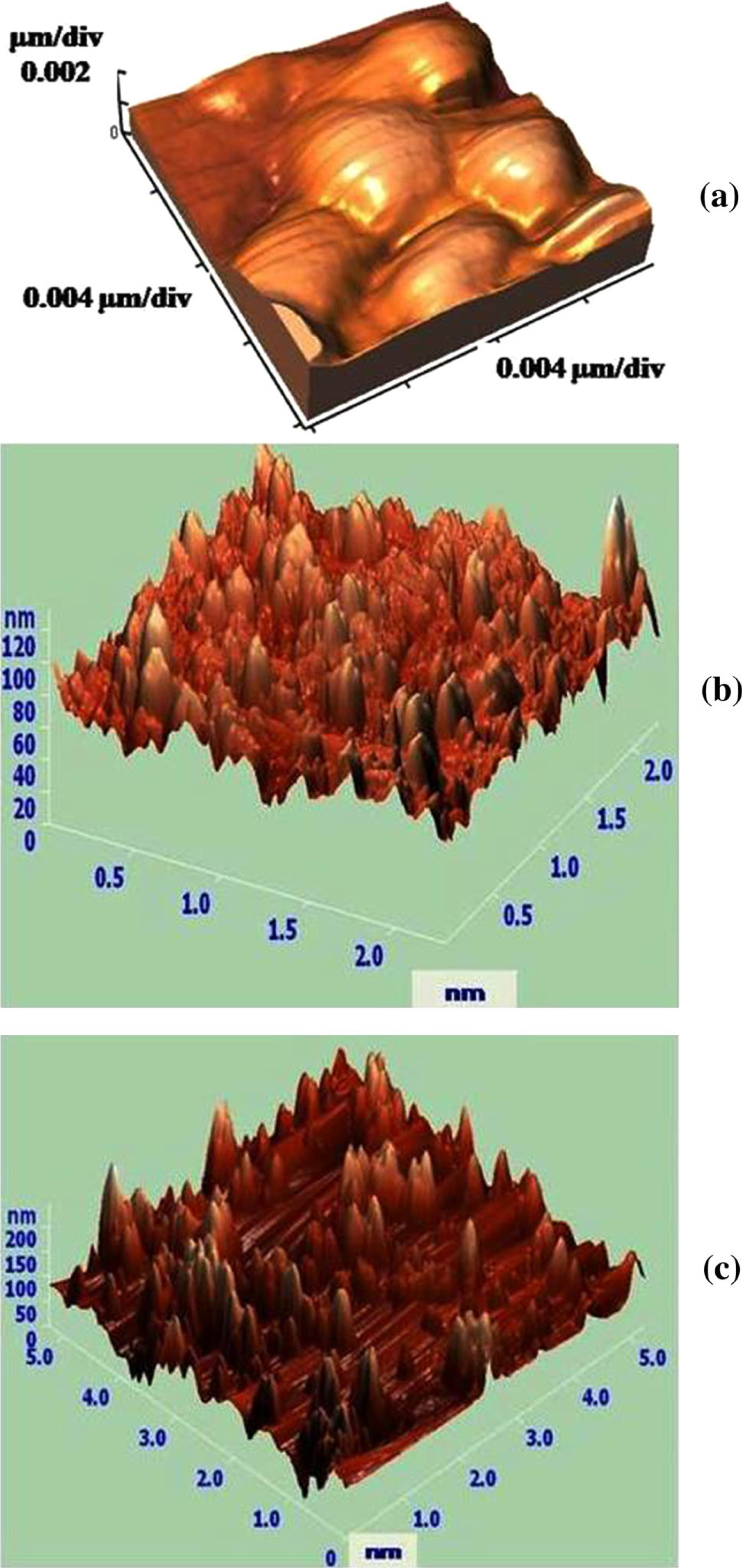
a, c. It can be clearly seen in the low magnification image that all the nanoparticles were present in monodisperse stage. To further validate the surface morphology drop-coated AFM, three dimensional images were taken in non-contact mode (Fig.
4
a, c). Result shows variability in morphological features of biosynthesized nanoparticles of Zn, Mg and Ti.
TEM micrographs of biologically synthesized nanoparticles at 50 nm scale bar
a
Zn,
b
Mg,
c
Ti AFM micrographs of biologically synthesized
a
Zn,
b
Mg,
c
Ti nanoparticlesFig. 3
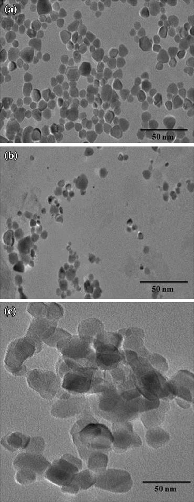
Fig. 4
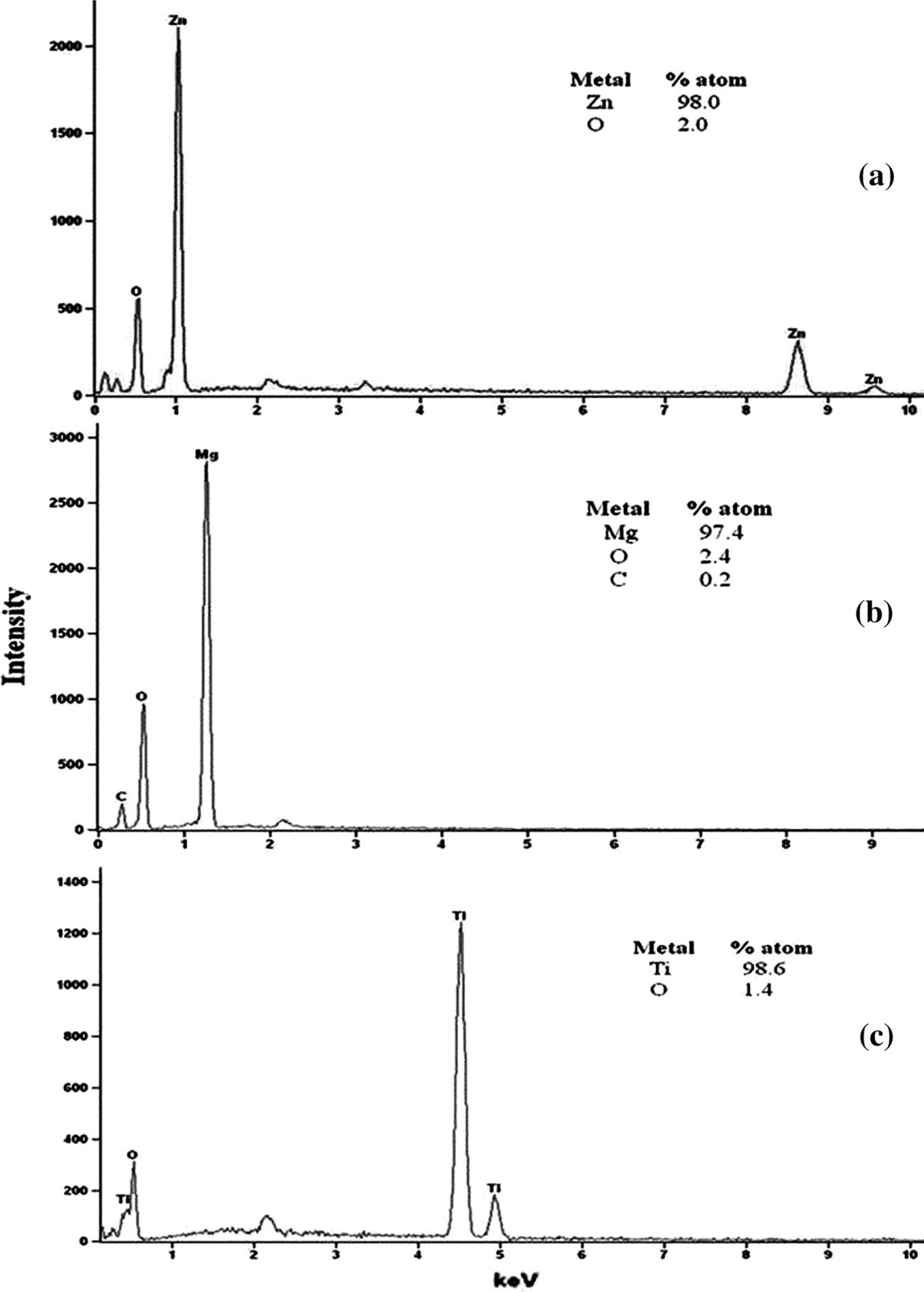
The elemental compositions of bio-transformed product containing Zn nanoparticles in the solution were confirmed by TEM equipped with energy dispersive X-ray spectroscopy (EDX). The results of TEM–EDX (Fig.
5
a, c) clearly show that the Zn nanoparticles were highly intense and the maximum intensity was found at 1 keV, whereas, Mg and Ti show maximum intensity at 1.3 and 4.6 keV, respectively. Results clearly exhibit the purity of biosynthesized metal nanoparticles.
EDX spectrums of biosynthesized
a
Zn,
b
Mg,
c
Ti nanoparticlesFig. 5
The present study approaches towards the eco-friendly biological synthesis of Zn, Mg and Ti nanoparticle using different precursor salt compounds by employing the cell-free enzymatic protein solution obtained by the secretion of fungal ball of mycelia. Fungal balls were developed for the recovery of higher protein contents used for nanoparticle biosynthesis. It is well reported that rhizosphere region exhibits more diverse niche for microbial population due to presence of organic contents. Production of metal nanoparticle from fungi has several advantages over bacteria, plant and other approaches i.e. physical, chemical and aerosol. However, development of simple and eco-friendly route would help in promoting further interest in the synthesis and application of Zn, Mg and Ti nanoparticles. Nature has provided us exciting possibilities for utilizing fungi as biological system for this purpose. In our previous report, Raliya and Tarafdar [ 19 ], we also obtained ZnO nanoparticle using the cell-free filtrate obtained from fungus of Aspergillus species which further supports the present research work.
To synthesize metal nanoparticles from fungal extracellular enzymes, precursor compound of each metal of interest was added. Selection of compounds was based on their ionic potential in water with fungal extracellular enzymes. Sulfates, chlorides, nitrates and oxides of the metal were selected for the synthesis of Zn, Mg and Ti nanoparticle on the basis of their ionic strength. It was found that the oxides of the metals have the best potential for reduction of metal into nanoparticles of that metal. A study was earlier reported by Kaul et al. [ 20 ] in which they show the synthesis of Mg and Fe nanoparticles in different chemical environments, supports the present investigation.
The majority of enzymes normally exhibit a strong dependence of activity on the pH of the medium [ 21 ], thus it is important to optimize the pH of the reaction medium in which nanoparticle synthesizes. In the biosynthesis of Zn, Mg and Ti nanoparticle concentration of hydrogen ion of the reaction medium plays an important role for nanoparticle size. Results showed in Table 7 clearly indicate that pH close to 5.5 is suitable for biosynthesis of Zn, Mg and Ti nanoparticles. It might be possible that pH specific catalytic activity of enzyme secreted from fungus and as the pH changes, catalytic efficiency of enzyme also altered. A similar study was reported by Kathiresan et al. [ 22 ] for the synthesis of silver nanoparticle using marine fungus Penicillium fellutanum , isolated from coastal mangrove sediment.
The biologically synthesized Zn, Mg and Ti nanoparticles present in the aqueous medium were quite stable, even up to 90 days for Zn and Ti nanoparticles whereas 105 days for Mg nanoparticles. This is an important aspect of biological nanoparticle synthesis, since the lack of sufficient stability of the nanoparticle preparation has to some extent impeded the development of the real-world application of biologically developed nanomaterial. A similar study was conducted for silver nanoparticle by Bhainsa and D’Souza [ 23 ]. The aggregation of Zn, Mg and Ti nanoparticle occurs after 90 and 105 days due to intermolecular interaction formed between synthesized nanoparticles and may be among the proteins, which encapsulate the nanoparticle.