We report the low temperature synthesis of CdE (E = S, Se and Te) semiconductor nanoparticles capped by anacardic acid, an environmentally benign naturally occurring capping ligand. The cadmium chalcogenide nanoparticles were synthesized by reacting the reduced chalcogen powder with cadmium chloride to form the corresponding bulk material which was then dispersed in tri-
n
-octylphosphine (TOP) and thermolysed in anacardic acid. The low temperature of the reaction and the use of a naturally occurring oil solvent make this a relatively green synthetic route. The optical properties of the anacardic-acid-capped cadmium chalcogenide particles show evidence of quantum confinement. The particles were characterized by X-ray diffraction and electron microscopy techniques.
Background
The ability to fabricate structures at the nanoscale level has advanced in the field of material science. Semiconducting quantum dots have received much attention because of their tunable properties [
1
–
5
]. There has been considerable progress in the synthetic protocols, techniques and sample analysis of these nanosized materials. Two broad approaches have emerged to fabricate materials at this level: a top-down approach which the size and dimensionality of solid matter has gradually been reduced, and a bottom-up approach in which quantum dots are viewed as enormously large molecules [
5
,
6
].
Cadmium chalcogenide (CdS, CdSe and CdTe) nanomaterials present enormous industrial interest in applications such as biomedical labeling reagents [
7
,
8
], photovoltaic and optoelectronic devices [
9
–
12
]. The unique optical properties of these nanomaterials are dictated by their structure, shape and size, and the possibilities for band gap tailoring and tunable luminescence [
2
,
13
,
14
]. However, despite a remarkable amount of research, morphological control in semiconductor nanoparticle synthesis is still quite difficult. For semiconductor nanoparticles, the size- and shape-dependent optical properties of these nanomaterials have been found to be directly reliant on the resulting morphology [
15
].
To date many fabrication methods have been developed to synthesize high quality cadmium chalcogenide nanoparticles [
14
,
16
]. This has provided a great opportunity of using these materials in ways that exploit their novel properties in their various potential applications. The high temperature thermolysis route is one of the pioneering routes to cadmium chalcogenide nanoparticles. In this method dimethyl cadmium was used as the cadmium source [
14
] and tri-
n
-octylphosphine chalcogenide (TOPE) as the chalcogenide source at high reaction temperatures. While much of the fundamental studies on these materials was on materials synthesized using this route, it was deemed not suitable for large scale production of nanomaterials due to the high reaction temperatures and the toxic and pyrophoric nature of the metal alkyl. To avoid these drawbacks, three alternative methods were developed. Firstly, design of low temperature processing methods [
17
,
18
], replacement of (CH
3
)
2
Cd as Cd-metal source by CdO [
16
,
19
] and other Cd metal salts [
20
,
21
] and lastly, the use of safe, cheaper, natural and more air stable coordinating solvents [
17
,
22
,
23
] such as olive oil have produced stable semiconductor nanoparticles.
Recently we reported the synthesis of lead chalcogenide nanoparticles capped by anacardic acid, a naturally occurring oil which is extracted from cashew nut shell liquid (CNSL) [
24
]. We have used the same synthetic procedure to synthesize cadmium chalcogenides (CdE) capped by anacardic acid. The potential of the work lies in using the greener solvent in the synthesis of cadmium chalcogenide nanoparticles at a relatively low temperature.
Methods
All chemicals used were of analytical grade. Selenium powder, tellurium powder, sulfur powder, sodium borohydride (NaBH
4
), cadmium chloride, deionized water, methanol, toluene and tri-
n
-octylphosphine (TOP) were purchased from Sigma–Aldrich. The anacardic acid was isolated from solvent extracted cashew nut shell liquid (CNSL), using the method described by Paramashivappa et al. [
25
] with slight modifications reported by Lucio et al. [
26
].
The chalcogen powder was reduced to the corresponding ion by adding 0.32 mmol of chalcogen powder to 20.0 mL of deionized water in a three neck flask at room temperature. A 0.79 mmol solution of sodium borohydride was added to the flask contents and the flask was purged with nitrogen flow to create an inert atmosphere. The reduction reaction was carried out for 2 h with continuous stirring and with the flow of N
2
gas. A 0.32 mmol solution of the cadmium salt was added to the reduced ion solution and stirred for 5 min, followed by the addition of excess methanol to form the bulk CdE (E = S, Se and Te) which was isolated by centrifugation and dispersed in 6.0 mL of TOP. The resultant CdE-TOP mixture was then injected into 6.0 g of anacardic acid, pre-heated to 140 °C. The reaction temperature was kept for 2 h. The reaction contents were cooled to room temperature, followed by the addition of excess methanol to flocculate the nanoparticles. The anacardic-acid-capped CdE nanoparticles were isolated after centrifugation. The resultant nanoparticles were dissolved in toluene solvent for further characterization.
A Varian Cary 50 Conc UV–Visible spectrophotometer and Perkin-Elmer LS 55 Luminescence spectrometer were used to measure the absorption and photoluminescence properties of the particles, respectively. The crystalline phase was identified by X-ray diffraction (XRD), at a scanning rate of 0.05 min
−1
in a 2θ range from 20° to 80°, using a Bruker AXS D8 diffractometer equipped with nickel filtered Co Kα radiation (
λ
= 1.5418 Å) at 40 kV. A JEOL 1010 TEM with an accelerating voltage of 100 kV, Megaview III camera, and Soft Imaging Systems iTEM software was used to determine the morphology. HRTEM was done with a JEOL 2010 transmission electron microscope operated at an accelerating voltage of 200 kV.
Results and discussion
Optical properties by UV–Vis and photoluminescence analysis
Quantum size effects have been observed experimentally for many nanocrystalline semiconductor nanoparticles [
27
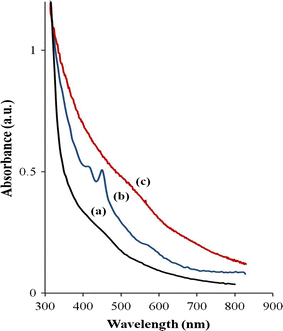
]. The absorption spectrum of a semiconductor nanoparticle provides an accessible and straight forward method for the evaluation of quantum size effects. Figure
1
shows the room temperature UV–Vis absorption spectra of the anacardic-acid-capped CdS, CdSe and CdTe nanoparticles thermolysed at 140 °C. The absorption band edge for the anacardic-acid-capped CdS is observed at 505 nm (2.46 eV), without any distinct excitonic features. The CdSe spectrum shows three distinct resolved electronic transitions, which could be assigned to the excitonic absorption features of nanosized CdSe [
28
] as predicted theoretically [
29
]. These transitions between any two electronic levels are seen as discrete peaks in the optical spectra, evidence of a quantum confined system [
30
]. Previous studies have also showed the same trend for CdSe nanoparticles thermolysed via the organometallic route [
31
]. There is an excitonic shoulder at 547 nm, a distinct excitonic peak at 450 nm and a band edge at 620 nm (2.0 eV). The anacardic-acid-capped CdTe shows a band edge at 720 nm (1.72 eV). All three samples show blue shifts in the absorption band edges in relation to their bulk band gaps implying the quantum confinement effect of these particles.
Fig. 1
UV–Visible absorption spectra of anacardic acid capped
a
CdS,
b
CdSe and
c
CdTe nanoparticles synthesized at 140 °C
Figure
2
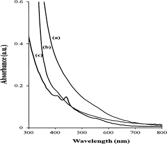
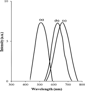
shows the combined absorption spectra of the 30, 60 and 120 min samples of the anacardic-acid-capped CdSe nanoparticles. There is no pronounced shift in the band gap as the reaction time increases from 30 to 120 min other than evolution of pronounced excitonic features observed for the sample collected after 120 min. This result revealed that the size of CdSe nanocrystals gradually increased as reaction time prolonged, probably because of the low temperature of reaction. The photoluminescence spectra of anacardic-acid-capped CdS, CdSe and CdTe nanoparticles synthesized at 140 °C is shown in Fig.
3
. For CdSe, the emission peak (637 nm) is red shifted to that of absorption maximum (620 nm) which can be attributed to a combination of relaxation into shallow trap states and the size distribution [
32
]. All three PL spectra justify highly monodispersed samples and narrow emission line widths indicating growth of nanoparticles with few electronic defect sites as a result of the efficiency of anacardic acid to electronically passivate the nanoparticles.
Fig. 2
UV–Visible absorption spectra of anacardic-acid-capped CdSe nanoparticles synthesized at 140 °C at different time intervals of
a
30 min,
b
1 h and
c
2 h
Fig. 3
Photoluminescence spectra of anacardic acid capped
a
CdS,
b
CdSe and
c
CdTe nanoparticles synthesized at 140 °C
TEM and HRTEM analysis
The morphology of the cadmium chalcogenide nanoparticles was studied by transmission electron microscopy (TEM) and high resolution TEM. Figure
4
a shows the TEM image of the anacardic-acid-capped CdS nanoparticles synthesized at 140 °C. The particles are anisotropic ranging from oblate to elongated shaped particles with an average size of 14.02 ± 2.4 nm. The CdSe nanoparticles and CdTe nanoparticles show similar morphology to that of CdS. The CdSe particles are slightly more elongated than the CdS particles, with an average length of 28.26 ± 3.8 nm and width of 8.3 ± 1.3 nm (Fig.
4
b). The CdTe particles (Fig.
4
c) were more oblate in shape with an average size of 14.90 ± 2.23 nm. The HRTEM image (Fig.
4
d) of a single particle shows distinct lattice fringes with the interplanar distance of 3.83 Å, corresponding to the (111) plane of cubic CdTe. The assembly of all the anacardic-acid-capped CdS, CdSe and CdTe nanoparticles is in a well ordered, mosaic-type arrangement of particles (Fig.
4
). There is almost an equal inter-particle distance of approximately 3 nm for the CdS, CdSe and CdTe particles as observed from the TEM images.
Fig. 4
TEM images of anacardic acid capped
a
CdS,
b
CdSe and
c
CdTe, and
d
HRTEM image of CdTe nanoparticles synthesized at 140 °C
XRD analysis
Figure
5
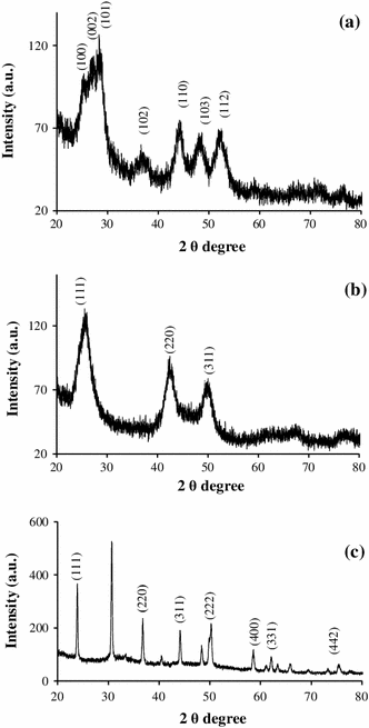
a shows the powder X-ray diffraction pattern of the anacardic-acid-capped CdS nanoparticles. The XRD pattern shows the presence of (100), (002), (101), (102), (110) and (103) reflection planes of the hexagonal phase of CdS. The average crystallite size was calculated to be 14.37 nm, using the Scherrer formula [
33
,
34
] corroborating the size determined by TEM.
Fig. 5
Powder X-ray diffraction spectra of anacardic acid capped
a
CdS,
b
CdSe and
c
CdTe nanoparticles
The X-ray diffraction peaks (Fig.
5
b) of the as-synthesized anacardic-acid-capped CdSe nanoparticles show three distinct peaks at 2θ values of 25.48°, 42.34° and 50.10° corresponding to the (111), (220) and (311) planes of cubic CdSe, respectively. The high intensity of (111) peak indicates that the elongated particles have a large number of (111) planes thus making that peak the dominant reflection in the first diffraction feature [
35
]. The anacardic-acid-capped CdTe (Fig.
5
c) also has a dominant cubic structure, which is also the dominant crystal phase of bulk CdTe. The reflections are indexed to the (111), (220), and (311) planes of the cubic CdTe [
36
]. The presence of the (222), (400) and (311) planes further supports the existence of cubic structure of CdTe.
Conclusions
Cadmium chalcogenide (CdE) nanoparticles have been successfully synthesized using anacardic acid as a coordinating solvent. The UV–Vis and PL studies confirmed that the CdE nanoparticles undergo quantum confinement effects. The TEM studies reveal a mosaic-like pattern for all three samples. The results reveal that anacardic acid could be an effective and potential capping agent in the synthesis of metal chalcogenide nanoparticles. The method used an inexpensive and non-toxic naturally occurring acid as a capping agent which proved to be a good choice for high quality, monodispersed nanoparticles.
Conflict of interest
The authors declare that they have no competing interests.
Author’s contributions
SM carried out the experiments and drafted the manuscript. VSRP carried out XRD, TEM and HRTEM analysis. EBM, FNN and NR developed the conceptual framework and supervised the whole work. All authors read and approved the final manuscript.
Bruchez et al. (1998) Semiconductor nanocrystals as fluorescent biological labels (pp. 2013-2016) 10.1126/science.281.5385.2013
Chan and Nie (1998) Quantum dot bioconjugates for ultrasensitive nonisotopic detection (pp. 2016-2018) 10.1126/science.281.5385.2016
Wang et al. (2005) Biological assembly of nanocircuit prototypes from protein-modified CdTe nanowires (pp. 243-248) 10.1021/nl0482682
Chen et al. (2009) An oleic acid-capped CdSe quantum-dot sensitized solar cell10.1063/1.3117221
Gao et al. (2000) Electroluminescence of different colors from polycation/CdTe nanocrystal self-assembled films (pp. 2297-2302) 10.1063/1.372177
Greenham et al. (1996) Charge separation and transport in conjugated-polymer/semiconductor-nanocrystal composites studied by photoluminescence quenching and photoconductivity (pp. 17628-17637) 10.1103/PhysRevB.54.17628
Brus (1984) Electron-electron and electron-hole interactions in small semiconductor crystallites: the size dependence of the lowest excited electronic state (pp. 4403-4409) 10.1063/1.447218
Murray et al. (1993) Synthesis and characterization of nearly monodisperse CdE (E = S, Se, Te) semiconductor nanocrystallites (pp. 8706-8715) 10.1021/ja00072a025
Manna et al. (2000) Synthesis of soluble and processable rod-, arrow-, teardrop-, and tetrapod-shaped CdSe nanocrystals (pp. 12700-12706) 10.1021/ja003055+
Peng and Peng (2001) Formation of high-quality CdTe, CdSe, and CdS nanocrystals using CdO as precursor (pp. 183-184) 10.1021/ja003633m
He and Gu (2006) Synthesis and characterization of monodisperse CdSe nanocrystals at lower temperature. Colloids and surfaces (pp. 111-116) 10.1016/j.colsurfa.2005.07.017
Dunpall et al. (2012) An in vitro assessment of the interaction of cadmium selenide quantum dots with DNA, iron, and blood platelets (pp. 995-1002) 10.1002/iub.1100
Boatman et al. (2005) A safer, easier, faster synthesis for CdSe quantum dot nanocrystals (pp. 1697-1699) 10.1021/ed082p1697
Maseko et al. (2010) The influence of the cadmium source on the shape of CdSe nanoparticles (pp. 1037-1040) 10.1016/j.matlet.2010.02.002
Sapra et al. (2006) Phosphine-free synthesis of monodisperse CdSe nanocrystals in olive oil (pp. 3391-3395) 10.1039/b607022a
Patel et al. (2011) Simple non-aqueous fabrication route for oleic acid capped luminescent cadmium sulphide quantum dots at relatively low temperature (pp. 61-65) 10.4236/snl.2011.13011
Mlowe et al. (2013) Lead chalcogenides stabilized by anacardic acid (pp. 263-268) 10.1016/j.mssp.2012.10.017
Paramashivappa et al. (2001) Novel method for isolation of major phenolic constituents from cashew (Anacardium occidentale L.) Nut Shell Liquid (pp. 2548-2551) 10.1021/jf001222j
Lucio et al. (2010) Synthesis and cytotoxicity screening of substituted isobenzofuranones designed from Anacardic acids (pp. 3480-3489) 10.1016/j.ejmech.2010.05.015
Bawendi et al. (1990) The quantum mechanics of larger semiconductor clusters (“quantum dots”) (pp. 477-496) 10.1146/annurev.pc.41.100190.002401
Ithuria and Dubertret (2008) Quasi 2D colloidal CdSe platelets with thicknesses controlled at the atomic level (pp. 16504-16505) 10.1021/ja807724e
Efros et al. (1996) Band-edge exciton in quantum dots of semiconductors with a degenerate valence band: dark and bright exciton states (pp. 4843-4856) 10.1103/PhysRevB.54.4843
Yoffe (2001) Semiconductor quantum dots and related systems: electronic, optical, luminescence and related properties of low dimensional systems (pp. 1-208) 10.1080/00018730010006608
Norris et al. (1996) Size dependence of exciton fine structure in CdSe quantum dots (pp. 16347-16354) 10.1103/PhysRevB.53.16347
Bawendi et al. (1992) Luminescence properties of CdSe quantum crystallites: resonance between interior and surface localized states (pp. 946-956) 10.1063/1.462114
Bandaranayake et al. (1995) Structural phase behavior in II-VI semiconductor nanoparticles (pp. 831-833) 10.1063/1.115458
Patterson (1939) The Scherrer formula for X-Ray particle size determination (pp. 978-982) 10.1103/PhysRev.56.978
Rogach et al. (1999) Synthesis and characterization of a size series of extremely small thiol-stabilized CdSe nanocrystals (pp. 3065-3069) 10.1021/jp984833b
Blokland et al. (2011) Exciton lifetime of CdTe nanocrystal quantum dots in high magnetic fields10.1103/PhysRevB.83.035304