Results and discussion
FT-IR spectrum resulting from CNTs
To study the formation of functional groups on the surface of CNTs, an FT-IR spectrum was prepared. Figure
1
illustrates the FT-IR spectrum resulting from CNTs before and after the process of acid treatment. Existing peaks in FT-IR spectrum show the formation of functional groups of O–C=O, C–O–O, C–O and C=O, respectively, at wavelengths of 1,210, 1,415, 1,475 and 1,720 (cm
−1
) [
26
,
27
]. These functional groups play an important role in primary nucleation and formation of MnFe
2
O
4
nanoparticles on the surface of carbon nanotubes. MnFe
2
O
4
/CNT nanocomposite powder is formed through strong chemical bonds between ions of iron atoms and functional groups located on the surface of carbon nanotubes. Due to the high level of carbon nanotubes and large-scale formation of such bonds, they can be applied as an appropriate support for nucleation and better distribution of MnFe
2
O
4
nanoparticles on their surface and formation of MnFe
2
O
4
/CNTs nanocomposite powder.
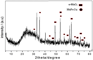
FT-IR spectrum resulting from CNTs before and after the process of acid treatment XRD spectrum for a synthesized sample of MnFe
2
O
4
nanoparticlesFig. 1
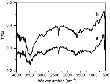
Fig. 2
Analysis of X-ray diffraction spectra
Figure 1 presents the spectra resulting from X-ray diffraction of the prepared samples. Existing peaks in (XRD) spectra within the range of 2θ = 5–80° suggest the formation of a two-phase structure from MnFe 2 O 4 and α-MnO 2 nanoparticles. The peaks in 2θ = 28.97, 34.06, 43.6, 53.59, 57.21, 64.5 and 75.2° (shown by circles) are related to MnFe 2 O 4 nanoparticles, which are related to the reflection of crystal plates of (220), (311), (400), (422), (511), (440) and (553), respectively [ 13 , 15 , 23 ]. On the other hand, existing peaks located at 2θ = 47.5, 50.59, 53.59, 60.39, 66.5, 68, 70.52 and 73.79° (shown by squares) are associated with α-MnO 2 crystal structure which are related to reflection of crystal plates of (510), (411), (440), (531), (002), (202), (541) and (312), respectively [ 24 ]. Moreover, the peaks being sharp represent the crystalline order and high crystalline structure of MnFe 2 O 4 nanoparticles. There were no additional peaks, due to the existing impurity in the prepared powder.
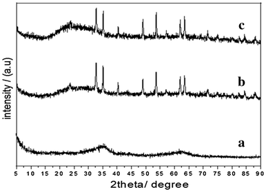
An XRD spectrum was prepared to study the effect of calcination temperature on MnFe
2
O
4
and MnFe
2
O
4
/CNT synthesized samples. Figure
3
presents the spectra resulting from XRD of MnFe
2
O
4
calcined samples at temperatures of 400, 600 and 800 °C. The average sizes of nanoparticles were determined according to the maximum peak of the XRD spectrum using the Scherrer equation [
27
]:
XRD spectrum for MnFe
2
O
4
nanoparticles calcined at:
a
400 °C,
b
600 °C and
c
800 °CFig. 3
Figure
4
shows the XRD pattern of MnFe
2
O
4
/CNT nanocomposite powder with a ratio of 1:1 calcined at temperatures of 400, 600 and 800 °C. It was observed that with increase in the calcination temperature, the peaks get sharper and higher. Using Scherrer equation, the average nanoparticle sizes at calcination temperatures of 400, 600 and 800 °C were 11.5, 34.5 and 46 nm, respectively.
XRD pattern of MnFe
2
O
4
/CNT nanocomposite calcined at temperatures of:
a
400 °C,
b
600 °C and
c
800 °CFig. 4
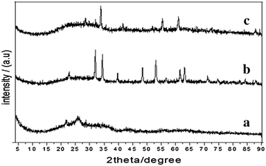
After comparing the two XRD spectra in Figs. 3 and 4 , for calcined samples at temperature of 400 °C and regarding the result of Scherrer equation, it is observed that the presence of carbon nanotubes’ support in the synthesis process increases the mean size of MnFe 2 O 4 nanoparticles. The increase in the size of nanoparticles may be due to existing CNTs in solution and the decreased required space for nucleation of MnFe 2 O 4 nanoparticles. By increasing the calcination temperature, CNTs prevent the formation of larger crystallite nanoparticles compared to the state of pure MnFe 2 O 4 (Fig. 3 b, c).
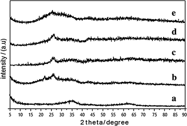
To study the effect of weight ratio of MnFe
2
O
4
to CNTs, the composite samples were prepared at a calcination temperature of 400 °C with ratio of 1:1, 1:4 and 1:8 of MnFe
2
O
4
to CNTs. The XRD spectrum resulting from the obtained composite powder samples is presented in Fig.
5
. According to the XRD spectra, it can be seen that in addition to the associated peaks of MnFe
2
O
4
nanoparticles, there is another peak in 2θ = 26.28° estimated to be the (0 0 2) plane of CNTs [
26
,
27
]. On comparing the XRD spectra in Fig. (
5
b, c), it is observed that by increasing the concentration of CNTs in nanocomposite, the intensity of the related peaks to MnFe
2
O
4
will reduce and their widths may increase. Therefore, the size of reduced nanoparticles and their values for ratios of 1:1, 1:2, 1:4 and 1:8 were, respectively, determined to be 11.5, 10.6, 8.9 and 2.3 nm. The decreased size of nanoparticles is due to reduced ionic concentration in solution, increased surface of CNTs and also increased required places for nanoparticles’ nucleation.
XRD pattern of MnFe
2
O
4
/CNT nanocomposite with ratios of:
a
1:0,
b
1:1,
c
1:2,
d
1:4 and
e
1:8Fig. 5
Figure
6
shows the SEM images of calcinated MnFe
2
O
4
nanoparticles at temperatures of 400, 600 and 800 °C. As shown in Fig.
6
a for calcinated MnFe
2
O
4
nanoparticles at 400 °C, nanocrystallites of MnFe
2
O
4
with average size of 3 nm are aggregated together to form larger crystallite with a relative size of 77 nm. On increasing the calcination temperature between 600 and 800 °C (Fig.
6
b, c), the aggregated particles are increased with an average value of 100 and 141 nm, respectively. The increased size of the particles is due to smaller crystallites sticking together. On the other hand, the geometric shape of crystallites changed from spherical to oval, due to sticking together of two spherical crystallites to form a longer structure.
SEM images of MnFe
2
O
4
nanoparticles calcinated at:
a
400 °C,
b
600 °C and
c
800 °C (In all images magnification is ×60k)Fig. 6
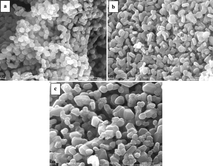
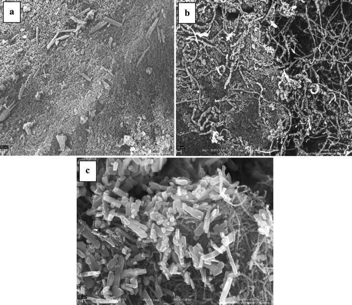
To study the effect of carbon nanotubes as a support at different calcination temperatures on the morphology of synthesized MnFe
2
O
4
/CNT nanocomposite powders, SEM images were prepared. Figure
7
shows the SEM images from calcinated MnFe
2
O
4
/CNTs nanocomposite samples (with 1:1 ratio) at temperatures of 400, 600 and 800 °C. At 400 and 600 °C (Fig.
7
a, b), crystallites have stuck together and formed irregular particles among the nanotubes. With increase in the calcination temperature to 800 °C (Fig.
7
c), it is observe that in addition to the formation of ordered strings from MnFe
2
O
4
/CNTs nanocomposite powder, some crystallites together form cubic-shaped structures. Therefore, increase in the calcination temperature causes the smaller aggregated nanoparticles to permeate into each other and form bigger particles [
28
].
SEM images of MnFe
2
O
4
/CNT nanocomposite calcinated at:
a
400 °C,
b
600 °C and
c
800 °C (In all images magnification is ×60k)Fig. 7